Bio Lab II Respiratory/Cardiovascular Body Systems
1/48
There's no tags or description
Looks like no tags are added yet.
Name | Mastery | Learn | Test | Matching | Spaced | Call with Kai |
|---|
No analytics yet
Send a link to your students to track their progress
49 Terms
Closed circulatory system
network of blood vessels (arteries, veins, capillaries) where blood is confined, enabling high-pressure, efficient transport of oxygen and nutrients to tissues
Main function of the Cardiovascular system
to deliver oxygen and nutrients and to remove carbon dioxide and other waste products
Blood Vessel Layers
Tunic intima (inner lining)
Tunic media (middle muscle layer)
Tunic externa (outer supportive layer)
Tunica Intima
provides a smooth, frictionless surface for blood flow and acts as a barrier that prevents clotting and regulates vessel diameter; endothelium
Tunic Media
thickest layer, containing smooth muscle cells and elastic fibers; controlled by sympathetic nervous system
Tunic Externa
outermost layer composed of connective tissue, primarily collagen fibers, which anchors the vessels to surrounding tissues and provides structural support
Arteries
muscular, elastic blood vessels that transport blood away from the heart, typically carrying oxygen-rich blood to tissues and organs
Arterioles
small-diameter blood vessels in the microcirculation that branch off from arteries and lead into capillaries
Capillaries
the smallest and most numerous blood vessels in the human body, acting as the crucial link between the arterial and venous systems
Venules
small, thin-walled blood vessels that collect deoxygenated blood from capillaries and transport it toward larger veins
Veins
blood vessels in the cardiovascular system that carry blood toward the heart
Lumens
the inner, open space or cavity within a tubular structure, organ, or cell organelle
Lumens of arteries vs veins
Arteries have a narrow, small lumen designed to maintain high blood pressure, while veins have a large, wide lumen to accommodate high-volume, low-pressure blood flow
arteries < veins
Arterial blood
the bright red, highly oxygenated blood transported by arteries from the heart to tissues, representing the oxygenated side of the circulatory system
Blood
specialized circulating connective tissue composed of cells suspended in plasma, responsible for transporting oxygen, nutrients, and waste throughout the body
Heart
fist-sized, muscular organ that serves as the primary pump in the cardiovascular system, continuously circulating blood throughout the body to deliver oxygen and nutrients while removing waste
Myocardium
the thick, middle muscular layer of the heart wall, responsible for the involuntary contraction and relaxation that pumps blood throughout the body
Heart Anatomy
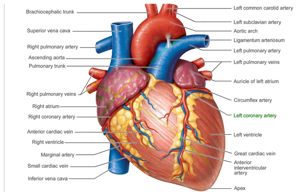
Coronary arteries
the blood vessels that branch from the aorta to supply oxygen-rich blood and nutrients specifically to the heart muscle (myocardium)
Cardiac veins
specialized blood vessels that remove deoxygenated blood and metabolic waste from the myocardium (heart muscle), returning it to the right atrium
Coronary sinus
the largest vein of the heart, located on its posterior aspect between the left atrium and ventricle
The 4 heart chambers
Right Atrium — receives deoxygenated blood from the body
Right Ventricle — pumps deoxygenated blood to the lungs for oxygen
Left Atrium — receives oxygen-rich blood back from the lungs
Left Ventricle — pumps oxygen-rich blood to the rest of the body
Pathway of Blood
Venae cavae
Right atrium
Tricuspid valve
Right ventricle
Pulmonary semilunar valve
Pulmonary artery/Lungs
Pulmonary veins
Left atrium
Bicuspid valve
Left ventricle
Aortic valve
Aorta Body
Intrinsic conduction system
network of specialized, non-contractile cardiac cells that initiate and distribute electrical impulses, setting the heart's rhythm and coordinating contraction
Sinoatrial (SA) node
the heart's primary pacemaker, a specialized bundle of myocytes located in the right atrium near the superior vena cava
Atrioventricular (AV) node
a critical component of the heart's electrical conduction system, located in the right atrium near the interatrial septum
Atrioventricular bundle
a specialized group of cardiac muscle fibers responsible for conducting electrical impulses from the atrioventricular (AV) node to the ventricles
Bundle Branches
specialized cardiac muscle fibers that conduct electrical impulses from the bundle of His to the Purkinje fibers, causing the ventricles to contract and pump blood
Purkinje fibers
specialized, fast-conducting cardiac muscle cells located in the inner ventricular walls (subendocardium) of the heart
Systole (contraction/top number)
the heart muscle contracts, forcing blood out of the ventricles into the aorta and pulmonary artery
Diastole (relaxation/bottom number)
the heart muscle relaxes, allowing the ventricles to fill with blood from the atria
Pulse
the rhythmic, palpable expansion and contraction of an artery caused by the ejection of blood from the heart's left ventricle during each cardiac cycle
Brachial artery
the primary blood vessel supplying oxygen-rich blood to the upper arm
measurements by health professionals are made on the pressure in large arteries like this one
Sphygmomanometer
medical device used to measure blood pressure by temporarily restricting blood flow, composed of an inflatable cuff, inflation bulb, and manometer; measures systolic and diastolic pressure
Renin
hormonal control
Factors that effect blood pressure
Temperature
Chemicals
Diet
Variations in Blood Pressure
normal
hypotension
hypertension
Normal blood pressure
140—110mm Hg systolic
80—75mm Hg diastolic
Hypotension
Low systolic (below 110mm Hg)
Often associated with illness
Hypertension
High systolic (above 140mm Hg)
Can be dangerous if it’s chronic
Lungs
the primary, paired sponge-like respiratory organs in air-breathing vertebrates, situated within the thoracic cavity
Alveoli
microscopic, balloon-like air sacs located at the end of the bronchioles in the lungs
Inspiration
the active physiological process of drawing air into the lungs, driven by the contraction of the diaphragm and external intercostal muscles
Expiration
the passive process of breathing out, where air containing carbon dioxide is expelled from the lungs
Spirometer
scientific instrument used in biology and medicine to measure the volume and speed of air inspired and expired by the lungs
Tidal volume (TV)
the normal amount of air that moves into and out of lungs with normal breath
Inspiratory reserve volume (IRV)
the amount of air that can be forcibly inhaled after a normal breath
Expiratory reserve volume (ERV)
the amount of air that can be forcibly exhaled after a normal breath
Vital capacity (VC)
the volume of air that can be forcibly exhaled after forcibly inhaling