Dermatology
1/132
There's no tags or description
Looks like no tags are added yet.
Name | Mastery | Learn | Test | Matching | Spaced |
|---|
No study sessions yet.
133 Terms
impetigo - managment
Advice and information
Usually heals completely and without scarring, serious complications rare
Hygiene measures (soap and water, topical 1% hydrogen peroxide cream, wash with antimicrobial emollient eg Dermol, avoid scratching, wash hands after touching or applying cream, avoid sharing towels, flannels etc.)
Stay away from school/work until lesions dry/scabbed, or for 48hrs after Rx started
Follow-up if no improvement after 7 days
Topical Rx (localised infection)
Fusidic acid tds-qds 5-7/7
Oral Rx (extensive or at risk of complications, bullous impetigo)
Flucloxacillin qds 5-7/7
Erythromycin or clarithromycin if penicillin allergy
Tinea corporis (ringworm)
common where?
common organisms
Infection on trunk legs or arms
Trichophyton rubrum
Microsporum canis (acute red patches with pustules)
Tinea corporis (ringworm)
presentations
Single or multiple lesions
Grow slowly
Erythematous, scaly with annular border
Can be central clearing
White lines in lesion when organism very inflammatory
Can be itchy
Tinea corporis (ringworm)
treatment
Treat associated tinea pedis (feet)
Topical antifungal cream
Terbinafine - ≥ 12 years
Miconazole - ≥ 2 years
Oral antifungal (extensive or pustular)
Terbinafine - ≥ 2 years
Tinea cruris
where
common organism
Groin, upper thigh and buttocks
Commonly bilateral
Trichophyton rubrum
Epidermophyton floccosum
Tinea cruris
presentation
Red scaly expanding patch
Diagnosis and treatment as for tinea corporis
can itch
Tinea cruris
treatment
Treat associated tinea pedis
Dry groin separate towel
Avoid occlusive or synthetic clothing
Lose weight
Add hydrocortisone for itching if needed
Tinea pedis
presentations
organism
Dry scaling lesion on the sole, heel and sides of foot
Blisters and pustules on sides of feet or instep
Moist peeling itchy skin in interdigital space (athlete’s foot)
Red scaly patches on the dorsum of foot
Trichophyton rubrum
Trichophyton interdigitale
Tinea pedis
treatment
Topical antifungal (terbinafine 2 weeks; miconazole 2-4 weeks now a single dose terbinafine film forming solution)
Systemic treatment if nail involved
Keep feet well ventilated
Prophylactic topical treatment once or twice a week
Tinea capitis
where
organism
Scalp
common in children
common in boys
Trichophyton tonsurans
Microsporum canis
Tinea capitis
presentation
Single or multiple patches of hair loss
Scaling erythema and pustules are variable
Scaling can occur without hair loss
Can be itchy
Can see black dot pattern
Can form a kerion: inflamed boggy mass – refer
Can Fluoresce under Wood’s light
Tinea capitis
treatment
Risk of scarring alopecia – systemic agents used
Oral antifungal
Terbinafine
Itraconazole (second line)
Griseofulvin (second line children)
At least twice weekly topical ketoconazole for first 2 weeks reduces transmission
Treat family members with shampoo too
Kerions use oral antifungal, can be for 12-16 weeks
Onychomycosis
causes
Dermatophytes – tinea unguium
Trichophyton rubrum
Trichophyton interdigitale
Yeasts
Moulds
Onychomycosis
clincial finding
Toenails more than fingers
Single or multiple nails affected
Tinea unguium associated with interdigital infection, tinea pedis or tinea manuum
Can be white or yellow opaque line on side of nail
Subungal hyperkeratosis: scaly thickening
Distal onycholysis: nail lifts and end crumbles
Superficial involvement: white powdery surface
Onychomycosis
treatment
Topical treatment
Low cure rate
Distal nail infection up to 50% nail
≤ 2 nails involved
Superficial infection
Amorolfine 5%: 6 months fingers 9-12months toes
Tioconazole solution twice daily for 6-12 months
Oral antifungal
Terbinafine 250mg once a day 6 weeks for fingernail 12-16 weeks for toenails
Treat associated tinea pedis
Keep nails short
Replace old footware - spores
Once or twice weekly topical antifungal to feet if recurrent
Pityriaisis versicolor
caused by what
where
Caused by the yeast-like fungus Malassezia furfur (Pityrosporum ovale )
Usually found on back and chest, can spread onto upper arms, neck and abdomen
asymptomatic sometimes itchy
Appears slowly, may take months to be noticed – not contagious
Pityriaisis versicolor
presenations
Oval lesions
Spread and become confluent
Fawn colour or pink in light skin
Pale in dark skin
Fine surface scale
Asymptomatic sometimes itchy
Fluoresce under Wood’s light
Pityriaisis versicolor
treatment
Small area
Ketoconazole shampoo lathered daily on skin and left for 5 minutes for 5 days
Do not use topical or oral steroids – can exacerbate the condition
Treatment failure or widespread
Itraconazole orally 200mg daily for 7 days also if widespread
Can take several months for skin to gain original colour sometimes a lot longer
Recurrent episodes
Continue ketoconazole shampoo once every 2-4 weeks for 6 months
Oral Candidiaisis
presentations
White patches that peel leaving raw skin
Smooth red patches on tongue with sore mouth
Oral candidiasis
treatment
Miconazole oral gel or nystatin suspension
Angular cheilitis
diagnosis/ Ix
Check FBC, ferritin and vitamin B12
Swab if think secondary bacterial infection
Angular cheilitis
presentation
Treatment
Painful cracking and fissures
Thick emollient for dribbling
Miconazole oral gel
With steroid if significant inflammation
Fucidin if staphylococcal isolated
Treat any oral infection and dentures at night
Candidal intertrigo
where
Between toes (athlete’s foot)
Web spaces of hands
Under breasts
Groin
Under abdominal skin folds
Napkin dermatitis
Candidal intertrigo
mangement
Swab or skin scrapings for diagnosis
Keep area dry and clean
Topical antifungal and topical steroid (Daktacort cream BD 2-4 weeks)
Consider Trimovate cream if significant inflammation
Pityriasis rosea
can minic what
secondary syphilis but this has lesions on soles and palms
Can be itchy
Reported to be associated with miscarriage in early pregnancy
Sometimes follows URTI
Pityriasis rosea
presentations
Herald patch appears first
Bright red with fine scale
Sharp border
2-5cm diameter
Few days (1-20) later multiple dull pink oval patches appear on trunk and limbs occasionally face in children
Runs along Langers lines parallel to ribs
Gives xmas tree distribution
Brown discolouration can last for few months more
Hyperpigmentation (black and pale skin) or less commonly hypopigmentation in black skin
Does not involve scalp, palms or soles, rarely the face in children
Consider secondary syphilis as differential
Pityriasis rosea
treatment
Use emollients and mild topical steroids for itch
Recurrence uncommon (1-3%) triggered by another infection
Inoculation herpes simplex
finger and face
Fingertips – whitlow; sometimes recurrent
Face - rugby players ‘scrumpox’
Hand, foot and mouth disease
where
cause
Acute viral illness characterised by vesicular eruptions in/around mouth and papulovesicular lesions on the extremities
Commonly due to coxsackie A16 virus (A10 and echovirus)
children <10yrs age, most common in <4 year olds
hand, foot and mouth disease
presentations
Prodrome may last 12-36hrs (low grade fever, anorexia, malaise, cough)
Sore mouth with or without fever before the rash appears
Ulcerative lesions in the mouth – hard palate, tongue, buccal mucosa
Macules to papules to vesicles to yellow-grey ulcer
May coalesce; tongue can be red and oedematous
Resolve 5-7 days
Erythematous, macules or papules appear on hands > feet
Can have a grey central vesicle
May be asymptomatic or painful
Lesions crust
Resolve 5-10 days
Buttock, groin and leg areas may also be affected
hand, foot and mouth disease
mangement
Reassurance and advice (hygiene, avoid close contact, do not pierce blisters, exclude from school initially while unwell)
Encourage oral fluid intake and soft diet
Symptomatic relief for painful oral lesions
Paracetamol
Oral ulcer gels
Saline mouth rinses
Benzydamine spray
Molluscum contagiosum
cause
Skin disease rather than rash as may only have a single lesion
Caused by a pox virus - 4 distinct classes, molluscum contagiosum virus type 1 (MCV-1) causes most
Molluscum contagiosum
presentations
Characteristic flesh-coloured, umbilicated pearly papules
Molluscum contagiosum
mangement
Reassure that it is a self-limiting condition
Avoid sharing towels, baths etc. advise not to scratch lesions
Treatment not usually recommended
Can be treated with trauma (squeezing or piercing after a warm bath)
Cryotherapy may be used in older children or adults
Warts and verrucae
caused by what
Small rough growths caused by infection of keratinocytes by certain strains of Human Papilloma Virus (HPV)
Warts
mangement
Pairing – Soak in warm water 5-10mins; pare away with disposable file or emery board; repeat 1-2 times per week
Salicylic acid
Duct tape
Cryotherapy
Curettage and cautery – filiform warts especially on the face; other warts; 30% recurrence rate
Laser treatment
Anogenital warts – usually seen in adults, consider GUM referral for full screening (treated with cryo or antimitotics eg podophyllotoxin)
Plantar warts - management
Difficult to treat
Pain is usually caused by pressure on thickened skin – paring
Exceptional circumstances:
Cryotherapy – not generally recommended – high failure rate, many cycles for cure, painful with blistering
Formaldehyde gel – applied once a day for 6 weeks; expensive
Scabies
causes
Caused by mite, Sarcoptes scabiei var. Hominis
Scabies
presentations
Itch effects trunk and limbs and is worse at night
In first episode itch appears 4-6 weeks after infestation, within hours of subsequent episodes
Generalised erythematous papular rash: hypersensitivity reaction; commonly on fingers, flexor surfaces wrist; axillae; abdomen; buttocks and genitals
Papules or nodules: shaft of penis, scrotum groin virtually pathognomonic in men
Itchy nipples with papular rash common; characteristic in women
Burrows: irregular tracks common on sides of fingers, in web spaces, borders of hands, wrists and feet; pathognomonic if found
Face and scalp rarely involved except infant and bed-bound elderly
Vesicles and pustules more common in infants especially on palms and soles
scabies
treatment
5% permethrin cream
scabies
Itch can persist for up to 6 weeks after eradication
treatemnt
Eurax
Hydrocortisone
scabies
Rash can persist for several weeks
treatment
Moderately potent steroid cream
Crusted scabies
treatment
Frequent applications of permethrin
Oral ivermectin single dose
lice
presentation
Scalp itch, usually not affect sleep
Bites especially around nape of neck
Inflammation on scalp from scratching or reaction to bites
Lymphadenopathy sometimes
hair lice
treatment
Topical insecticides
4% dimeticone lotion (Hedrin)
Isopropyl myristate and cyclomethicone (Full Marks solution)
Coconut, anise and ylang ylang spray (Lyclear Spray Away)
0.5% malathion aqueous liquid
Pubic lice
treatment
Topical insecticide: 0.5% malathion; 5% permethrin
Treat eyelashes with simple eye ointment
Viral Exanthems
what is it?
presetnation
Widespread rash
Febrile child especially if >72 hours or high >39o
Cough
Rash, even if child getting better
Not eating
Vomiting and/or diarrhoea
Confirmation of diagnosis
Roseola Infantosum
what is it?
common cause
presentations
sixth disease; erythema subitum
Caused by human herpesvirus (HHV) 6B and 7
Sudden high fever (around 39 ° C); lasts for 3-5 days
Sore throat
Coryza
Cough
Swollen eyelids
Lymphadenopathy in the neck
Mild diarrhoea and/or vomiting
Appetite loss
Malaise
Many HHV infections are asymptomatic; some do not have rash
Roseola infantosum
rash charateritis
Develops as fever settles (3-4 days)
Some other symptoms may persist
Blanching rose-pink papules and patches
Some may have a white surround
Starts on and spreads over trunk, to limbs
Tends to spare the face and feet
Lasts ~2 days
Roseola infantosum
mangement
Bed rest
Fluids – little and often
Antipyretics :
Paracetamol
Ibuprofen
Antibiotics?
Do not give antibiotics
Contagious from ~2 days before the fever until 1-2 days after the fever has settled, symptoms may continue
Erythema Infectiosum
caused by what?
presentation
fifth disease; slapped-cheek syndrome
Caused by Parvovirus B19
Fever (around 38 °C)
Coryza
Headache
Myalgia
Mild nausea and/or diarrhoea
Pruritus especially soles
rash
Arthralgia:
Adults, especially women
Small joints of hands and feet
Symmetrical pain/stiffness for 1-3 weeks, sometimes months
Erythema Infectiosum
rash apperance
Slapped-cheek rash appears 5-7 days after start of prodrome illness
Lace-like maculopapular rash on trunk and limbs after a further 1-2 days; can be itchy
Rashes fade after 1-2 weeks
Trunk rash can recur with heat, exercise, stress for a few weeks
Erythema infectiosum
Investigations and management
Serology: pregnancy; at risk complications
Rest
Fluids
Paracetamol/Ibuprofen:
Antipyretic
Myalgia
Antihistamine and/or emollients
Itch
Contagious from start of illness until the rash appears – children
Healthy adults – not need to stay off work if symptoms controlled – avoid pregnant women
Erythema infectiosum
complication: transient aplastic crisis
Sx
Tx
Erythropoiesis stops
Associated with:
Chronic haemolytic conditions - Sickle cell; thalassaemia
Immunodeficiency
Shortness of breath
Fainting
Pale
Tired
Confused
Transfusions
Immunosuppressants stopped – if possible
Erythema infectiosum
complication in pregancy
Foetal disease
Hydrops fetalis
No evidence of long term abnormalities if survive
Intrauterine transfusions
Chicken pox
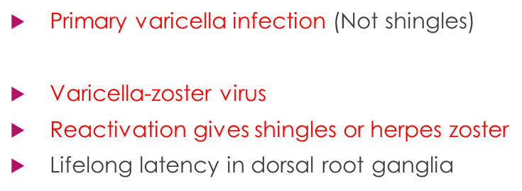
cause
reactivations gives what?
Chicken pox
clinical features
Fever
Rash
After 1-2 days:
Coryza
Headache
Nausea
Myalgia
Itch
Malaise
Appetite loss
Chicken pox
rash appearce

Chicken pox
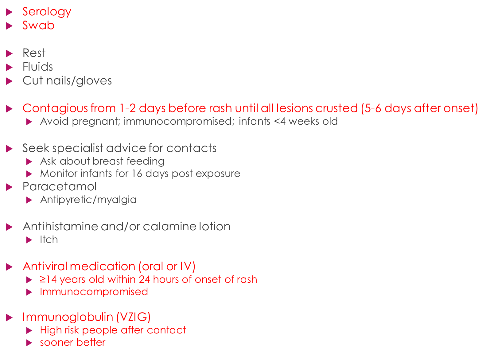
ix and Mangement
chicken pox
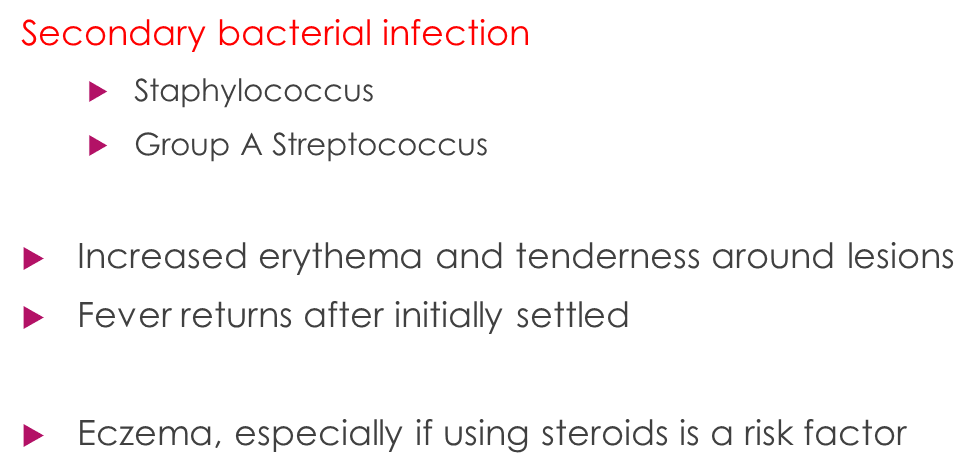
complications - secondary bacterial infection to what?
Measles
cause
Also called first disease
Morbillivirus of paramyxovirus family
Measles
clincila features
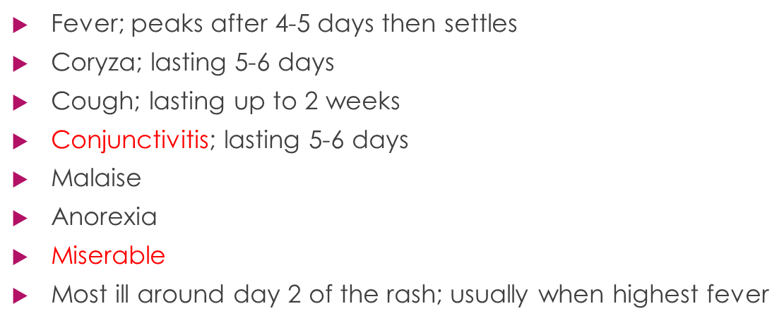
measels rash appearnce

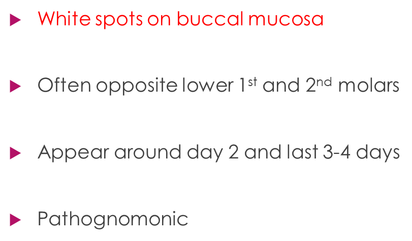
Koplik’s spots
what is it
seems in what and where
measles - Dx
IgM antibodies
PCR – oral fluid, urine, throat swab
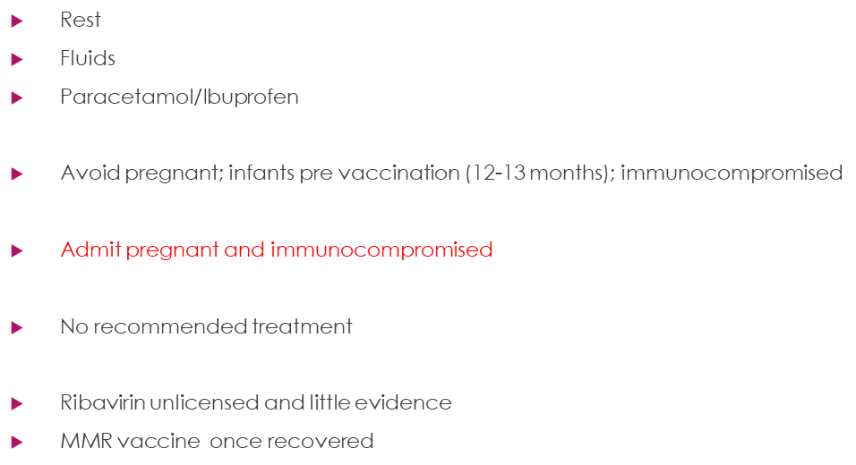
measels - Tx
Mumps
cause
Sx
Virus from paramyxovirus family
More common in winter and spring
Fever
Headache
Anorexia
After 24 hours
Earache
Ipsilateral parotid gland pain and swelling over 2-3 days
Contralateral parotid swelling within 2 days
Unilateral ~25%
Usually settles within 1 week
Munps
mangement
Rest
Fluids; avoid citrus juices
Paracetamol/ibuprofen
Contagious for up to 7 days after onset of parotid swelling
Rubella
cuase
Virus of the togavirus family
rash
Children
Asymptomatic
Mild Fever
Adults
Fever
Headache
Sore throat
Coryza
Conjunctivitis
Malaise
Lymphadenopathy
Occipital; posterior auricular; cervical
Tender and can be all over the body
May appear before the rash
Can last >2 weeks after rash settled
Arthralgia
Rubella rash
presentations
May be first sign in children
Maculopapular rash starting behind ears
Spreads to face; neck; then trunk; extremities
Non-confluent
Lasts 3-5 days
May be itchy in adults
Difficult to distinguish from other viral infections
Rubella
Mangement
Rest
Fluids
Paracetamol/ibuprofen
Contagious for up to 1 week after rash appears (6 days); moderately contagious
Avoid pregnant women: congenital rubella syndrome (CRS)
Vaccination history of contacts
No extra risk if immunocompromised
Rubella in pregnancy
Refer urgently to obstetrics if ≤ 20 weeks gestation
No treatment to prevent CRS
Other viral pathogens may be isolated:
Parvovirus B19; Varicella-zoster; Herpes simplex; Cytomegalovirus – also associated with congenital infection – seek specialist advice
Congenital rubella syndrome
complications/presentations
Microcephaly
Learning disability
Deafness
Eye abnormalities
Congenital heart disease
Can develop type 1 DM and thyroid problems
Aggravating factors for psoriasis
Streptococcal infection — strongly associated with guttate psoriasis
Medications — eg lithium, antimalarials, beta-blockers, NSAIDs, ACEI, antibacterials eg tetracycline and penicillin).
Sun exposure — usually beneficial, but may exacerbate psoriasis in a around 10% of patients
Trauma — Koebner phenomenon
Hormone changes (eg puberty, post-partum, menopause)
Psychological stress
Excessive alcohol intake
Smoking
Obesity
Other: HIV infection and AIDS, environment (for example change in climate),
Key features of Psoriasis
symmetrically distributed
well-defined
red, scaly plaques and/or papules
Common sites: scalp, elbows and knees BUT any part of the skin can be involved
Psoriasis
Dx and Ix
PASI Score (Psoriasis Area and Severity Index)
PGA (Physician’s Global Assessment) score
DLQI (Dermatology Life Quality Index)
PEST(Psoriasis Epidemiology Screening Tool) to assess for joint involvement
BP
Weight/BMI
Blood sugars
Lipid Profile
how is Severity of psoriasis measured?

Psoriatic - mangement
Topical Therapy
Creams, lotions, or gels are suitable for widespread psoriasis
Ointments are suitable for areas of skin with thick scale
Lotions, solutions, or gels are suitable for hair-bearing areas
Phototherapy
Systemic Therapy: Eg Methotrexate, Ciclosporin, Acitretin
Biologics: see PASI and DLQI scores - >10: eg, infliximab, interleukin antagonists
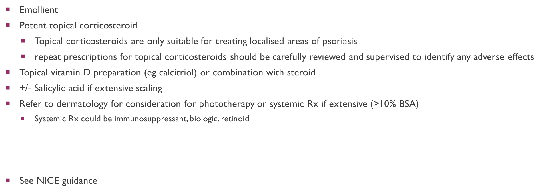
Chronic plaque psoriasis of trunk and limbs
tx
Management of scalp psoriasis
¡Important to remove thick scale in order for Rx to work
¡Topical potent corticosteroid
¡Coal tar shampoos (but not to be used alone if scalp psoriasis severe)
¡Vitamin D analogues
¡Combination prep if poor response or refer on if further treatment advice required
Management of facial/flexural/genital psoriasis
¡Emollient
¡Short term mild or moderate topical steroid (may cause irritation and greater risk of adverse effects)
¡Topical Rx alone unlikely to be effective if extensive disease (>10% of body involved)
¡(Topical calcineurin inhibitors – off licence)
Management of guttate psoriasis
¡If not widespread:
¡Reassure, self limiting condition which usually resolves in 3-4/12
¡Topical Rx as for trunk and limb psoriasis
¡Treatment of underlying infections
¡Refer for consideration of phototherapy if >10% BSA involvement
Management of nail psoriasis
¡Generally very difficult to treat
¡No Rx required if mild disease
¡Mainly advice:
¡Keep nails short — this avoids exacerbating onycholysis (detachment of the nail from the nail bed) and reduces the accumulation of material under the nail
¡Avoid manicure of the cuticle — this may provoke paronychia (infection of the nail bed); Avoid prosthetic nails
¡Refer to dermatologist if severe or having major functional impact
Lichen planus
cause
Pruritic eruption commonly associated with hepatitis C
Human herpes virus and VZV also implicated as triggers (and stress/anxiety)
Characterised by purple papules, polygonal and peripherally located (distal extremities)
Lichen planus
Clinical presentation (forms)
Actinic (sun-exposed areas)
Annular (tends to affect axillae)
Hypertrophic (tends to be on shins, looks like warts)
Atrophic
Erosive
Linear
Pigmented (oval grey/brown flat marks on trunk or limbs)
Vesicular/bullous (rare, usually affects lower legs)
Scalp (patchy alopecia)
Can also affect genital area
Typical Clinical presentation of lichen planus
Papules shiny, violaceous, polygonal
Varying size 1mm to >10mm diameter
Discrete or arranged in lines or circles
May have characteristic fine white lines over surface – Wickham striae
Clinical presentation of lichen planus
Mucous membrane involvement common, may affect people without cutaneous LP
Lesions most commonly on tongue and buccal mucosa
White or grey streaks forming a reticular or linear pattern on a violaceous background
Ulcerated oral lesions may have higher incidence of malignant transformation in men
Lesions may also be found on conjunctivae, larynx, oesophagus, tonsils, bladder, vulva, vagina and throughout the gi tract
Genital involvement common in LP
Annular lesions on glans in men
Vulval involvement more variable, from papules to severe erosions; dyspareunia, burning and pruritus common
Nail findings in about 10% (eg onycholysis)
Cutaneous lesions may be accompanied by ones on scalp (lichen planopilaris)
lichen planus
management
Self limiting disease, usually resolves in 8-18 months (some types can last for years though)
Mild cases – no Rx if no irritation
Moderate cases - potent topical steroids (0.05% clobetasol propionate) – withdraw as rash and itch begin to clear
Steroid sparing creams eg tacrolimus for more delicate areas
More severe or widespread cases (esp with scalp, nail and mucous membrane involvement) – more intensive Rx
Systemic steroids
Oral or topical retinoids
PUVA/UVB therapy
Methotrexate, hydroxychloroquine (esp. if hair/nails affected)
For oral lesions – steroid gel, mouthwash
Pemphigus vulgaris
mainly in who?
Potentially fatal blistering disease
Occurs in all races but commoner in Ashkenazi Jews
Onset usually in middle age; 50-60yrs
Pemphigus vulgaris
non-itchy flaccid bullae (particularly involving trunk)
Bullae rapidly denude, leaving erythematous, weeping, painful erosions
Mucosal involvement common
Pemphigus vulgaris
treatment
Emollients, wound care
Topical Steroids - Potent steroids in combination with oral
Systemic Steroids (mainstay of treatment) - Prednisolone 1-1.5 mg/kg/day
Adjuncts: Other immunosuppressants used as steroid-sparing agents (eg azathioprine, mycophenolate)
Bullous pemphigoid
features
Can be very itchy
Often starts with pruritus, urticaria and erythematous lesions
Large tense bullae develop
Appear anywhere on the skin, often involves trunk, limbs, flexures
Localised B.P can also occur. Less common and usually limited to lower extremeties
Mucosal involvement uncommon but can be present
Patients generally appear well, despite extensive blistering
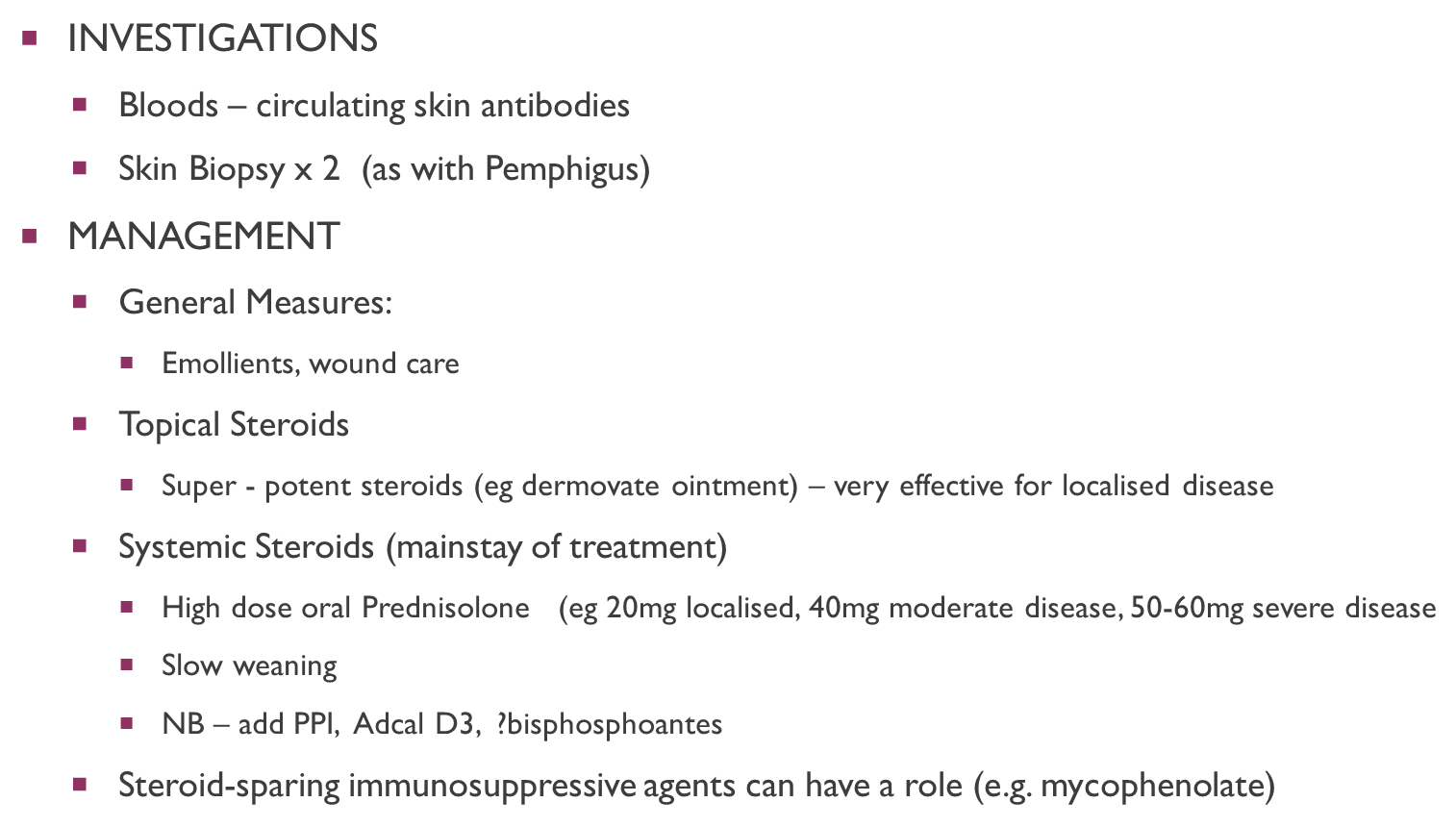
Bullous pemphigoid
Investigation & MANAGEMENT
Dermatitis herpetiformis
associated with what?
gluten enteropathy
M > F
Dermatitis herpetiformis
Clinical features
Symmetrical distribution
Extensor surfaces (knees, elbows, scalp, buttocks)
Small (papules / vesicles) that are intensely itchy and often grouped
Crusted erosions left behind from scratching
Remissions and exacerbations common
Dermatitis herpetiformis
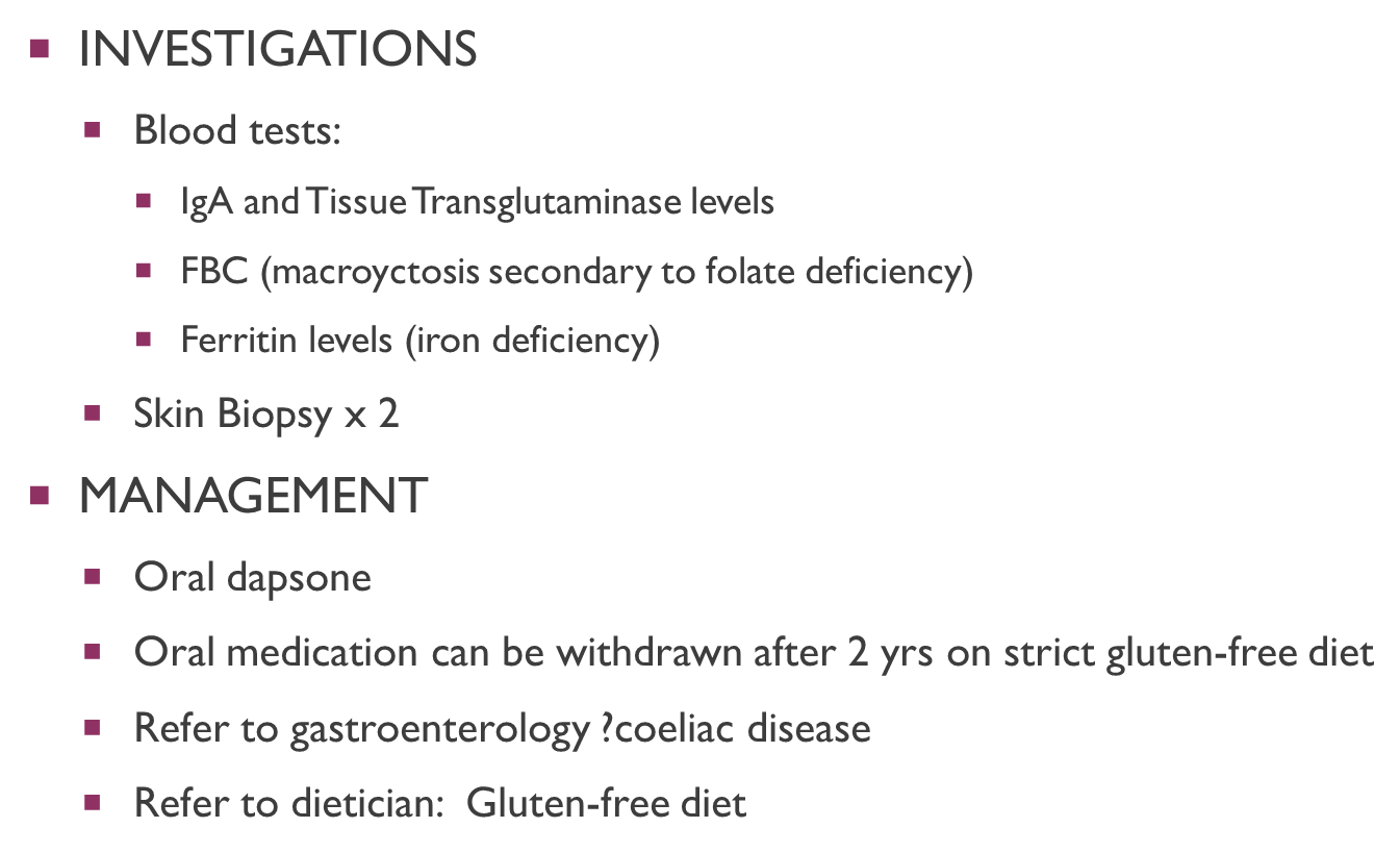
Investigation & management
Epidermolysis bullosa
what is it?
A group of genetically determined skin fragility conditions characterised by blistering of the skin and mucosae following mild mechanical trauma
Often appears at or shortly after birth
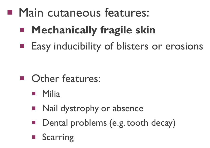
features of Epidermolysis bullosa
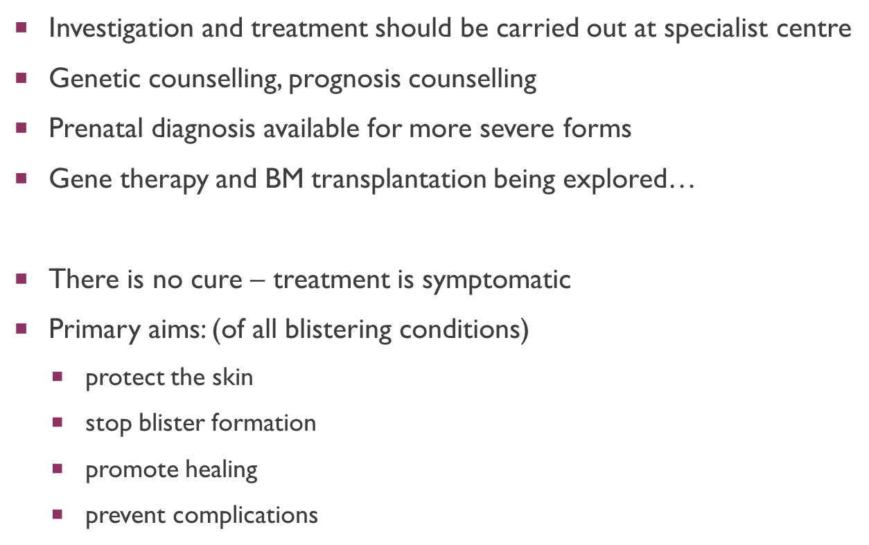
management of Epidermolysis bullosa
Actinic Keratoses
where?
presentations
size
Localised areas of sun damage
Present as scaly/rough areas on an erythematous base
Common on sun exposed sites
Men > women
Backs of hands, forehead, nose, upper lip,
usually < 1 cm across