Defense Against Disease/Viruses Test
1/44
There's no tags or description
Looks like no tags are added yet.
Name | Mastery | Learn | Test | Matching | Spaced | Call with Kai |
|---|
No analytics yet
Send a link to your students to track their progress
45 Terms
Pathogen
Disease-causing organisms and viruses
Semmelweis discovered...
handwashing reduces infection
John Snow discovered...
cholera is caused by pathogens; outbreaks occur after natural disasters and water main breaks
What qualities of skin make it a defense against pathogens?
It's a physical and chemical barrier
Has sebaceous glands that secrete lactic and fatty acids, creating acidic conditions on the skin, preventing bacterial growth
Produces lysozyme enzymes that kill bacteria by digesting the cell wall
What qualities of mucus make it a defense against pathogens?
It is a chemical barrier
Soft areas of skin (nose, trachea) are kept soft by mucus
Pathogens are trapped in the mucus. The mucus has lysozyme enzymes that kill bacteria by digesting the cell wall
Explain the process of blood clotting
1. Damage to blood vessel exposes collagen fibers
2. Platelets and damaged tissue release clotting factors, initiating a chain reaction that forms a blood clot
3. Clotting factors convert inactive prothrombin to active thrombin
4. Thrombin is an enzyme that converts soluble fibrinogen to insoluble fibrin
5. Fibrin forms a mesh around wounds, trapping blood cells, to form a semi-solid clot
6. Clot dries to form a scab, preventing blood loss, stopping pathogens from entering the body
When can clotting be bad?
In vessels, when it creates an unnecessary occlusion that blocks the blood pathway
Describe factors of the adaptive immune system
It's a specific, targeted response to pathogens that produces specific antibodies for specific antigens
Includes the production of memory cells, which increase effectiveness of immune response against future attacks
Describe factors of the innate immune system
It is a non-specific response to a broad range of pathogens
Does not change over a lifetime
Includes physical and chemical barriers and phagocytes (white blood cells that engulf and digest pathogens)
Phagocyte
white blood cells that engulf and digest pathogens

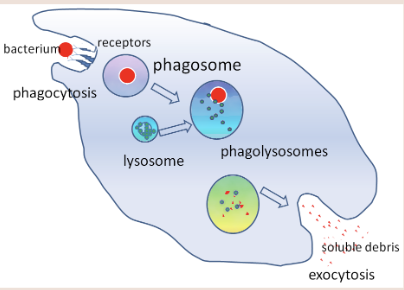
Explain how phagocytes defend the body against infection.
They are attracted to sites of infection, leaving the bloodstream with ameboid motion
A vacuole fuses with lysosomes to form a phagosome
Enzymes from the lysosomes that digest the engulfed pathogen
Soluble debris of the pathogen exists the body via exocytosis
Lymphocytes
group of white blood cells which recognize and target pathogens
located in lymph nodes and circulate in bloodstream
people have lots of B-lymphocytes that make specific antibodies
Explain the process of activating antibodies from T-lymphocytes
Helper T-lymphocytes activate B lymphocytes, B-lymphocytes produce antibodies (Helper T-lymphocytes —> B lymphocytes —> antibodies)
Antigens (location and purpose)
glycoproteins or other proteins found within the plasma membranes of cells; located on the surface of pathogens
stimulate the immune system to produce specific antibodies
How does an antibody work? (how does it kill antigens, future, self)
It has a receptor protein that can bind to a specific antigen. Once bound, it tells the body to destroy the whole compound. Antibodies stay in our bodies to fight future infections.
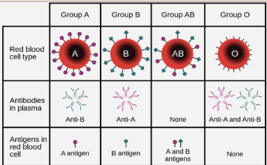
The immune system does NOT produce antibodies against antigens recognized as self (ex. person with type A blood and therefore has type A antigens will not produce B antibodies because that would kill them)
What type of antigens and antibodies does each blood type have?
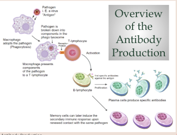
Explain how B-lymphocytes are activated by a pathogen entering the body
A pathogen enters the body. The pathogen is engulfed by a phagocyte.
The antigen is attached to an MHC protein. The phagocyte presents the antigen by moving it onto the plasma membrane using the MHC protein
Helper T-cells have specific receptor proteins in their plasma membrane that recognize and bind to antigens presented by phagocytes.
The phagocyte passes a chemical signal to the helper T-lymphocyte to activate the B-lymphocyte.
Inactive B-lymphocytes have (specific) antibodies on their plasma membranes. If the antigens on the Helper T-lymphocytes match the antibodies present on the B-lymphocyte, they bind.
The B-lymphocyte is activated by this interaction, producing plasma B-cells and memory cells.
B-cells undergo mitosis to form…(what things)
B-cells undergo mitosis to form plasma B-cells that form antibodies (clonal selection)
B-cells undergo mitosis to form memory cells which remain in the bloodstream after an infection has left the body to ensure future protection
How is HIV transmitted?
Sexual intercourse, sharing of hypodermic needles, blood transfusions, mother to baby during birth process or through breast milk
What does HIV and AIDS stand for?
Human Immunodeficiency Virus
Acquired ImmunoDeficiency Syndrome
How does HIV cause AIDS?
HIV infects and kills CD4 cells (helper T-lymphocytes). Since CD4 cells produce antibodies, there are now less antibodies to fight pathogens, increasing risk for infection.
What are antibiotics? How do they work? Do they have an effect on viruses? What about animals?
chemicals that inhibit bacterial growth. they destroy the cell wall of bacteria, and when cells don’t have a cell wall, they die.
NO. Antibiotics interfere with the metabolism of prokaryotic living cells, and viruses are NOT alive. Animals are eukaryotic, so antibiotics don’t greatly affect them.
How do bacteria become resistant to antibiotics?
Bacteria overproduce and compete for resources. The presence of antibiotics is a selective pressure that determines which bacteria survive. Bacteria with the favorable trait of antibiotic resistance will live on, but those without the trait will die. Natural selection determines antibiotic resistance!
Define Zoonoses. Examples?
infectious diseases that transfer from other species to humans
Covid (human to animal contact)
Tuberculosis (human to animal contact)
Rabies (bites)
Japanese encephalitis (mosquito vectors)
Vaccines have either…
antigens or nucleic acids (DNA or RNA) that code for antigens
Define herd immunity
If a sufficient percentage of a population is immune to a disease, transmission is greatly impeded.
Epidemic v pandemic
Epidemic = within national borders
Pandemic = across national borders
Percent difference
final-initial/average x 100
Percent change
final-initial/initial x 100
Virus
submicroscopic infectious agent that replicates only inside the living cells of an organism. infects ALL life forms. it is non-living.
Common features of viruses
Small fixed size, typically 20 to 200 nm in diameter [Bacteria are 2000 to 3000 nm]
Genetic material in the form of DNA or RNA
A protective protein coat, known as a capsid, surrounding the genetic material
No cytoplasm
Few or no enzymes
What are the shapes of viruses and examples?
Icosahedral (influenza), helical (Ebola), and bacteriophage (T4 bacteriophage)
Differentiate enveloped versus non-enveloped viruses
Enveloped have a lipid bilayer that surrounds their capsid, making them more susceptible to environmental changes as their lipid enveloped can be easily disrupted
Non-enveloped lack a lipid bilayer, so their capsid (made of tough protein layer) is outermost. This is more stable when facing environmental changes.
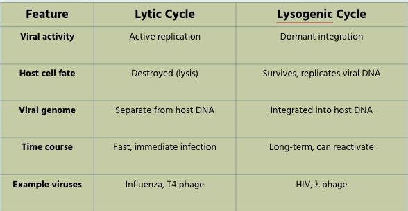
Describe the lytic cycle of the lambda bacteriophage. What are examples of viruses with the lytic cycle?
Attachment: The bacteriophage lambda attaches to receptors on an E. coli host cell. The capsid remains outside the bacterial cell.
Penetration: The bacteriophage lambda injects its DNA into the E. coli cell.
Phage DNA Replication: Endonuclease enzymes produced by the virus degrade the E. coli chromosomes, allowing the bacteriophage to hijack the host cell.
Phage Assembly: Since the E. coli cell is now hijacked, the host cell’s metabolism assembles phage DNA and capsids into many new bacteriophage lambdas.
Host cell Lysis: Enzymes are produced, which damages the E. coli cell wall. The cell lyses (bursts) releasing many bacteriophage lambda, which can attach to and infect other E. coli cells.
Note: During the lytic cycle, the bacteriophage lambda DNA remains separate from the host cell’s DNA. The DNA from the bacteriophage lambda takes control of the cell’s metabolism.
Ex. Common cold viruses, Influenza virus, Covid
Describe the lysogenic cycle of the lambda bacteriophage. What are examples of viruses with the lysogenic cycle?
Attachment: The bacteriophage lambda attaches to receptors on an E. coli cell. The capsid remains outside the bacterial cell.
Penetration: The bacteriophage lambda injects its DNA into the E. coli cell.
Prophage formation: The phage DNA is incorporated into the E. coli chromosome, to form a prophage. The infected bacterium is known as a lysogen.
E. coli reproduction: The infected E. coli lysogen (infected cell) reproduces. When the E. coli reproduces, the lambda bacteriophage DNA is replicated along with the bacterial chromosome. All offspring of the infected bacteria will contain the lambda bacteriophage DNA.
Induction: An E. coli lysogen is triggered to enter the lytic cycle. The prophage (lambda bacteriophage DNA) is excised from the bacterial chromosome.
Phage DNA Replication begins, leading to lysis and the release of many copies of the lambda bacteriophage to infect other E. coli cells.
Herpes simplex virus
HIV
Temperate bacteriophages (like λ phage in E. coli)
Compare and contrast lytic and lysogenic cycles
Outline the evidence for the origin of viruses and why there is no common ancestor
All viruses are obligate parasites, that require host cells for replication.
Viruses use the same genetic code as all organisms.
Virus first hypothesis
The Virus First Hypothesis proposes that viruses existed before cells, as they are much simpler than cells. The ancestors of modern viruses could have provided the raw material for the first cells.
Strength: Virus genomes have genes that are not present in cells.
Weakness: All modern viruses can only replicate using cells, suggesting that viruses could not have existed before cells
Escape Hypothesis
The Escape Hypothesis suggests viruses evolved from sections of DNA or RNA that escaped from cells.
Strengths: Modern bacterial cells exchange genetic material, suggesting a possible escape mechanism for genetic material. The hypothesis would explain the diversity of viruses if genetic material escaped many times.
Weaknesses: Most of the genes and proteins found in viruses are not found in cells.
Regressive Hypothesis
The Regressive Hypothesis suggests viruses were once small cells that parasitized larger cells. The genes not required for their parasitism have been lost over time.
Strengths: The existence of giant viruses which have similar genetic material to parasitic bacteria.
Weaknesses: The smallest cellular parasites do not resemble viruses in any way.
How do antigens change shape?
If the genetic material of the virus mutates, this can cause the shape of the protein coat, including the antigens, to change.
Some viruses have an unstable genome and mutate very rapidly for the following reasons:
Viruses have a very high replication rate, increasing the chance of a random mutation occurring.
Viruses do not have a proofreading mechanism during replication, making it more likely that a mutation happens. This is particularly true of RNA viruses such as the influenza virus and HIV.
Immune systems don’t make antibodies against mutated viruses, since they can’t recognize them as threats
What is HA and NA?
HA (hemagglutinin) and NA (neuraminidase) are surface proteins found on the influenza virus capsid. Both proteins act as antigens for the immune system.
Influenza hemagglutinin (HA) is integral to the virus’s infectivity. Hemagglutinin is a Class I Fusion Protein, having multifunctional activity as both an attachment factor and membrane fusion protein. (Wikipedia)
Viral neuraminidase (NA) is a type of neuraminidase found on the surface of influenza viruses that enables the virus to be released from the host cell. Neuraminidases are enzymes that cleave sialic acid groups from glycoproteins. (Wikipedia)
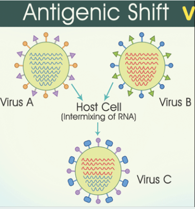
Antigenic shift
Antigenic shift is an abrupt major change to the HA and NA surface proteins of the influenza virus.
SCARIER THAN ANTIGENIC DRIFT
Antigenic shift occurs when an organism is infected with two different strains of the influenza virus.
When the viruses are being synthesised by cells, the genetic material from the two virus strains can recombine, resulting in novel combinations of the HA and NA genes.
The resulting virus produces combinations of HA and NA surface proteins (antigens) not recognised by the immune system, and may result in a pandemic.
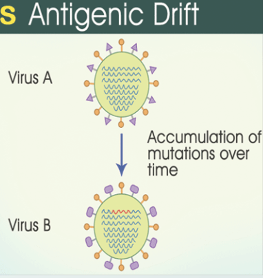
Antigenic drift
Antigenic drift is a gradual process.
The influenza virus has a high replication rate resulting in mutations. This leads to the gradual accumulation of mutations in the genes coding for the HA and NA surface proteins (antigens).
The mutations cause the shape of the HA and NA antigens to change over time, resulting in new strains of the influenza virus which the immune system no longer recognises.
A person who is immune to the original virus will not be immune to the mutated virus, due to the different-shaped antigens.