Chapter 5-Part 1-Skeletal Systems
1/95
There's no tags or description
Looks like no tags are added yet.
Name | Mastery | Learn | Test | Matching | Spaced |
|---|
No study sessions yet.
96 Terms
The skeletal system is made up of
bones, joints, and related connective tissues.
206 bones in the adult skeleton.
two major divisions:
the axial skeleton
appendicular skeleton.

axial skeleton
80 bones that include
the bones of the skull
hyoid bone
auditory ossicles
vertebral column
sternum
ribs.
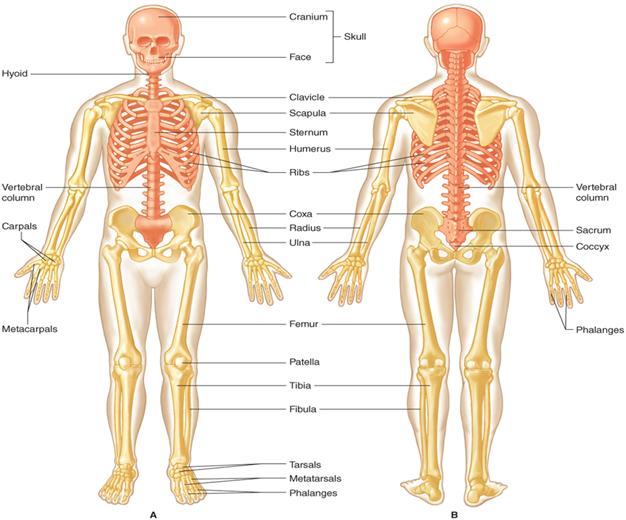
appendicular skeleton (general)
has 126 bones
includes
the pectoral and pelvic girdles
upper and lower extremities.
upper extremities (the arms) are attached to the axial skeleton by the pectoral girdle
lower extremities (the legs) are attached to the axial skeleton by the pelvic girdle.
Functions of the Skeletal System
Protection: skull protects the brain from injury and the ribs and vertebra protect the heart and lungs. The bones of your vertebral column (spine) protect your spinal cord, and the bones of your cranium (skull) protect your brain.
Support: allows us to sit, stand, and move by the attachment of muscles to bones.
Storage: bone acts as a reservoir for over 99 percent of the calcium in our body.
Production: white and red blood cells are produced by red bone marrow contained within bone.
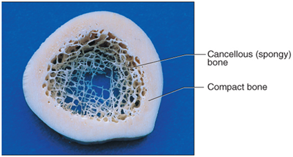
Bone is
a living connective tissue with two types:
Cancellous (spongy) bone
Compact bone

Cancellous (spongy) bone
has spaces filled with red bone marrow and supports shifts in weight distribution.
This type of bone is found in the end of long bones, the middle of the flat and irregular bones.

Compact bone
denser than spongy bone so that it can withstand compressive forces.
It is found under the periosteum and in the diaphysis of long bones, where it provides support and protection.
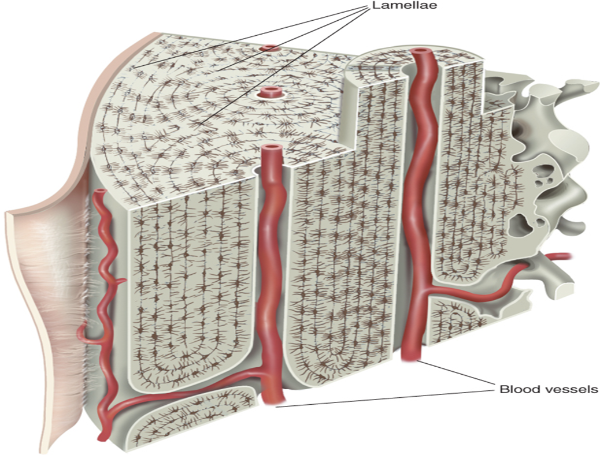
has a unique arrangement of cells and canals
Osteons are fundamental unit of compact bone
Osteons
(Haversian systems) are the fundamental unit of compact bone
is a long, cylindrical tube consisting of
concentric rings of calcified matrix called lamellae
surround a central canal or Haversian canal, which contains blood vessels, nerves, and lymphatic vessels.
Within the lamella are lacunae where osteocytes can be found.
Blood vessels and nerves are contained within the central canal which is lined with a membrane called the endosteum.
Smaller canals, called canaliculi, run perpendicular to the central canal and carry blood vessels and nerves to the lamellae and lacunae
tubes inside tube
have collagen fibres on different layers in diff directions to resist torison stress
central cannals of blood vessels and nerves
Bone Cells
are crucial to the function of bones.
There are four types of bone cells
Osteogenic cells
Osteoblasts
Osteocytes
Osteoclasts

Osteogenic cells
are non-specific cells in the periosteum.
They are undifferentiated and develop into osteoblasts.

Osteoblasts
immature bone cells, responsible for building new bone by forming a soft matrix of protein and carbohydrate molecule with collagen fibers.
Hard mineral crystals are deposited in the matrix, mineralizing the bone matrix.
When osteoblasts are trapped within the calcified matrix, their structure and function changes, and they become osteocytes.
When there is not as great a demand for calcium elsewhere in the body, osteoblasts will be more active keeping calcium within bone.
Increased demand for calcium, osteoclastic activity will increase. Decreased demand for calcium, osteoblastic activity will decrease.

Osteocytes
mature bone cells and are the most common type of bone cell.
Each osteocyte is located in a space called a lacuna and is surrounded by bone tissue.
Osteocytes maintain the mineral concentration of the matrix via the secretion of enzymes.
Bone is made of 35% organic material and 65% inorganic material.
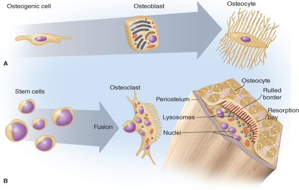
Osteoclasts
tear down bone for remodeling and cause the release of calcium.
Osteoclasts develop from monocytes and macrophages.
tear down bone
are under the control of parathyroid hormone that is secreted by the parathyroid glands.
Bone remodeling is a balance between tearing down and building up bone.
The majority of calcium is stored in bone and so when the body requires more, osteoclastic activity will increase to release calcium into the bloodstream.
Increased demand for calcium, osteoclastic activity will increase. Decreased demand for calcium, osteoblastic activity will increase.

Bone Growth and Development
Early stages of embryonic development, the embryo’s skeleton consists of fibrous membranes and hyaline cartilage.
By the sixth or seventh week of embryonic life, the actual process of bone development, ossification (osteogenesis), begins.
Bones grow through a process called ossification or osteogenesis.
There are two types of ossification:
intramembranous
endochondral.
ossification or osteogenesis.
Bone growth
two types
Intramembranous ossification
endochondral ossification
Intramembranous ossification
The flat bones of the face, most of the cranial bones, and the clavicles (collarbones) are formed by this type ossification.
Bones begin as tough, fibrous membranes that are replaced by a bone matrix that is produced by osteoblasts.
Calcium and other minerals are deposited within the matrix and then it calcifies.
The matrix is made up of calcium, phosphate, collagen fibers, and water.
The collagen fibers give bone its flexibility and the minerals give bone its strength.
Other factors essential to bone growth include
vitamin A for
stimulation of osteoblasts,
vitamin C for
collagen synthesis,
growth hormone,
insulin
thyroid hormone.
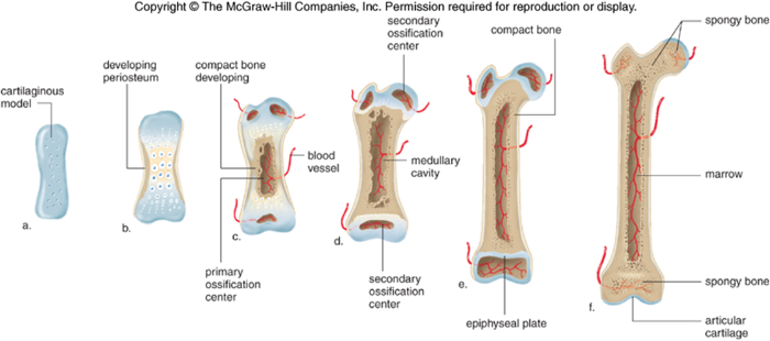
Endochondral ossification
Bones develops by replacing hyaline cartilage.
Cartilage does not become bone.
cartilage serves as a template to be completely replaced by new bone.
common in long bones such as the femur.
Eventually, the osteoblasts form a bone “collar” around the diaphysis of the cartilage model and eventually will replace the cartilage model.
The area where ossification first begins is called the primary ossification center.
Each end or epiphysis of the long bone will begin to ossify later in the process and so are called secondary ossification centers.
Red bone marrow
produces RBC
yellow bone marrow
stores energy as fat
synarthroses (funtional joint clasiffication)
non moving joint
e.x cranium
Amphiarthroses (funtional joint classification)
partially moving joints
Diarthroses (functional joint classification)
fully movable, found in limbs
Joint or articulations
junctions or articulations between bones
bones of joints do not touch each other during movement.
more abundant then bones
meeting place between 2 or more bones
needed for skeletal muscle movement
two types
structural
what kind of material bind the bones at joint
Fibrous
Cartilaginous
synovial
functional
how much can joint move/flex
junctions or articulations between bones
Fibrous (structural joint classification)
are connected together with short connective tissue fibers abundant with collagen that do not allow much, if any, movement.
Examples cranial bones and facial bones.
Fibrous joints in the skull are called sutures
connect bones with dense fibres
e.x cranium sutures
immovable joint
Cartilaginous joints (structural joint classification)
are connected together by a disc of cartilage
are slightly movable.
Joints between vertebrae are cartilaginous joints.
unite bone with cartilage
dont move much
come in 2 types
synchondroses
symphyses
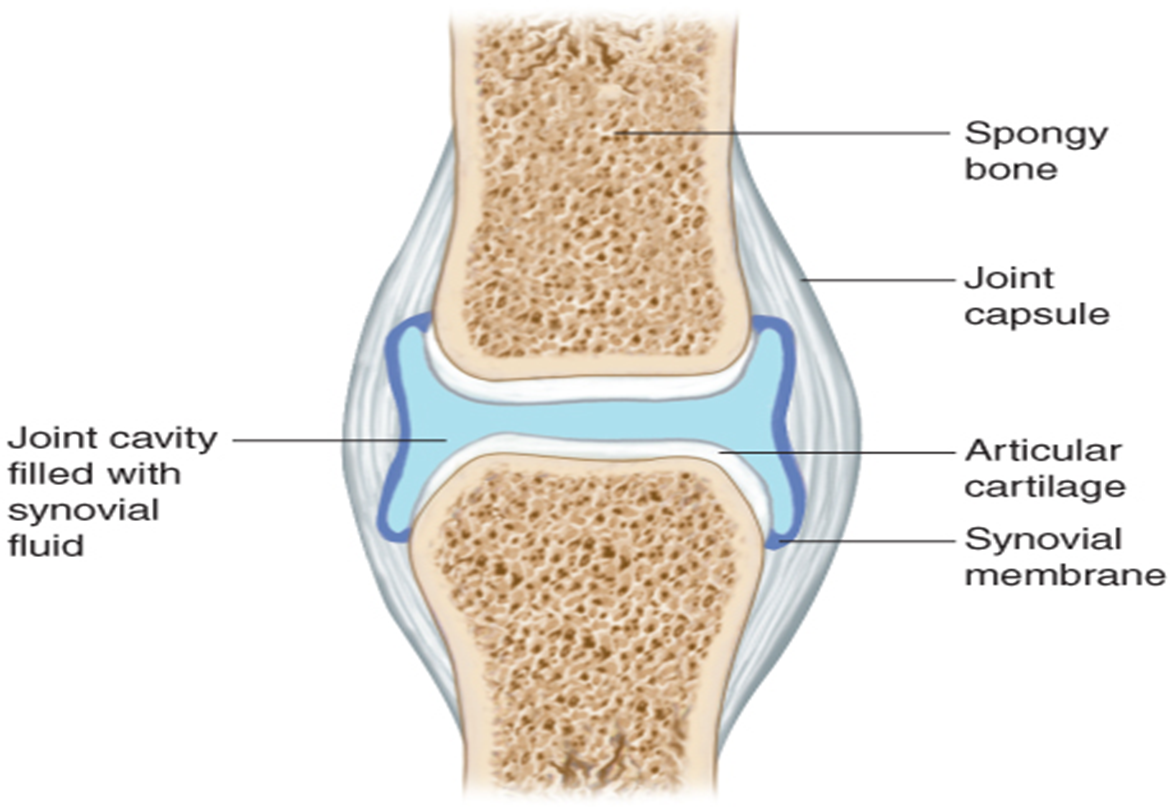
synovial (structural joint classification)
The most numerous type of joint in the body, and the most movable one, is the synovial joint.
Covered with hyaline cartilage
is surrounded by a fibrous joint capsule lined with a synovial membrane.
secretes a slippery fluid called synovial fluid,
acts as a lubricant to allow the bones to move easily across each other.
Example of synovial joints are
elbows, knees, shoulders, and knuckles.
free moving
most jonts
use fibres and cartilage tissue
bones joined are separated by synovial fluid filled joint cavity
allow bones to rub on each other without friction and create flexible movment
Ligaments
Bones are also attached to each other by dense connective tissue fibers called
Angular movements (permitted by synovial joints)
Flexion
bending joint toward body
Extension
bending joint away from body
Adduction
the movement of a limb or other part toward the midline of the body or toward another part.
Bringing arms down to sides of bodyu
Abduction
the motion of a limb or appendage away from the midline of the body
lifting arms arm side of arms to T-pose
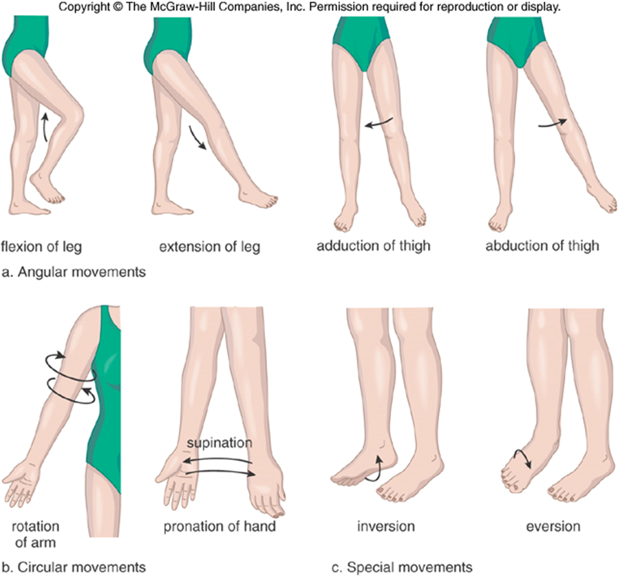
Circular movements (permitted by synovial joints)
Circumduction
movement of a limb or extremity so that the distal end describes a circle while the proximal end remains fixed
Rotation
the twisting movement of a limb or body part around its own longitudinal axis
Supination
facing up/in
Pronation
facing down/out

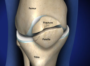
Knee Joint
connects femur and tibia
patella, knee cap protect joint
is a synovial joint
Articular cartiliges
material cover the end of bones in joints
1 inch thick
white shiny and rubbery
allow bone surface to slide without damage
facilitates motion and absorb shocks
on all bone joints
Medial and lateral collateral ligments
support knee joints
most important part
are connective tissue
prevent knee from moving to far in the side direction
ACL and PCL (anterior and posterior cruciate ligament)
control front to back motion of knee
control stability of knee
ACL keeps tipia from sliding to far forward
PCL keeps tipia from sliding to far back
lateral and medial meniscus (on tibia superior)
cartilage of knee
gasket distruputing weight, and help ligaments with knee stability
Tendon connect muscle to bones connect muscle to joint
popitial artey supplies blood to leg, essential for its surrival
Special movements (permitted by synovial joints)
Inversion and eversion
Inversion: tilt the sole of the foot towards the midline of body
Eversion: tilt the sole of the foot away from midline
Elevation and depression
Elevation: move up the horizonal resting
Depression: move down the horizontal resting
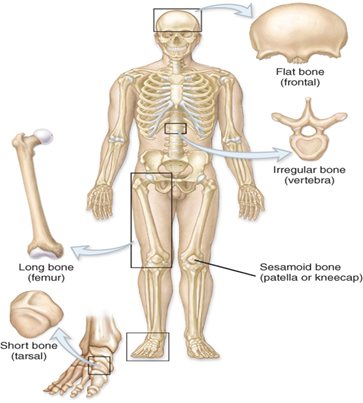
Flat bones
are thin and consist of a layer of spongy bone between two layers of compact bone.
serve as points of attachment for muscles and often protect internal organs.
include the
sternum (breastbone)
ribs
scapulae (shoulder blades)
cranial (skull) bones.

Long bones
are greater in length than width.
mostly made up of compact bone, but their epiphyses or ends are mostly spongy bone.
include the
femur
tibia
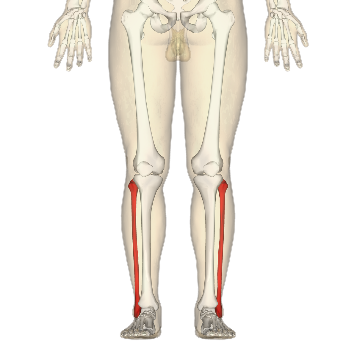
fibula
humerus
radius
ulna, and phalanges.
Found in the arm (femur, tibia, fibula) and legs (femur, tibia, fibula), as well as in the fingers (metacarpals, phalanges) and toes (metatarsals, phalanges).
function as levers - they move when muscles contract.
A long bone has two parts:
diaphysis and the epiphysis.

Diaphysis (part of long bone)
has hollow medula cavity with yellow marrow
makes up the majority of the length of the long bone.
It is a tubular structure with a thick collar of compact bone surrounding a space called the medullary cavity, which contains yellow marrow.
In children, this is the location of red bone marrow and is responsible for hematopoiesis
production of blood cells
10 to 15 million erythrocytes per second.
In adults, red bone marrow is replaced by yellow bone marrow
except for in a few locations such as the
skull
ribs
vertebra
pelvis.
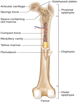
epiphyses (part of long bone)
ends of bone
houses red bone marrow
house spongy bone
is the wider section at each end of a long bone
an expanded area consisting of a thin layer of compact bone surrounding more abundant cancellous (spongy) bone.
Red marrow fills the spaces in the spongy bone.
Covering each epiphysis and the narrow area that contains the epiphyseal plate (growth plate is articular cartilage of the hyaline type.
It is smooth and aids in the almost frictionless movement of joints and acts as a shock absorber.
medullary cavity (part of long bone)
In the center of long bones
initially filled with red bone marrow and is involved in hematopoiesis or blood cell formation.
has a delicate membranous lining called the endosteum, where bone growth, repair, and remodeling occur.

epiphyseal plate or growth plate
Between the epiphysis and diaphysis of a long bone
Between 18 and 25 years of age, the growth plate which is made of hyaline cartilage is replaced by bone, becoming the epiphyseal line, and the bone is no longer capable of growing in length.
periosteum
surrounds the diaphysis.
made up of dense fibrous connective tissue
has nutrient blood vessels, nerves and lymphatic vessels that nourish compact bone.
responsible for “growing pains” during adolescence.
Articular cartilage
layer of hyaline cartilage where bones join together.
Short bones
are cuboidal in shape.
consist mostly of spongy bone with just a thin outer layer of compact bone.
provide stability and support as well as some limited motion.
include the
carpals (wrist bones) except for the pisiform
tarsal (ankle) bones except for the calcaneus (heel bone). Short bones

Irregular bones
have a variety of shapes
include the
vertebrae that support the spinal cord and protect it from compressive force
coxal (hip bones)
some facial bones,
calcaneus (heel bone)

Sesamoid bones
is a small round bone that have their name because of their resemblance to sesame seeds.
These bones form in tendons (the sheaths of tissue that connect bones to muscles) where a great deal of pressure is generated in a joint.
protect tendons by helping them overcome compressive forces.
Examples
tendons associated with the feet, hands, and knees (patella).
Bone markings
Every bump, groove, and hole on a bone has a name.
two main types of bone markings
projections
those that grow out
an area of a bone that projects above the surface of the bone.
These are the attachment points for tendons and ligaments.
depressions
those that indent the bone
is an opening or groove in the bone that allows blood vessels and nerves to enter the bone.
The axial skeleton
forms the vertical, central axis of the body and includes all bones of the skull, ribs, sternum, and vertebra.
protects the
brain
spinal cord
heart
lungs.
serves as the attachment site for muscles that move the
head, neck, and back, and for muscles that act across the shoulder and hip joints to move their corresponding limbs.
consists of 80 bones that lie along the longitudinal axis of the body.
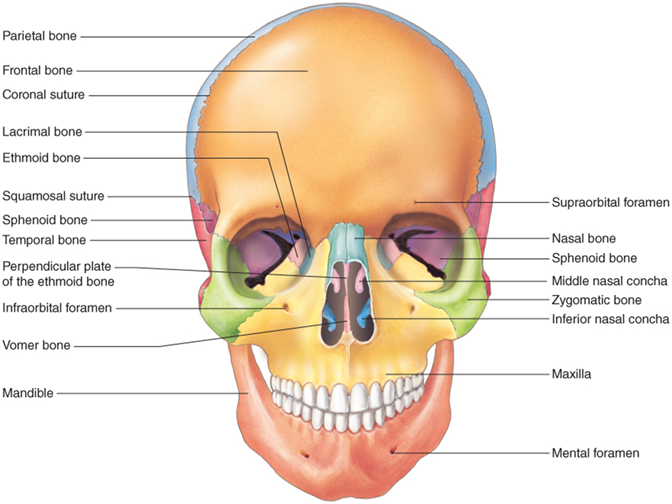
The skull (part of axial skeleton)
supports the face and protects the brain.
into two types:
cranial bones
facial bones.
In the adult, the skull consists of 22 individual bones,
21 of which are immobile and united into a single unit.
The 22nd bone is the mandible (lower jaw), which is the only moveable bone of the skull.
8 cranial bones form the top, sides, and back of the skull
14 facial bones form the face
3 ossicles found in each middle ear
Fontanelles
the soft spots on a baby's head where the skull bones have not yet fused
Not all the sutures have been formed creating “soft spots”
tough membranes that have not yet ossified felt on an infant’s skull.
fontanelles allow the bones of the infant’s skull to be “molded” as the infant moves through the birth canal.
There are 6 fontanelles in the infant skull:
one anterior
one posterior
two anterolateral,
two posterolateral
The posterior fontanelle is also located in the midline and is seen between the two parietal bones and the occipital bone. The posterior fontanelle is the first to close or ossify and this about 2 months after birth. By about 2 years of age, all the fontanelles have closed to form sutures that are immovable joints between bones of the skull.
anterior fontanelle
is located midline between the two parietal bones and the frontal bone.
It is the largest of the fontanelles and ossifies 18 to 24 months after birth.
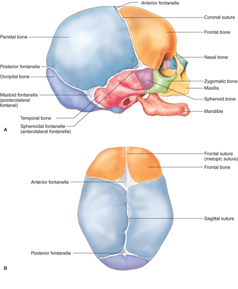
posterior fontanelle
located in the midline and is seen between the two parietal bones and the occipital bone.
The posterior fontanelle is the first to close or ossify and this about 2 months after birth.
By about 2 years of age, all the fontanelles have closed to form sutures that are immovable joints between bones of the skull.

Frontal bone (cranial bones)
is a single bone that forms the anterior portion of the cranium.
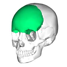
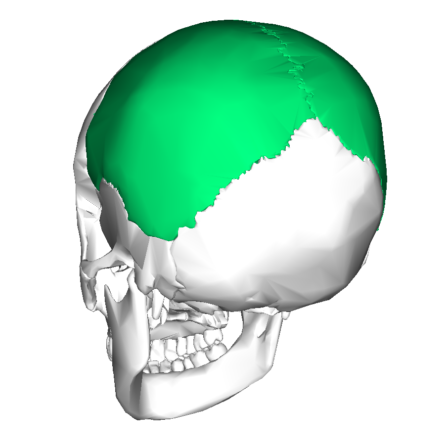
Parietal bones (cranial bones)
are paired bones that form most of the top and sides of the skull.

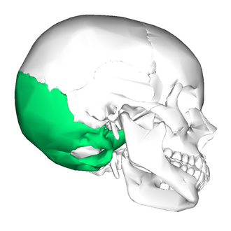
Occipital bone (cranial bones)
is a single bone that forms the back of the skull.
foramen magnum
The large opening at the base of the occipital bone
the and the spinal cord runs through it to allow for attachment to the brain stem.
occipital condyles
Two rounded elevations on the base of the occipital bone
on either side of the foramen magnum. They articulate with the first vertebra.
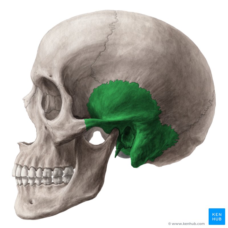
Temporal bones (cranial bones)
are paired bones on each side of the head that form the lower sides of the skull.
Through each temporal bone runs a canal called the external auditory meatus or ear canal.
Just behind each ear on the temporal bone is a bony prominence called the mastoid process.
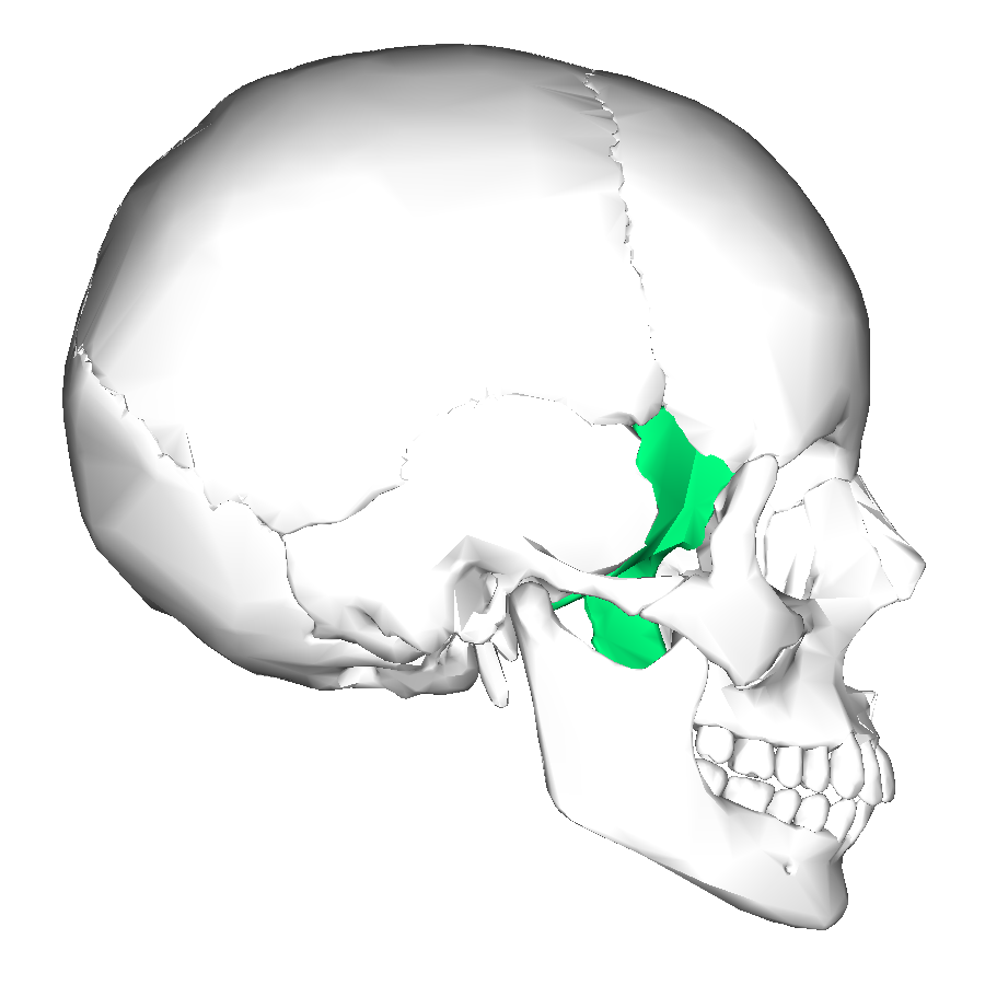
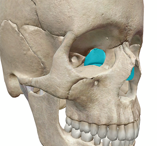
Sphenoid bone (cranial bones)
is an unpaired bone that is the most complex bone of the skull.
It is shaped somewhat like a butterfly
forms part of the floor of the cranium.
In the center of this bone is a deep depression called the hypophyseal fossa
where the pituitary gland is located.
Ethmoid bone (cranial bones)
is an unpaired bone located between the sphenoid bone and the nasal bones.
Together, the ethmoid bone and sphenoid bone form
part of the floor of the cranium.
With the vomer bone, the ethmoid bone forms the nasal septum.
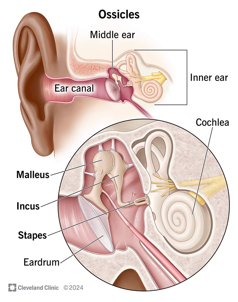
Auditory ossicles (cranial bones)
are the smallest bones of the body and not usually considered part of the skull:
the malleus (hammer)
incus (anvil),
stapes (stirrup).
They are found in the middle ear and are involved in hearing, and are connected to each other by synovial joints
The mandible (Facial bones)
single bone
it is the lower jawbone and the largest and strongest facial bone.
only movable bone in the skull other than the ossicles.
It articulates with the temporal bone in front of the external auditory meatus.

Maxillae (Facial bones)
are paired bones that have fused to form the upper jawbone of the facial skeleton.
The maxillae articulate with every bone of the face except the mandible.

Zygomatic bones (Facial bones)
are paired bones that are more commonly called the cheekbones.

Nasal bones (Facial bones)
are paired small bones that form the bridge of the nose.
The rest of the nose is cartilage.
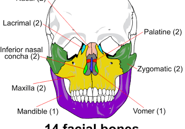
Palatine bones (Facial bones)
are paired bones form the posterior aspect of the hard palate as well as the lateral wall of the nasal cavity
small part of the floor of the orbit.

The vomer (Facial bones)
is single bone that forms the inferior part of the nasal septum.

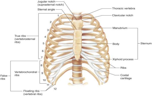
The rib cage (part of axial skeletal structure)
formed by sternum and 12 pairs of ribs.
The sternum is also called the breastbone and is actually made up of three bones.
From most superior to inferior they are the
manubrium,
the body,
xiphoid process.
Anteriorly,
the clavicles and most of the ribs attach to the sternum.
Posteriorly
the ribs articulate with the vertebra.
The first seven pairs of ribs are called true ribs
they attach directly to the sternum
through pieces of cartilage called costal cartilage.
The next five pairs of ribs are called false ribs
their costal cartilages attach indirectly to the sternum or do not attach at all.
Rib pairs 11 and 12 are called floating ribs
costal cartilages do not attach to the sternum anteriorly or to any other structure.
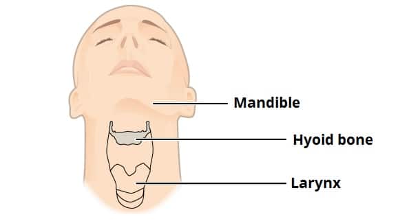
The hyoid bone
a small U-shaped bone located in the upper neck near the level of the inferior mandible, with the tips of the “U.”
independent bone is not attached to another bone and thus is not part of the skull.
Muscles attach to the hyoid bone to form the angle between the chin and the neck.
Movements of the hyoid are coordinated with movements of the tongue, larynx, and pharynx during swallowing and speaking.
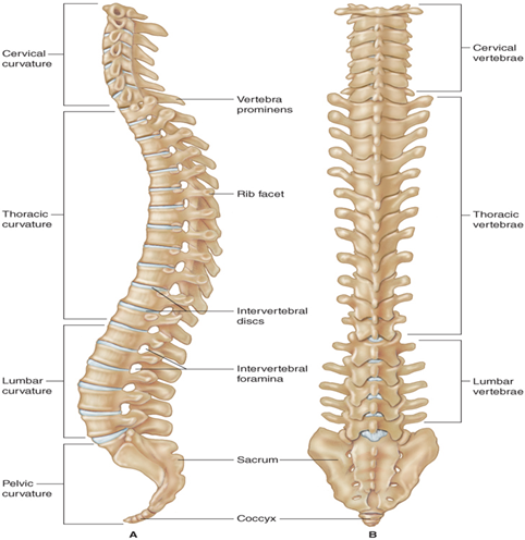
The spinal column (curvature; part of axial skeleton)
four major curvatures to the spine.
Cervical vertebrae
Thoracic vertebrae
Lumbar vertebrae
pelvic survature
Relative to the anterior surface of the body,
the thoracic and sacral curves are concave (CURVE INWARD)
called primary curvatures because we are born with them.
The cervical and lumbar curves are convex (CURVE OUTWARD) relative to the anterior surface of the body.
called secondary curvatures because we are not born with them, but instead they are developmental.
The cervical curve begins to develop as the infant begins to lift and hold its head up.
The lumbar curve starts to develop when the infant begins to stand upright

Cervical vertebrae C1-C7
Located in the neck, are the smallest and lightest of all the vertebrae.
The first cervical vertebra is called atlas or C1
When you turn your head from side to side (saying “no”), your atlas is pivoting around your axis.
the second is called the axis or C2.
The other 5 cervical vertebrae are noted as C3 to C7.
All cervical vertebrae have
three foramina
two transverse foramina
The vertebral artery and the corresponding vein and nerve fibers travel through the transverse foramina
and a single vertebral foramen.
The spinal cord passes through the vertebral foramen which is quite large in this region.

Thoracic vertebrae
Posterior attachments for the 12 pairs of ribs.
The thoracic vertebrae have long, sharp, spinous processes that you can feel when you run your finger down someone’s spine.
The thoracic vertebrae are denoted as T1 to T12.

Lumbar vertebrae
The most massive and sturdy of the vertebrae because they support much of the weight of the body.
When they are charted, you use the abbreviations L1 to L5.
Sacrum
A triangular-shaped bone that actually consists of five fused vertebrae.
The sacrum, when abbreviated, is noted as S1 through S5.
between 2 hipbones, and pelvis
Coccyx
a small, triangular-shaped bone
made up of three to five fused vertebrae.

The appendicular skeleton
consists of 126 bones
is made up of the upper and lower limb bones,
the bones of the hands and feet,
the bones that anchor the limbs to the axial skeleton (the pectoral and pelvic girdles)
Pectoral girdle (part of the appenidcualr skeleton)
the bones of the shoulder
include the
clavicles
the collar bone
slender in shape and each articulates (joined) with the sternum and scapula)
scapulae
The shoulder blades
are thin, triangular shaped flat bones located on the dorsal surface of the rib cage.
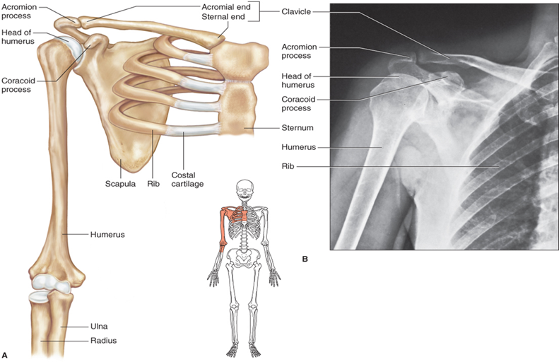
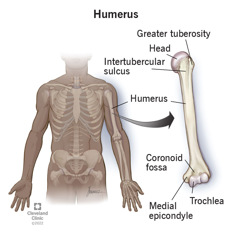
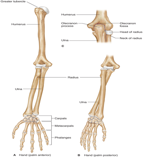
The humerus (part of the appenicular skeleton)
is located in the upper part of the arm.
Proximally it articulates with the scapula and distally with the radius and the ulna.
The head of the humerus articulates with the glenoid fossa of the scapula.
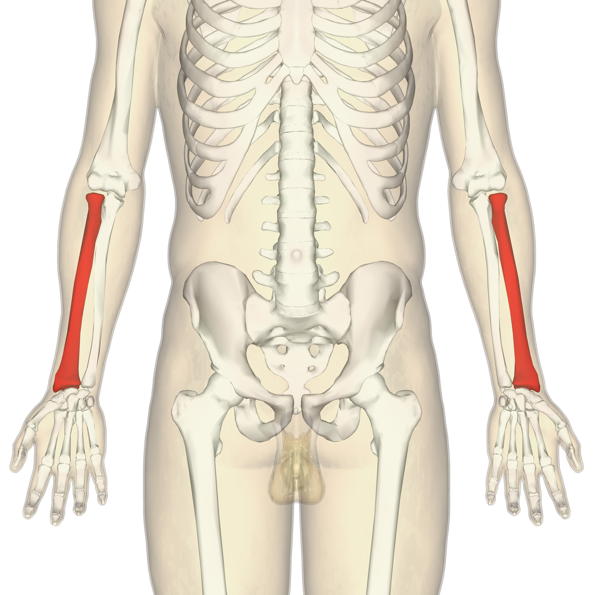
The radius (part of the appenicular skeleton; upper limb, or arm bones, includes the humerus, radius, and ulna.)
is the lateral bone of the forearm and when the body is in anatomical position,
it is on the same side of the arm as the thumb.
Proximally it joins with the humerus and the ulna and distally with the carpal bones.
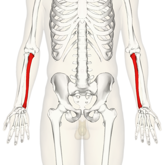
The ulna (part of the appenicular skeleton; upper limb, or arm bones, includes the humerus, radius, and ulna.)
is the medial bone of the lower arm.
The proximal end of the ulna joins with the humerus to form the elbow joint.
Distally, it also joins with the radius and some of the carpal bones of the wrist.
The bones of the hand (apppendiuclat skeleton)
include carpals, metacarpals, and phalanges.
The 8 carpal bones make up the wrist.
Each hand has 5 metacarpal bones that make up the palm of the hand.
There are 14 phalanges, the bones of the fingers, in each hand - 3 for each finger and 2 per thumb.
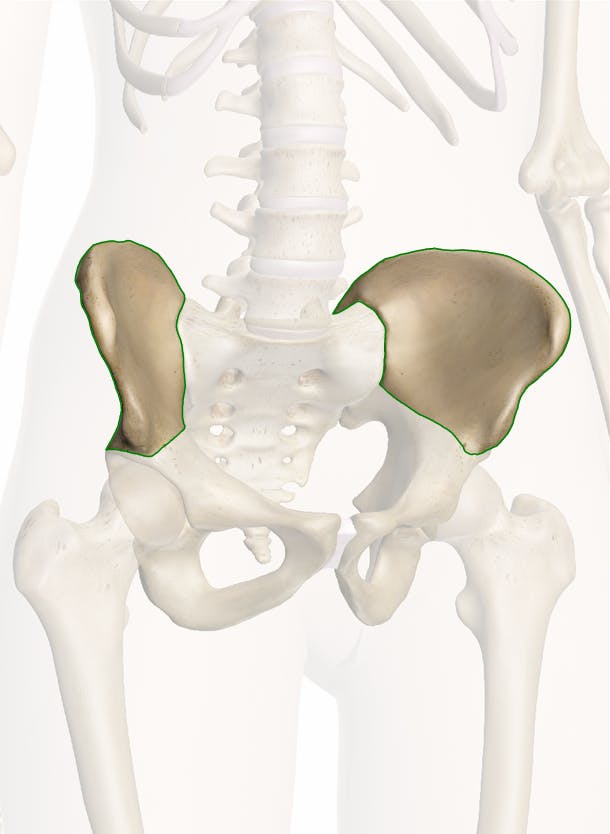
pelvic girdle
consist of
Coxal bones
ilium
ischium,
pubis
Lower extremities
The ilium (part of Coxal bones; and appenidcualr skeleton)
is the most superior part of a coxal bone
when you put your hands on your hips, you are touching the ilium.
The hip projection or ridge is the iliac crest
The ischium (part of Coxal bones; and appenidcualr skeleton)
forrms the lower part of a coxal bone
when an individual is sitting, he or she is sitting on the ischial tuberosities.
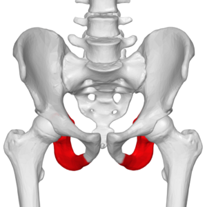
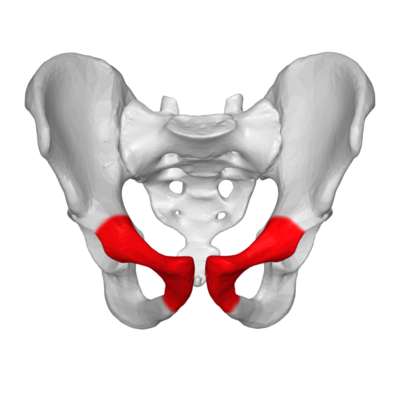
Pubis (part of Coxal bones; and appenidcualr skeleton)
The two pubic bones form the front of the coxal bone and come together at the pubic symphysis.
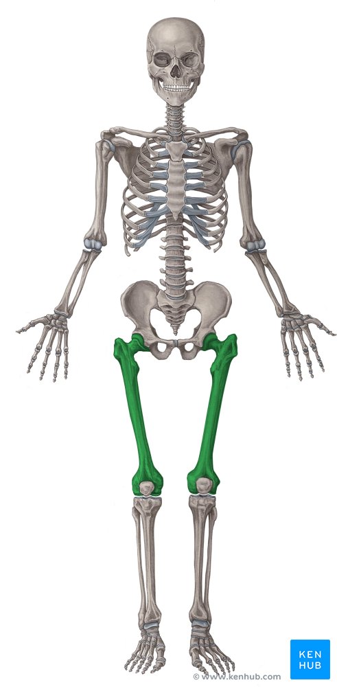
The femur (Lower extremities or leg; appendicular skeleton)
is the thigh bone
the longest and heaviest bone in the body.
Its proximal end joins with the hipbone at the hip socket or acetabulum.
The distal end of the femur attaches to the tibia and the patella or kneecap.
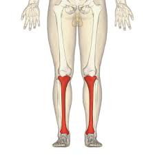
The tibia (Lower extremities of leg; appendicular skeleton)
is the medial bone of the lower leg and is commonly called the shinbone.
Its proximal end joins with the femur and fibula
its distal end attaches to the ankle bones.
The fibula (Lower extremities of leg; appendicular skeleton)
is the thinner, lateral bone of the lower leg
joins with the ankle bones at its distal end.
The patella or kneecap (Lower extremities of leg; appendicular skeleton)
is considered a sesamoid bone because of its resemblance to a sesame seed.
The bones of the foot include
the tarsals
calcaneus, or heel bone
the metatarsals
Five metatarsal bones form the front of the foot.
the phalanges.
The bones of the toes are called phalanges.
Each foot contains 14 phalanges
2 for each big toe
3 in each of the other toes.
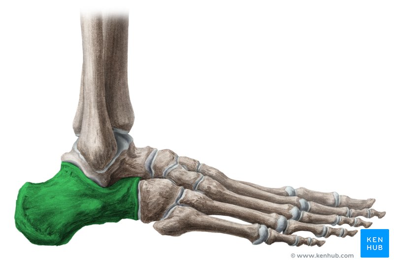
The calcaneus, or heel bone (bonies in foot; appendicular skeleton)
is the largest tarsal bone.
The talus bone sits on top of calcaneus and articulates with the tibia and fibula.
Five additional tarsal bones help make up the posterior part of the foot.
Osteoarthritis (OA)
Affects the weight-bearing joints of the hips and knees
Cartilage between the bones and the bones themselves begin to break down.
Occurs in elderly individuals due to the aging process
Joint stiffness, aching, and pain
Common Treatments:
anti-inflammatory drugs
intra-articular steroid injections
transplanting harvested cartilage cells
surgical scraping of the joint,
joint replacement
Rheumatoid Arthritis (RA)
Chronic systemic autoimmune disease
Synovial membrane is destroyed causing edema and congestion.
Scar tissue forms, bones atrophy, and visible deformities become apparent.
Moderate to severe pain in the affected joints
Common Treatments:
anti-inflammatory drugs
heat or cold treatments
cortisone injections
immunosuppressive drugs
low-impact aerobic exercise
warm water exercises
Gout (Gouty Arthritis)
Type of arthritis associated with high uric acid levels in the blood
Crystalline deposits in the joints, kidneys, and various soft tissues
Acute or chronic joint pain, commonly in the great toe
Common Treatments:
Pain medications
anti-inflammatory drugs
dietary modifications
Osteoporosis
Bones become more porous over time and a decrease in bone density is the result.
caused by
Hormone deficiencies
a sedentary lifestyle
a lack of calcium and vitamin D in the diet,
bone cancers,
corticosteroid excess,
smoking
excess alcohol consumption
the use of steroids
In later stages of the disease may experience
pathological fractures,
back and neck pain
a loss of height over time,
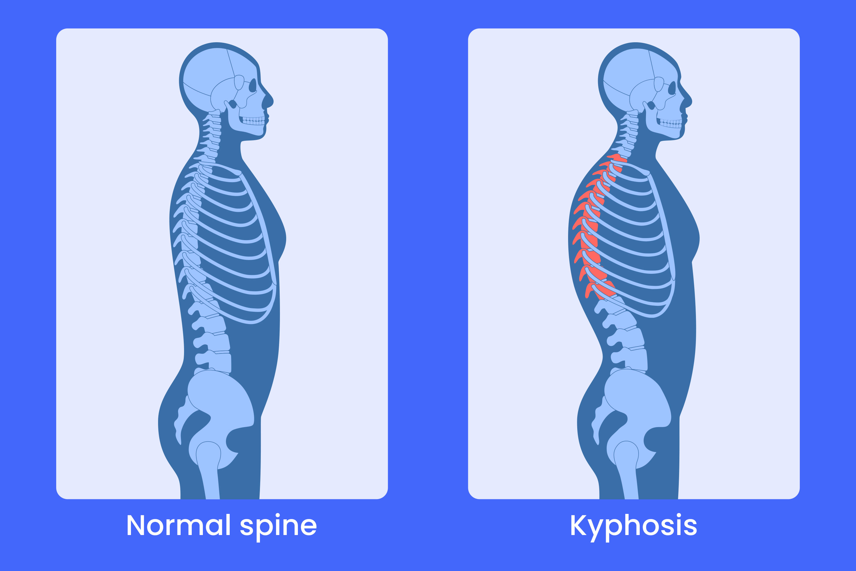
kyphosis
is an excessive forward curve of your spine
Common Treatments: include
medications
to prevent bone loss relieve bone pain,
hormone replacement
therapy,
lifestyle changes to prevent bone loss
moderation or elimination of the use of alcohol
cessation of smoking
Bursitis
Inflammation of a bursa, the fluid-filled sac that cushions tendons, elbow, knee, shoulder, and hip
Overuse and trauma to joints
Joint pain, swelling, and tenderness in the structures surrounding the joint
Common Treatments:
Rest
pain medications
steroid injections
aspiration of excess fluid from the bursa
antibiotics if caused by infection
Carpal Tunnel Syndrome
Occurs when the median nerve in the wrist is excessively compressed by an inflamed tendon
Overuse of the wrist
Weakness, numbness, pain, or a tingling sensation in the hand, wrist, or elbow
Common Treatments:
Wrist splints
pain medications
steroid injections
surgery to reduce the pressure on the affected nerves
Adolescent kyphosis
Growth retardation or improper development of the epiphyses as a result of rapid growth
No symptoms may occur, may be mild pain, tiredness, or tenderness or stiffness of the thoracic spine
Common Treatments:
Exercise,
a firm mattress
back brace
Adult kyphosis
Degenerative disc disease
Pain, back weakness, and fatigue
Common Treatments:
Spinal fusion or grafting,
Harrington rods may also be surgically inserted
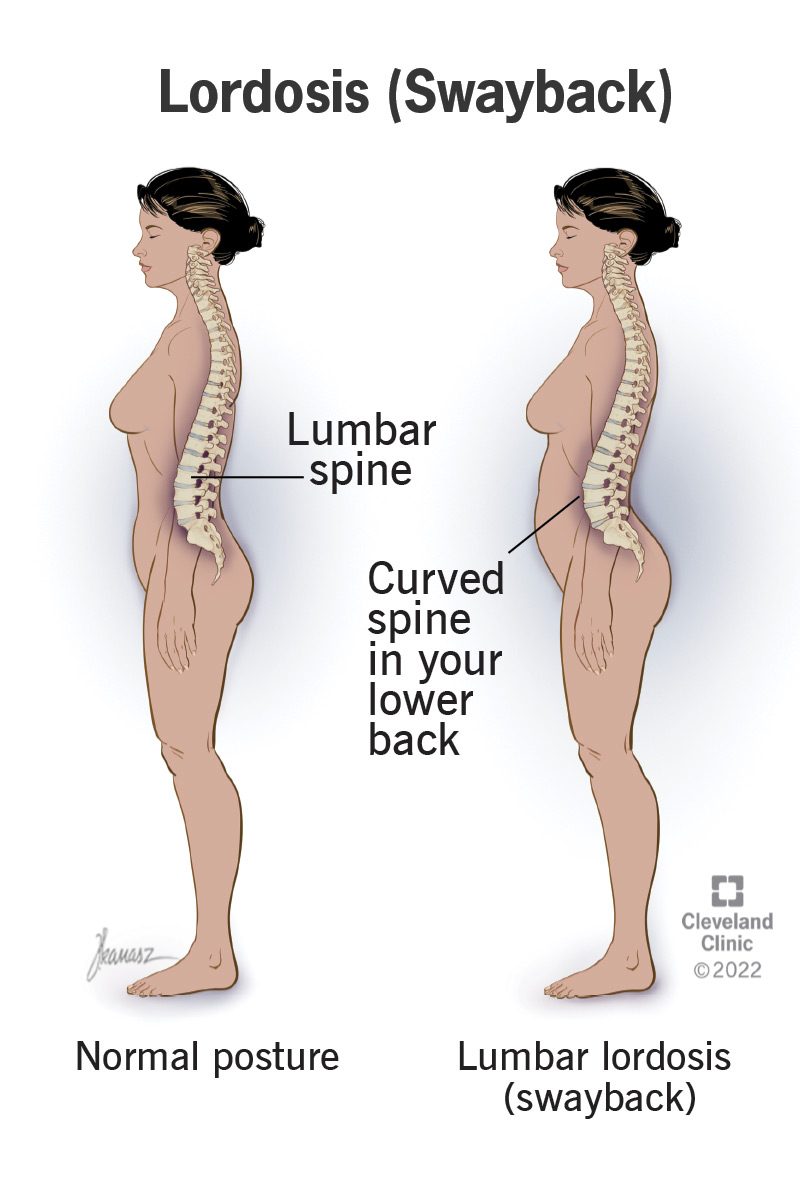
Lordosis
Exaggerated inward (convex) curvature of the lumbar spine
cause by
Obesity,
tuberculosis of the spine
poor posture
pregnancy, osteoporosis,
continual wearing of high heels
Mild to moderate pain
Preventing or treating the underlying cause
Scoliosis
Abnormal, S-shaped lateral curvature of the thoracic or lumbar spine
Develop prenatally when vertebrae do not fuse together or result from diseases that cause weakness of the muscles that hold vertebrae together
Back pain
Common Treatments:
Back braces
surgery to correct spinal curves
physical therapy to strengthen the spine
Laboratory Tests for skeletal systems
Serum calcium levels
Serum phosphate levels
Serum alkaline phosphatase (ALP)
Bone marrow examination
Microbial analysis
Synovial fluid analysis
Uric Acid
Anti Nuclear Antibodies
Rheumatoid factor
HLA-B27 genetic test
Vitamin D