Spinal Cord Segments and Nerve Roots (Week 1, Mod 8)
1/24
Earn XP
Description and Tags
Name | Mastery | Learn | Test | Matching | Spaced | Call with Kai |
|---|
No analytics yet
Send a link to your students to track their progress
25 Terms
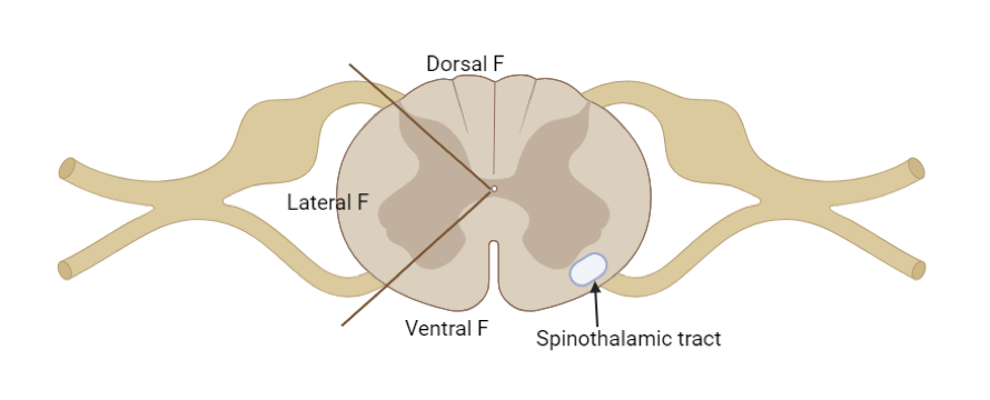
What are the funiculi of the spine?
Each of the three regions of WHITE MATTER in the spine; divided into a dorsal funiculi, lateral funiculi, and ventral funiculi
divided by the points of penetration of the spinal roots (see image)
What are the fasciculi of the spine?
Collection of nerve fibers WITHIN the funiculi
What is a “pathway” in relation to the spine?
Sequential tracts separated by synapses that are all involved in one neural function
What is the definition of a “spinal cord segment”? How many do canines and felines have, and what groupings are they divided into?
Spinal cord segment = a spinal cord section that gives rise to one pair of spinal nerve roots.
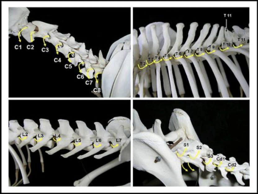
In dogs and cats it consists of between 35 and 38 segments (divided into: 8 cervical, 13 thoracic, 7 lumbar, 3 sacral and between 4 and 7 caudal)
Spinal nerve roots continues with spinal nerves.
C8 T13 L7 S3
What is unique about the spinal cord segments of the CERVICAL spine? What does this mean for the orientation of its spinal nerves?
There are 8 cervical segments, but only 7 vertebrae
This causes C2 - C7 spinal nerves to exit the vertebral canal CRANIAL to its associated vertebrae… all other spinal nerves, including those associated with C8, exit the vertebral canal CAUDALLY
C1 actually leaves through the LATERAL vertebral foramen… just had to be different
Describe the relationship between the growth of the neural tube, and the growth of the vertebral column… what does this result in later in adulthood?
As an embryo, the two grow at the same rate… but as the embryo develops, the neural tube slows down while the vertebral column continues, resulting in different lengths of the vertebrae and the spinal cord later into adulthood
Because of this, the cauda equina is developed
A group of dorsal and ventral roots of spinal nerves in the lumbo-sacral vertebral canal, stretch past the spinal cord to innervate the rest of the vertebrae
Looks like a horse tail
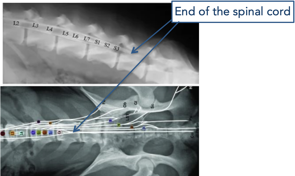
What are the 3 different aspects of the cauda equina?
Conus Medullaris: a caudal elongation of the spinal cord (long triangle shaped)
Filum terminale: Thin cord of fibrous tissue that attaches the conus medullaris to the caudal vertebrae
The nerves themselves
What are the “intumescences” of the spinal cord? How many of them are there per individual?
There are TWO: a cervical intumescence and a lumbar intumescence
THIS IS WHERE THE BRACHIAL AND LUMBOSACRAL PLEXUSES CONNECT TO THE SPINE
Are enlargements of the spine; have an increased amount of white matter and cell bodies here
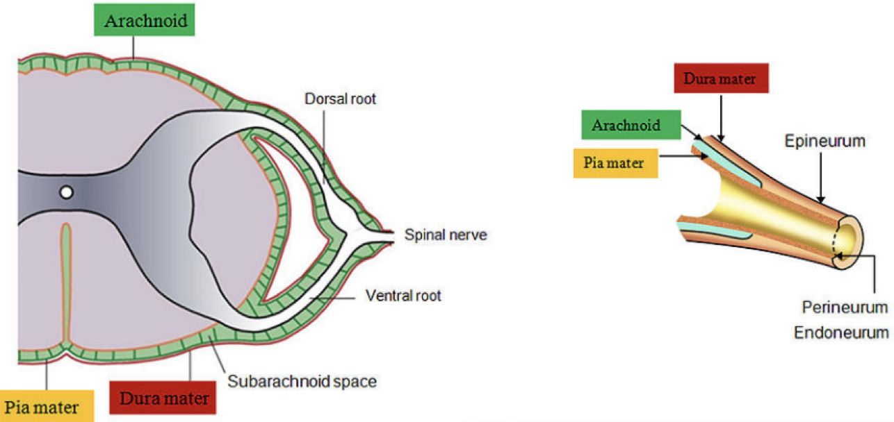
What are the 3 layers of the meninges? from out to in
Dura mater = tough
Arachnoid membrane = spider web-like appearance
Pia mater = tender
Describe the aspects of the dura mater…
The external layer
Dense connective tissue
Separated from periosteum and surrounded by epidural space (Fat & blood vessels)
Free of attachments to the vertebrae except:
At the level of C1-C2: adhered to periosteum of the vertebrae (Intracranial: adhered to cranial wall)
Caudally combines with filum terminale.
Describe the arachnoid membrane…
Delicate collagenous connective tissue
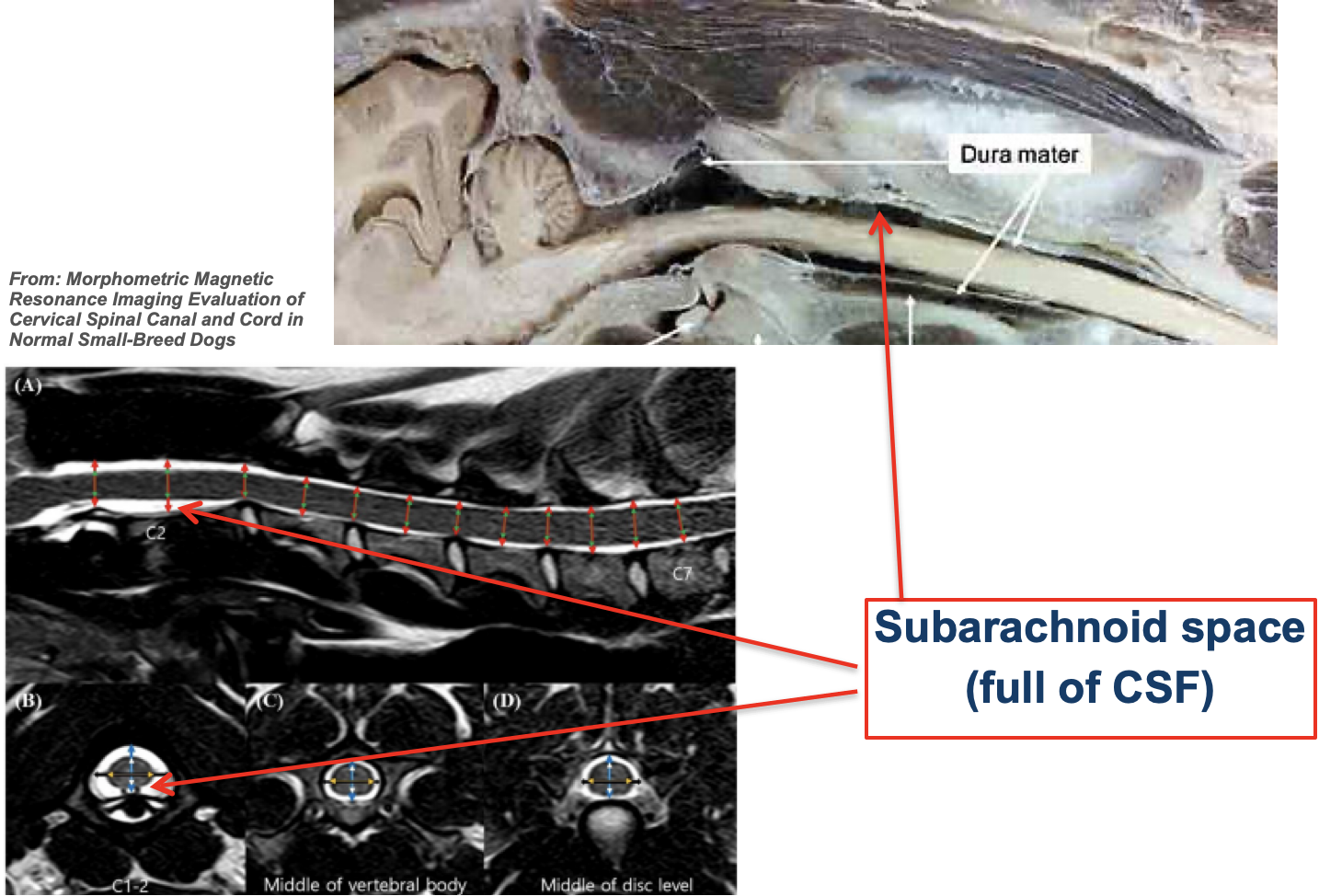
In close contact with the dura mater because the pressure of the cerebrospinal fluid (CSF), that occupies the subarachnoid space, pushes the arachnoid outwards.
Numerous fine filaments which blend with pia mater - spider’s web appearance
Describe the pia mater… what are denticulate ligaments?
Inner layer surrounding the spinal cord and roots.
Thin layer of connective tissue (thicker than arachnoid).
The pia mater is thickened bilaterally along the lateral margin of the spinal cord, forming denticulate ligaments → this ligament attaches to the arachnoid and dura laterally.
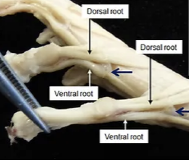
Describe the roots of the spinal nerves… how are they oriented in relation to the spinal segment? How many are there per segment?
Each spinal segment has two roots on EACH side…
Dorsal root → divides into rootlets
Ventral root → divides into rootlets
The dorsal and ventral roots meet on the OUTSIDE of the vertebrae to form a spinal nerve
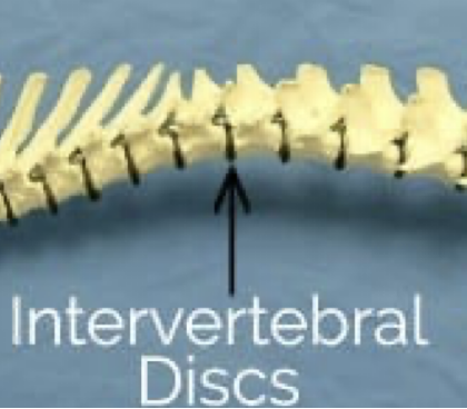
Pass through the intervertebral foramen between each vertebrae (see image)
This can also be an area where nerves can get PINCHED
What happens to the meninges as the spinal nerves extend out from the roots?
The ROOTS are surrounded by meninges, but once they elongate as spinal nerves, the dura mater becomes the epineurum that will protect the nerves
Does NOT contain CSF
What is unique about the dorsal roots from the ventral roots of the spine?
DORSAL ROOT GANGLIONS
Each dorsal root contains a spinal ganglion (aggregation of cell bodies) OUTIDE OF THE CNS
Will see a bulging at the dorsal root; collection of neurons
Is the exception; all neurons are INSIDE of the CNS EXCEPT for these!!
How do the dorsal and ventral roots communicate with each other in the spine?
Dorsal root: Afferent axons enter the cord with sensory information
Ventral root: Efferent axons exit the spinal cord with motor information
Think ascending → dorsal → afferent…… descending → ventral → efferent
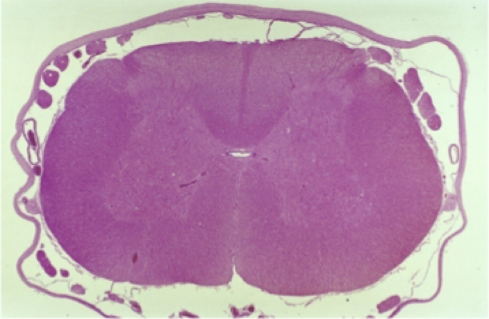
Describe the parenchyma of the spinal cord… where are gray matter and white matter in relation to each other?
White matter → on the OUTSIDE
Gray matter → on the INSIDE
Has a butterfly shape; each side communicate with each other via the gray commissure (surrounds central canal of spine)
Has a dorsal horn → responsible for SENSORY, and a ventral horn → responsible for MOTOR
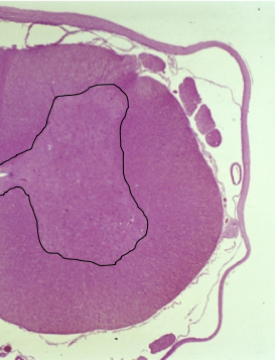
What can be seen at the center of a transverse histological section of the spine?
The central canal
Lined with EPENDYMAL cells and FILLED WITH CSF
White matter of the spine has 3 different sections… What are these called, and what are the functions of each?
Are the funiculi of the spine… divided into the:
Dorsal funiculus
Hold the ASCENDING sensory tracts (proprioceptive, tactile, and nociceptive sensations)
Lateral funiculus
ALSO hold ascending sensory tracts (all the same as ^^ except + thermal)
Ventral funiculus
Holds the DESCENDING motor tracts (facilitate extensor muscle activity)
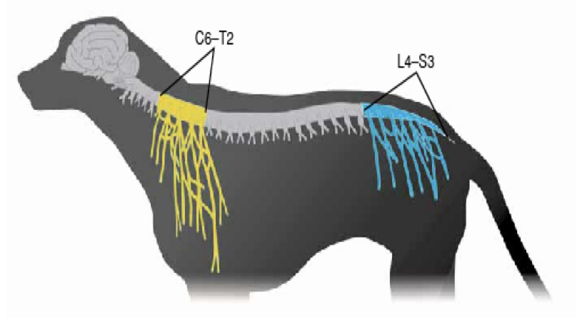
Where can the brachial plexus be found in relation to the spine? (name the exact vertebrae)
C6 - T2
What are 5 major nerves that branch off from the C6-T2 segment of the spine?
1) Musculocutaneous
2) Axillary
3) Radial
4) Median
5) Ulnar
How can we test for damage to the nerves in the C6-T2 segment?
Flexor (withdrawal) reflex
sensory: varies with area stimulated
motor: musculocutaneous, axillary, median, ulnar, radia
Where is the lumbosacral plexus in relation to the spine?
L4 - S3
What are 3 major nerves that come from the L4-S3 junction?
1) Femoral
2) Sciatic
3) Pudendal
How can we test for damage in the L4-S3 segment?
Flexor (withdrawal) reflex
sensory & motor: sciatic nerve
Patellar reflex
sensory & motor: femoral
Perineal reflex
sensory & motor: pudendal