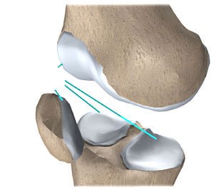
Lab Practical 2
5.0(1)
Card Sorting
1/145
Earn XP
Description and Tags
Study Analytics
Name | Mastery | Learn | Test | Matching | Spaced |
|---|
No study sessions yet.
146 Terms
1
New cards
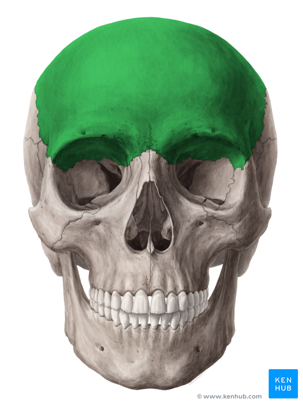
Frontal Bone
Forehead
2
New cards
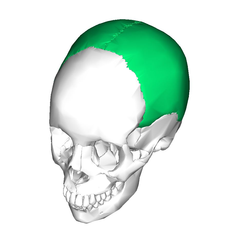
2 Parietal Bones
Top back of skull, joined by sutures
3
New cards
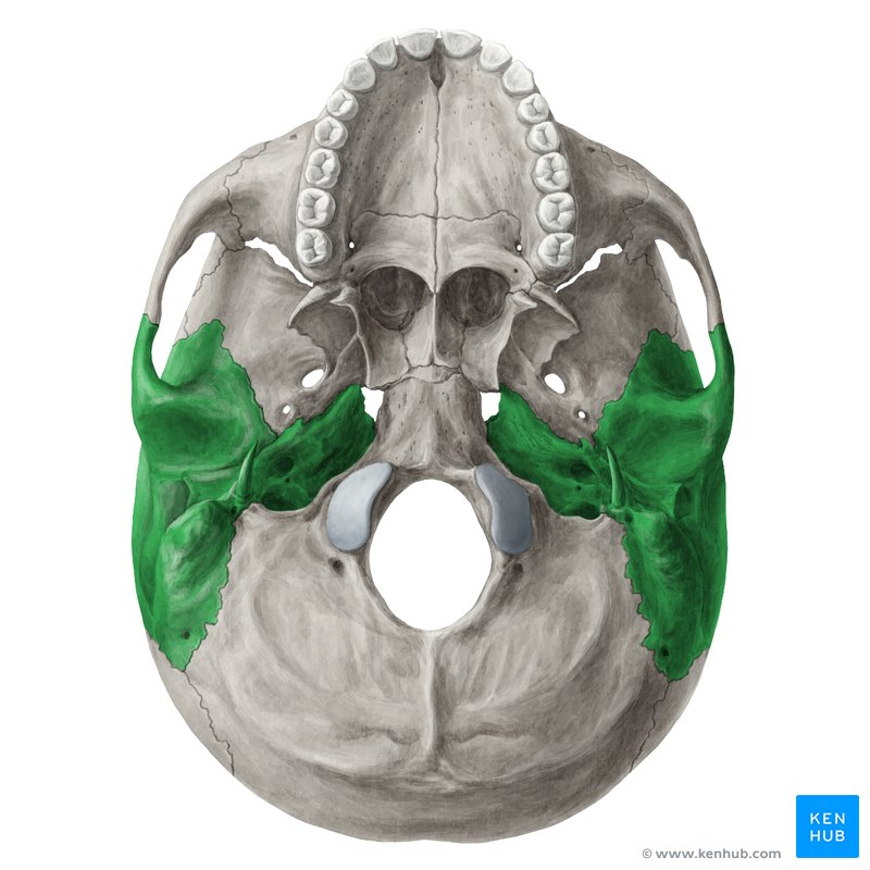
Temporal Bones
Side of the skull
4
New cards
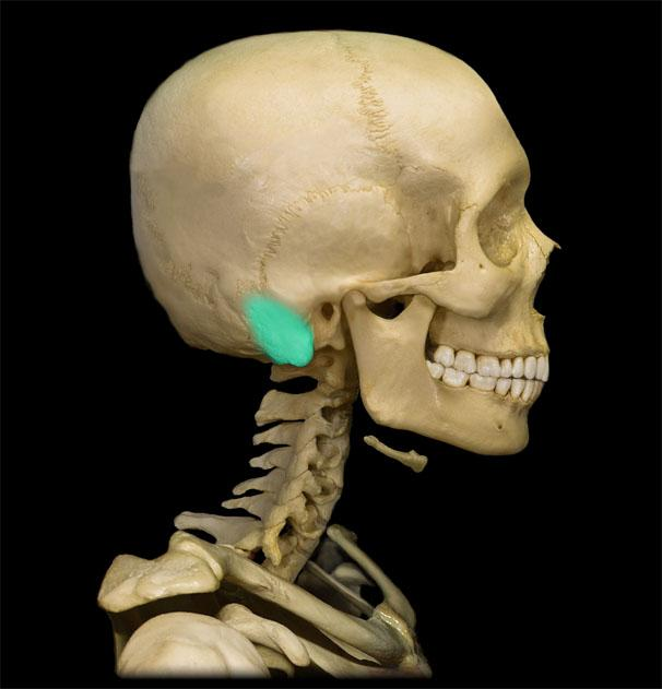
Mastoid Process
Projection at the base of the skull (Part of Temporal Bones)
5
New cards
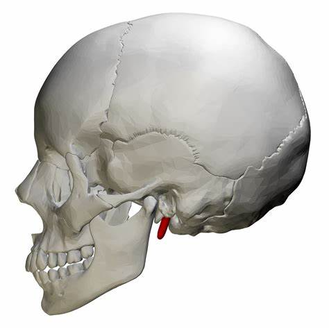
Styloid Process
Needle-like projection (Part of Temporal Bones)
6
New cards
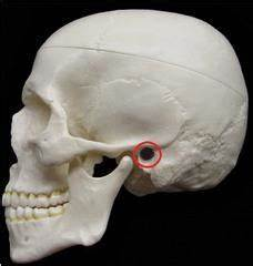
External Auditory Canal
Canal behind the jaw (Part of Temporal Bones)
7
New cards
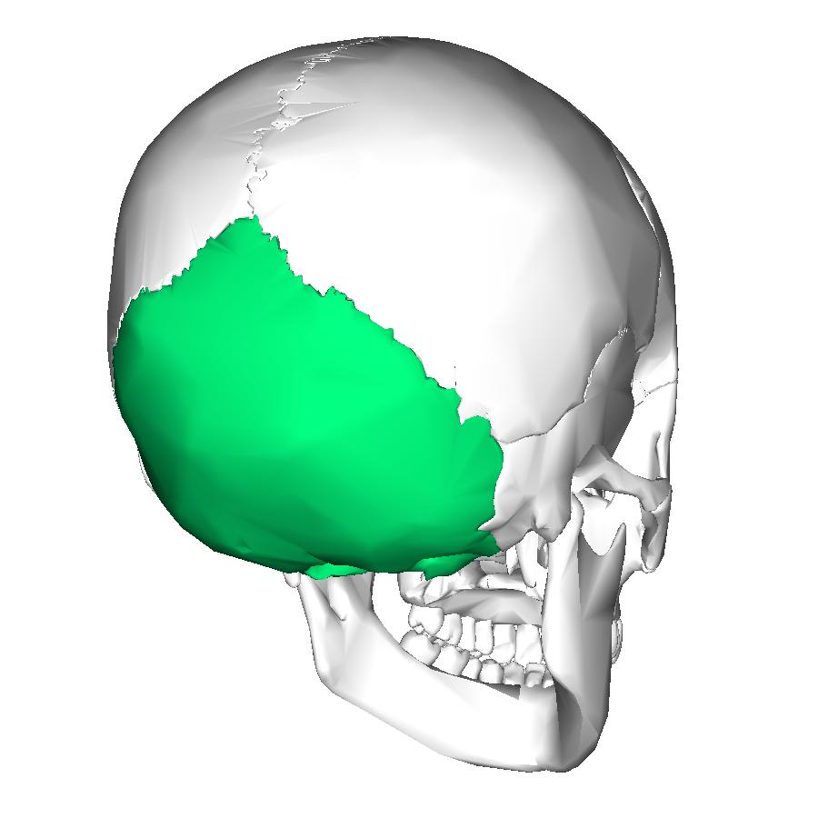
Occipital Bone
Back bottom of skull
8
New cards
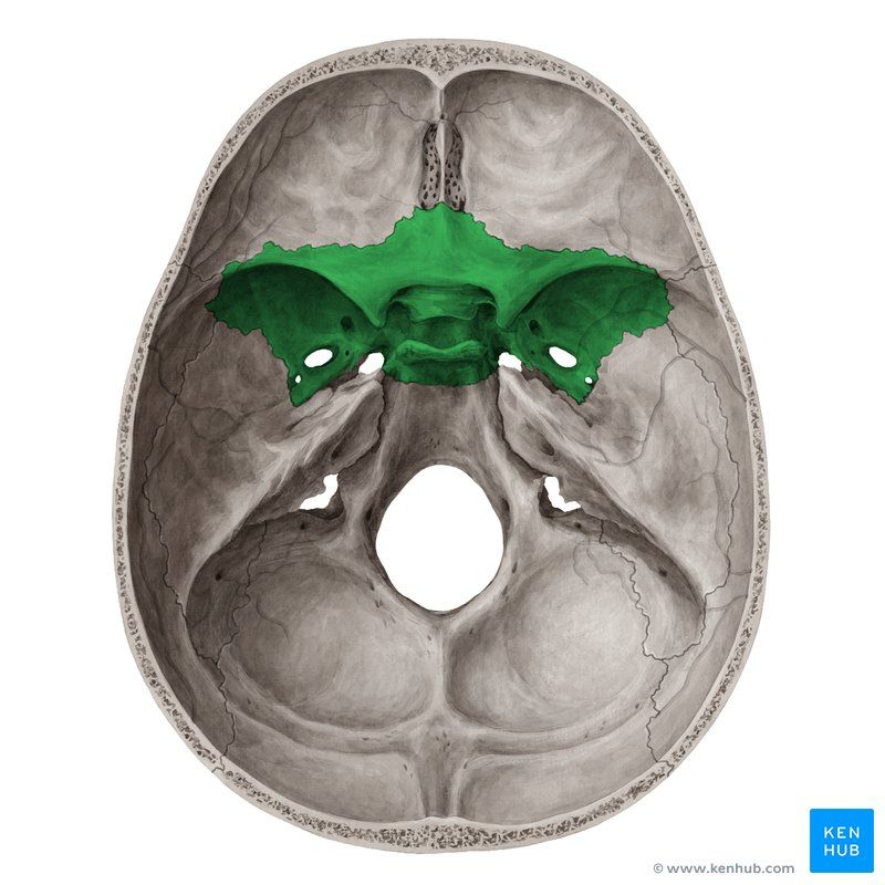
Sphenoid Bone
Looks like a butterfly, behind the jaw from frontal view
9
New cards
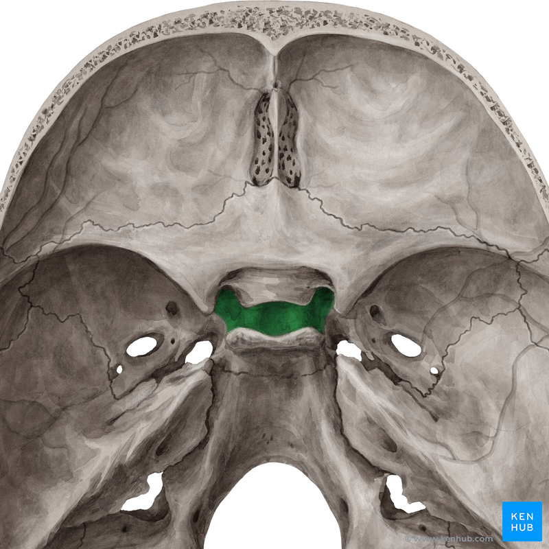
Sella Turcica
Part of the sphenoid bone
10
New cards
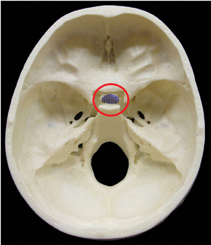
Hypophyseal Fossa
Deepest part of Sella Turcica (Part of Sphenoid)
11
New cards
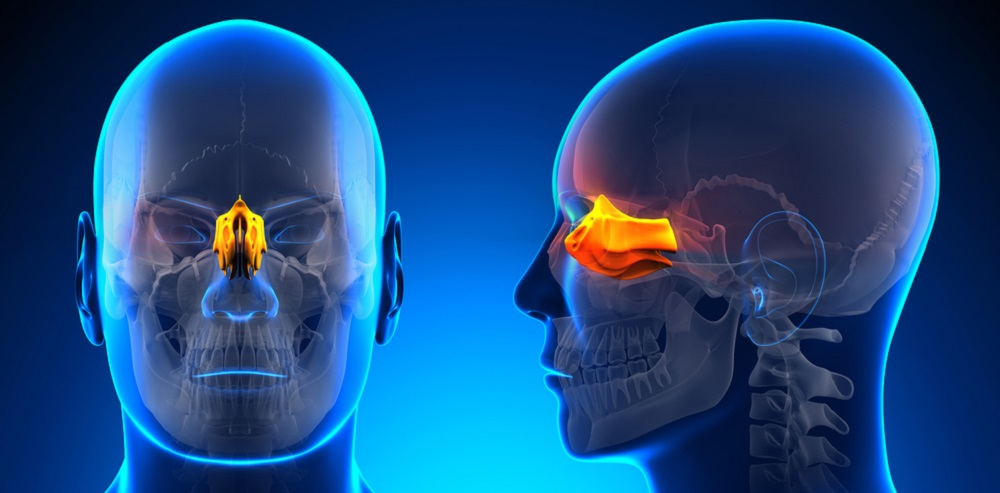
Ethmoid Bone
Behind the nasal bone
12
New cards
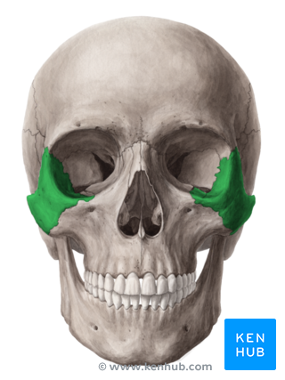
Zygomatic Bones
Cheek Bones
13
New cards
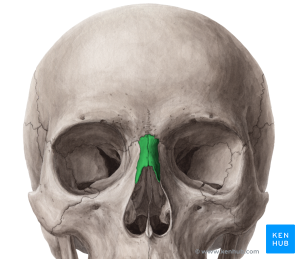
Nasal Bones
Located right above nasal cavity
14
New cards
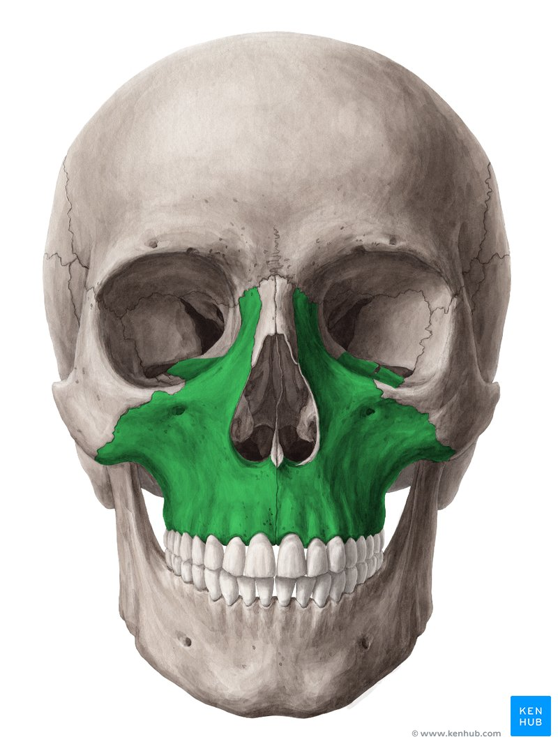
Maxillae
\
15
New cards
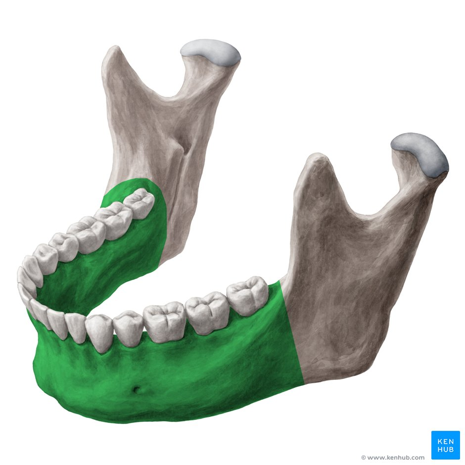
Mandible
Bottom part of jaw (Can be referred to as the body)
16
New cards
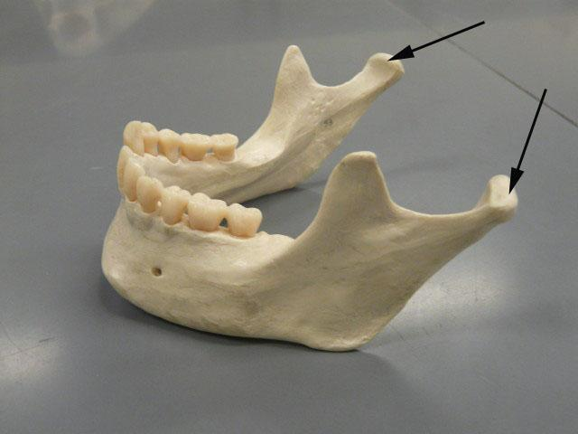
Mandibular Condyle
Where the jaw attaches to the skull (Part of Mandible)
17
New cards
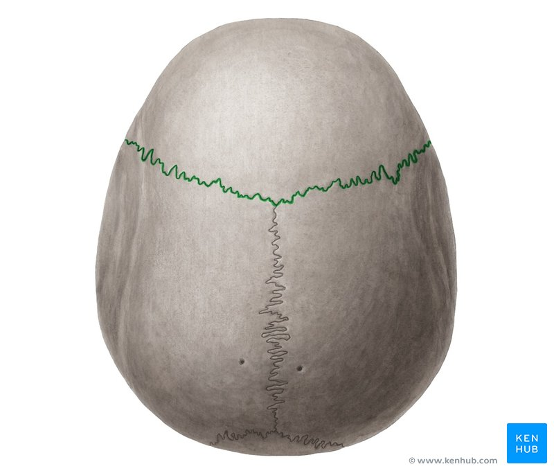
Coronal Suture
Horizontal (Top View) (Between frontal and parietal bone)
18
New cards
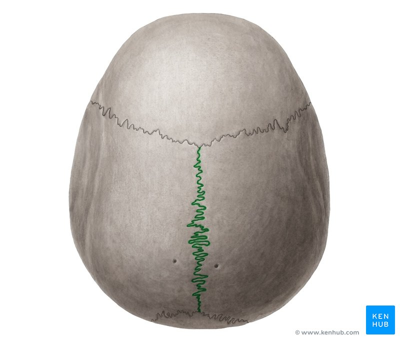
Sagittal Suture
Vertical (Top View) (Between 2 parietal bones)
19
New cards
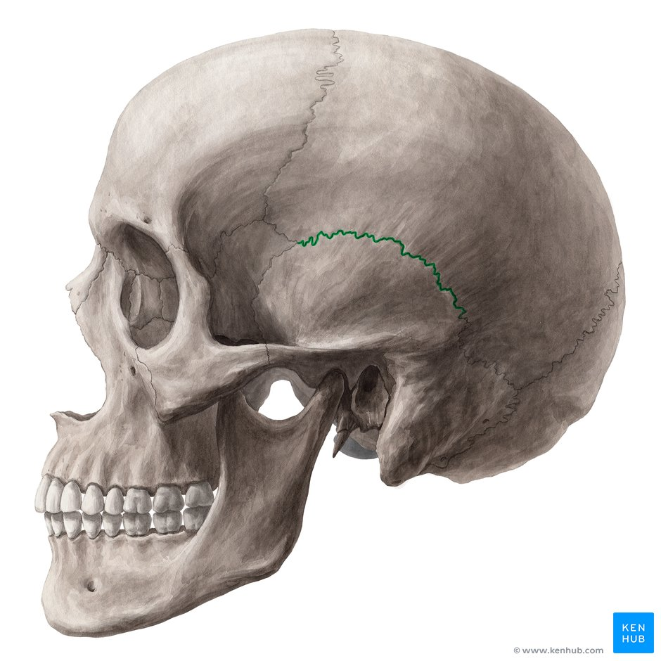
Squamous Suture
Between temporal and parietal bone
20
New cards
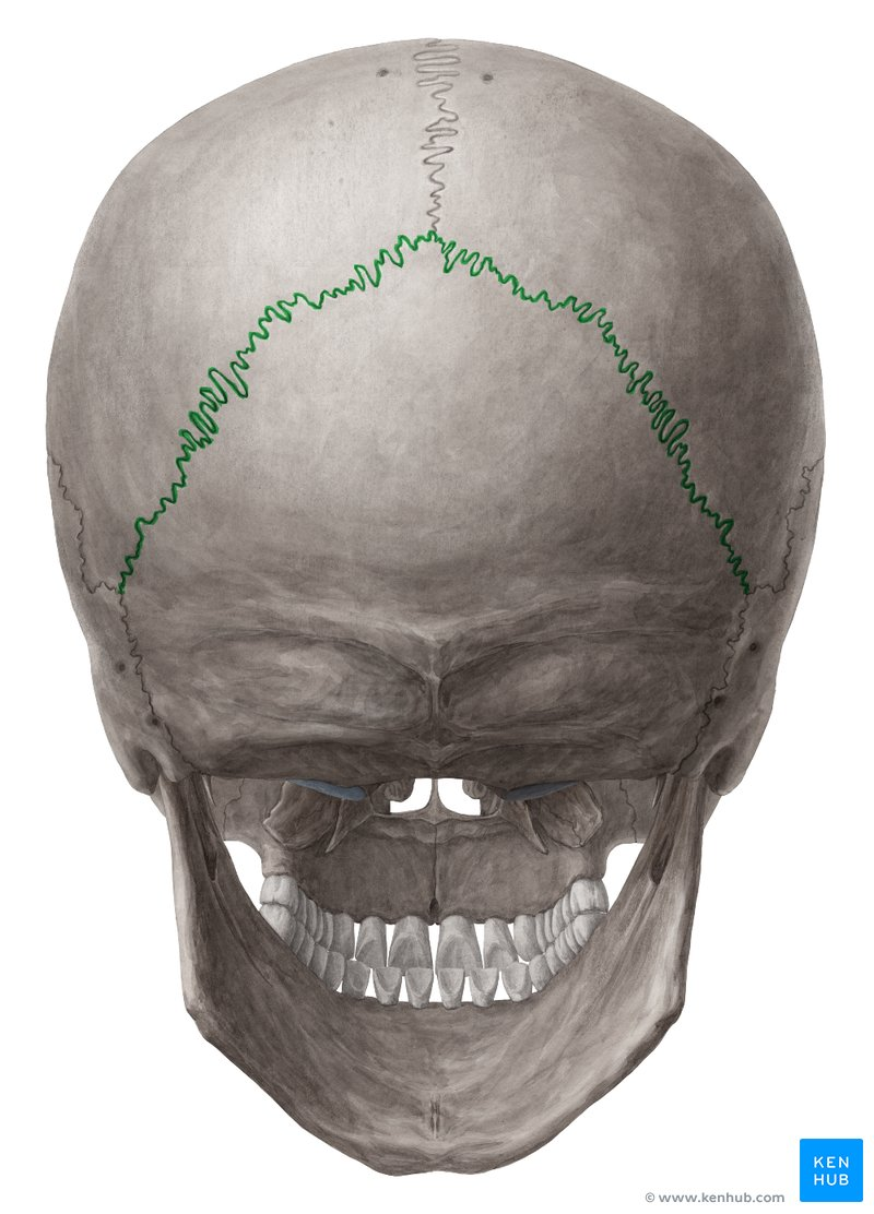
Lambdoid Suture
Between Occipital and Parietal
21
New cards
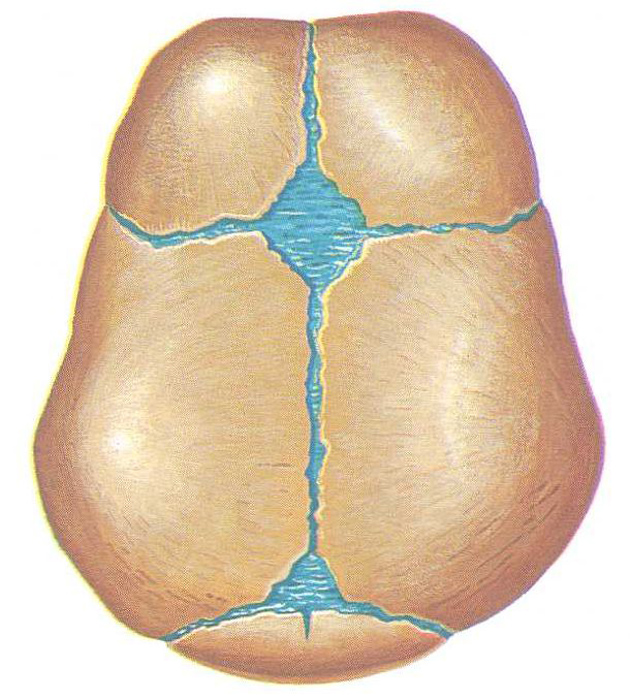
Fetal Skull- Anterior Fontanel
Top Opening in Fetal Skull
22
New cards
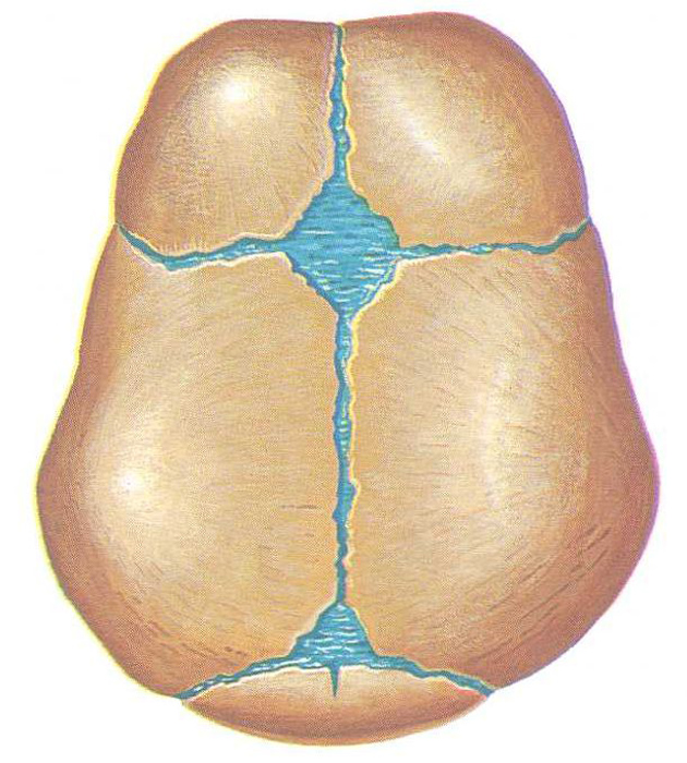
Fetal Skull- Posterior Fontanel
Bottom Opening in Fetal Skull
23
New cards
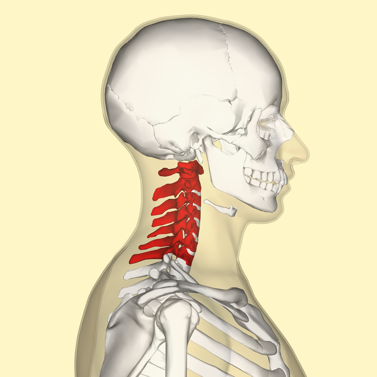
7 Cervical Vertebrae (C1-C7)
Numbered in order from top to bottom
24
New cards
C1= Atlas and C2= Axis
C1 and C2 Cervical Vertebrae are known respectively as…
25
New cards
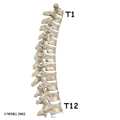
12 Thoracic Vertebrae (T1-T12)
26
New cards
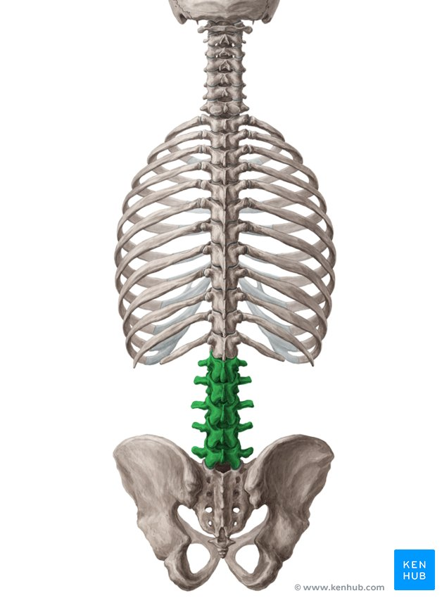
5 Lumber Vertebrae (L1-L5)
Top to Bottom (L1-L5)
27
New cards
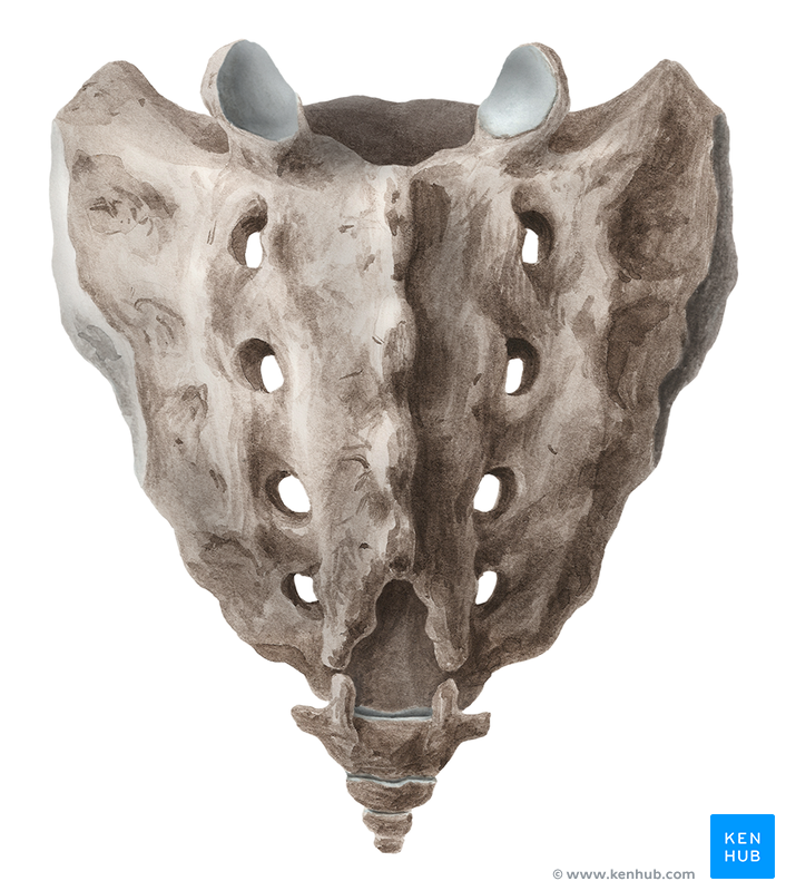
Sacrum (5 Fused Vertebrae)
Last bone of spinal cord
28
New cards
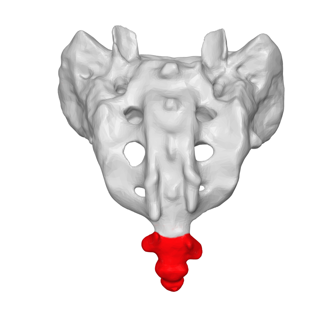
Coccyx (4 Fused Vertebrae)
Bottom of Saccrum
29
New cards
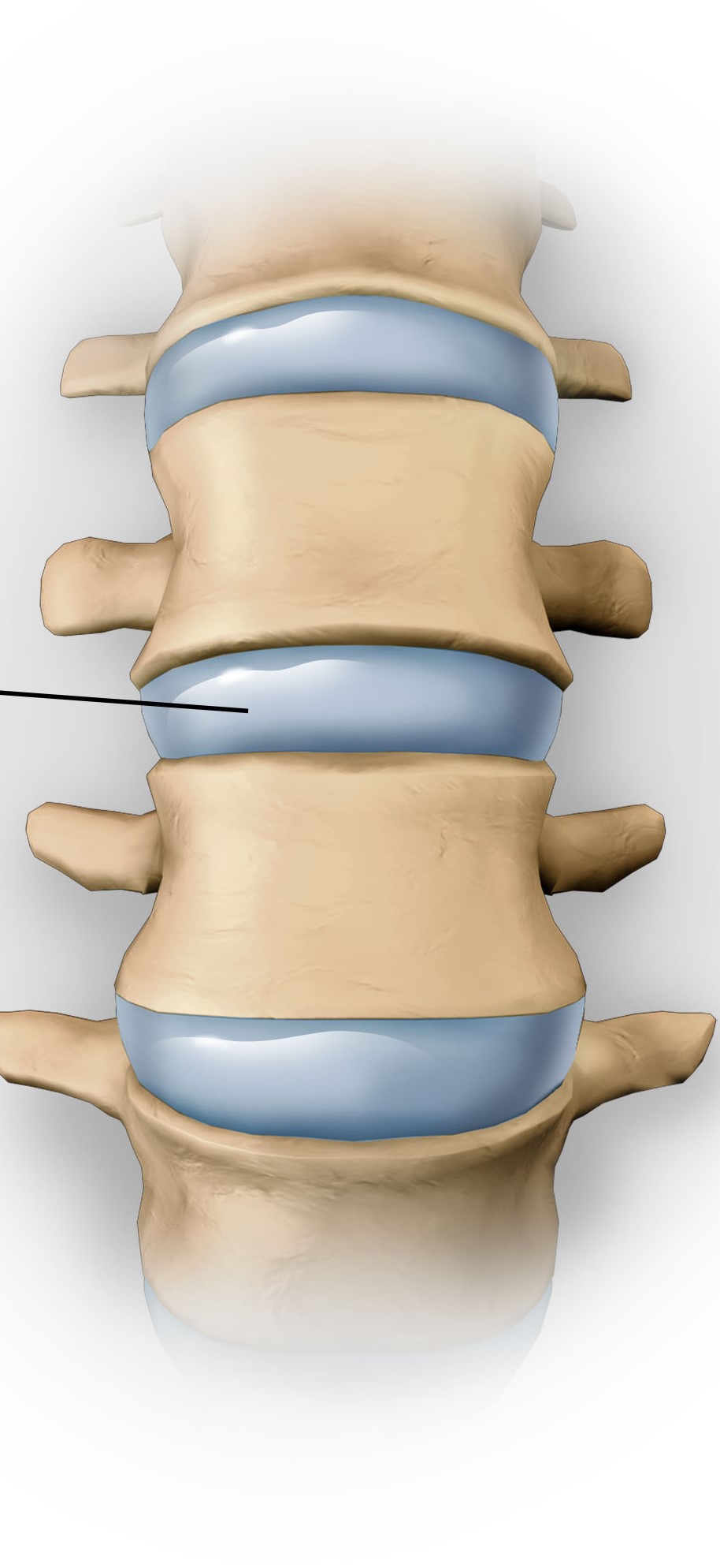
Intervertebral discs
Cushions between the bones of the spine that absorb shock and allow movement
30
New cards
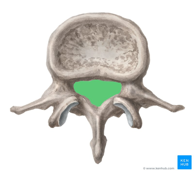
Vertebral Foramen
Opening in vertebrae
31
New cards
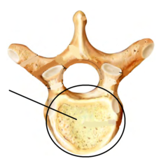
Vertebral Body
32
New cards
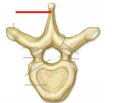
Spinous Process
Spiked part of Vertebrae
33
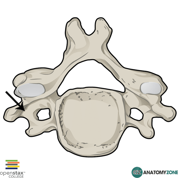
New cards
Transverse Process
Small bony projection off the right and left side of each vertebrae
34
New cards
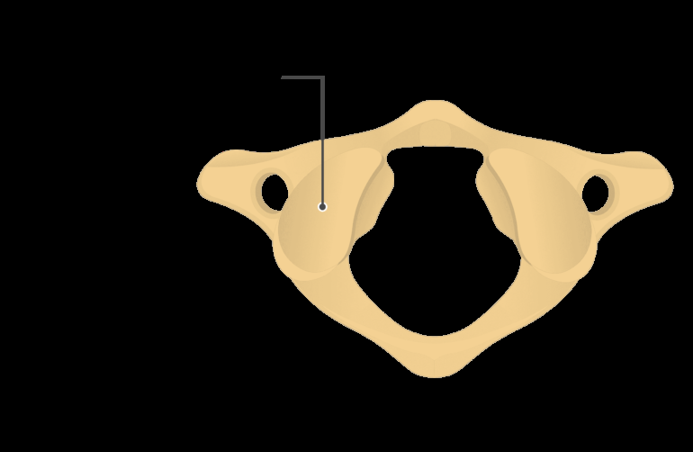
Superior Articular Surface
35
New cards
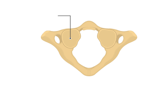
Inferior Articular Surface
\
36
New cards
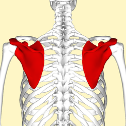
Scapula
37
New cards
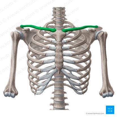
Clavicle
Medial (Sternal)
Lateral (Acromial)
Lateral (Acromial)
38
New cards
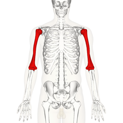
Humerus
39
New cards
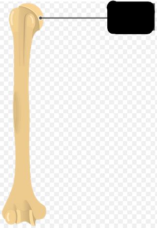
Head of Humerus
40
New cards
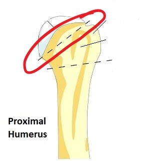
Anatomical Neck of Humerus
41
New cards
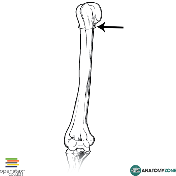
Surgical Neck of Humerus
42
New cards
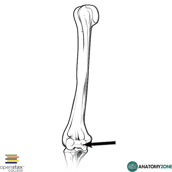
Trochlea of Humerus
Longer projection of the bottom of the humerus
43
New cards
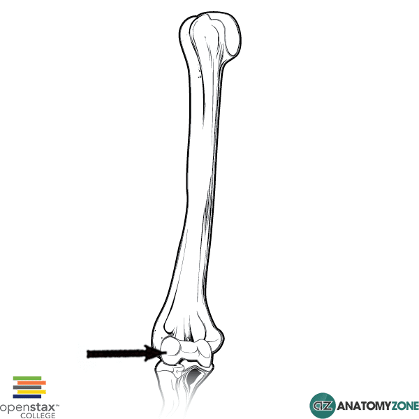
Capitulum
Shorter projection of the bottom of the humerus
44
New cards
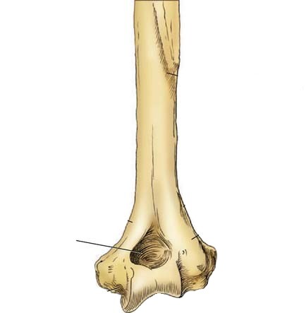
Olecranon Fossa
Deep triangular depression on the **posterior side** of the humerus
Superior to the Trochlea
Superior to the Trochlea
45
New cards
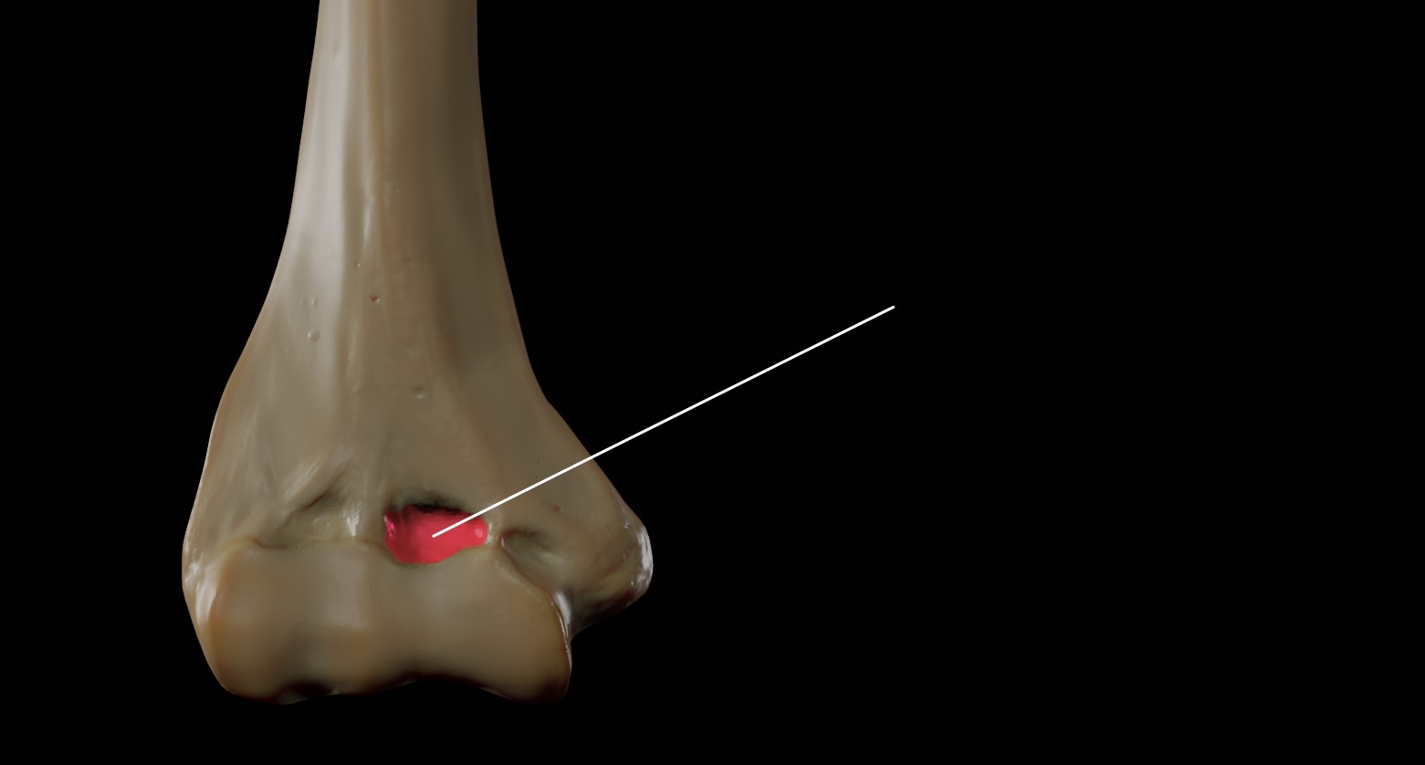
Coronoid Fossa
Slight depression at the distal end of the humerus on the **anterior side**
Superior to the Trachlea
Superior to the Trachlea
46
New cards
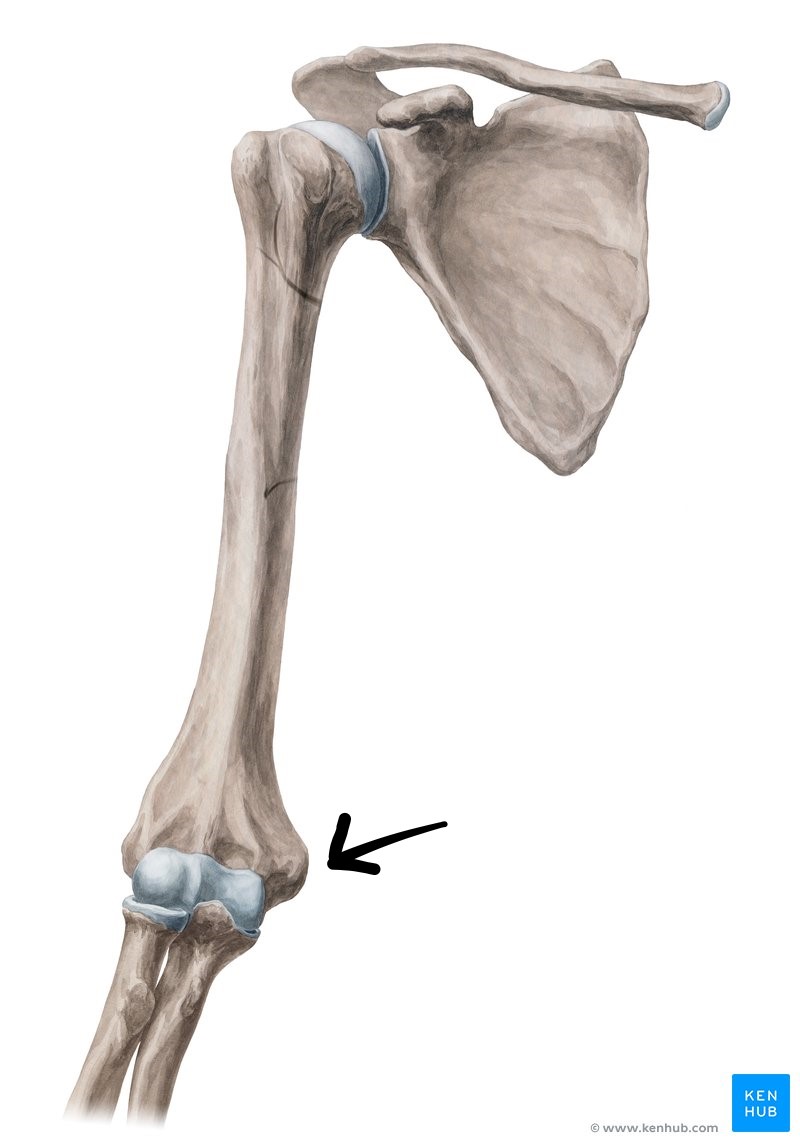
Medial Epicondyle of Humerus
47
New cards
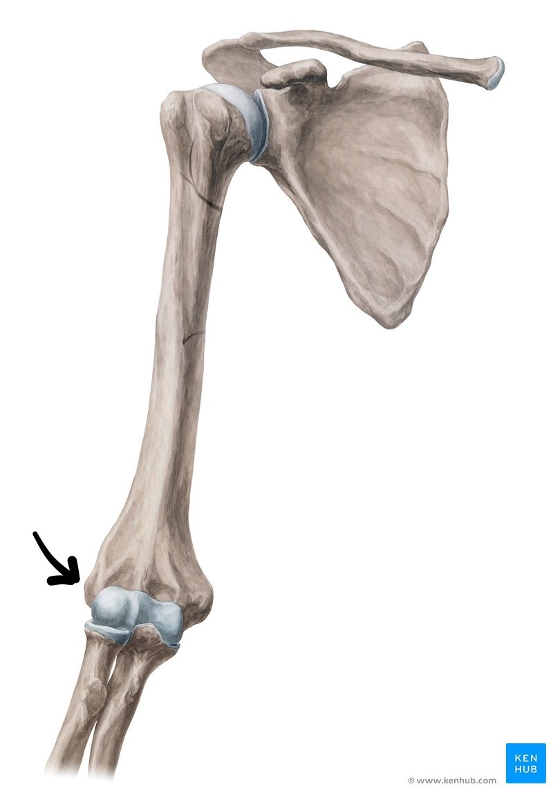
Lateral Epicondyle of Humerus
48
New cards
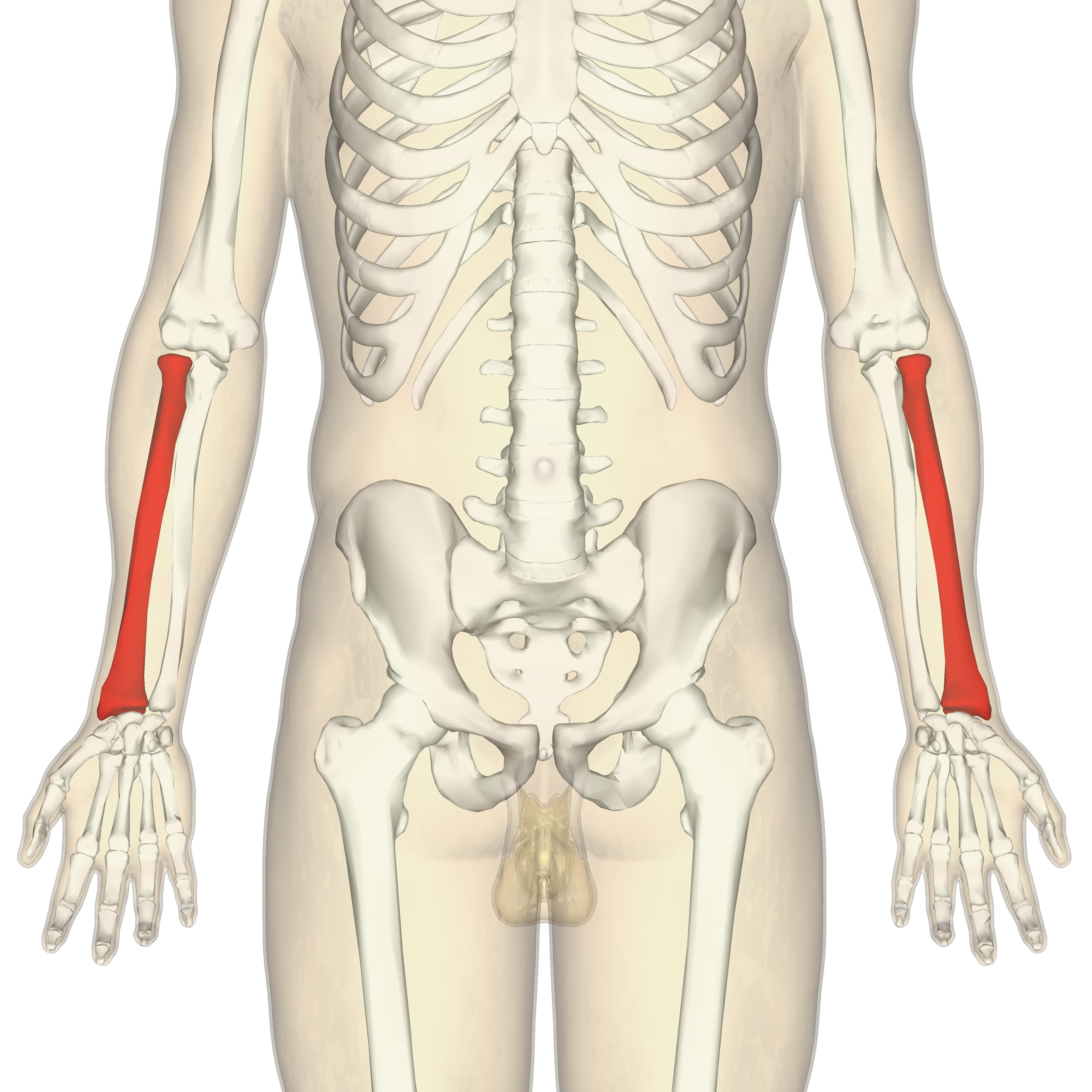
Radius
49
New cards
Head of Radius
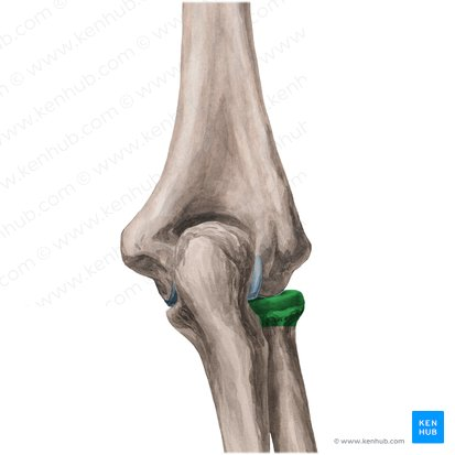
50
New cards
Neck of Radius
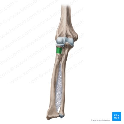
51
New cards
Trochlea of Radius
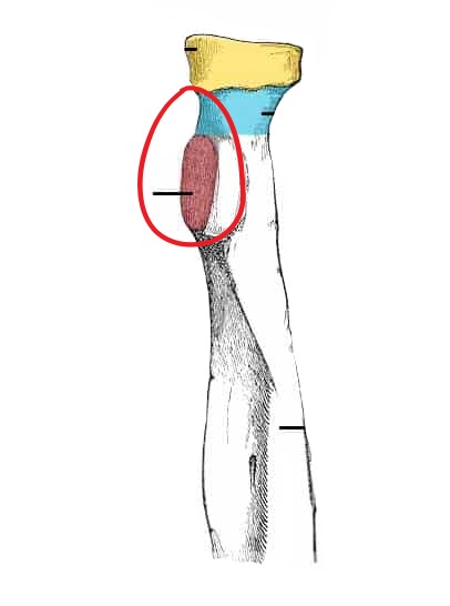
52
New cards
Styloid Process of Radius
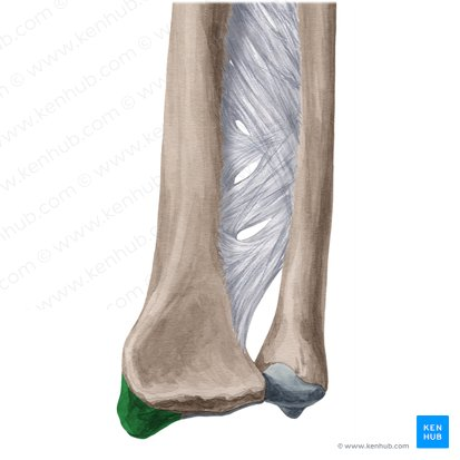
53
New cards
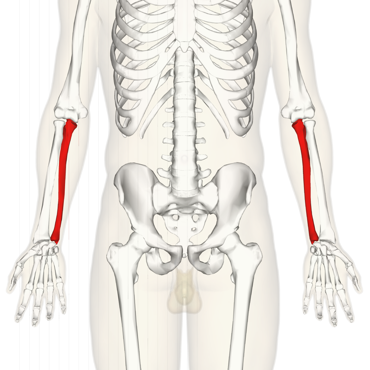
Ulna
54
New cards
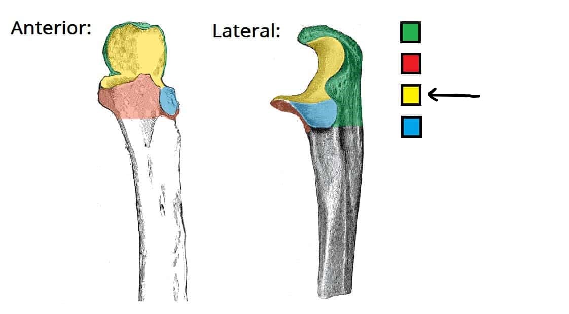
Trochlear Notch of Ulna
55
New cards
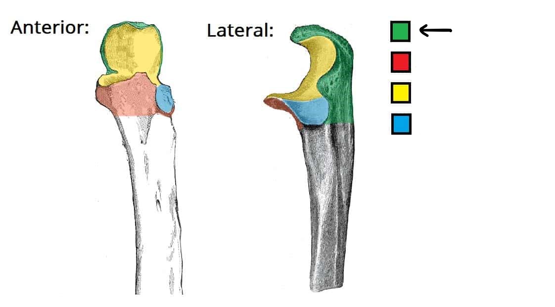
Olecranon of Ulna
56
New cards
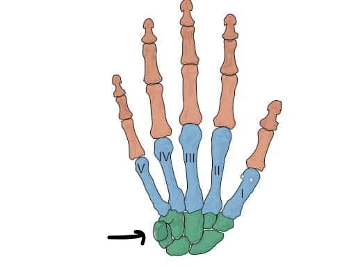
Carpals
57
New cards
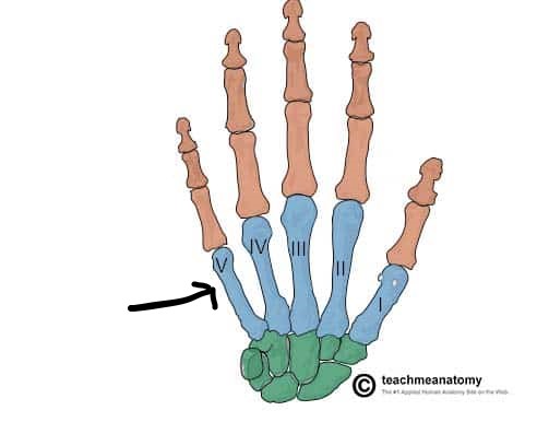
Metacarpals
Numbered I-V from Thumb to Pinky
58
New cards
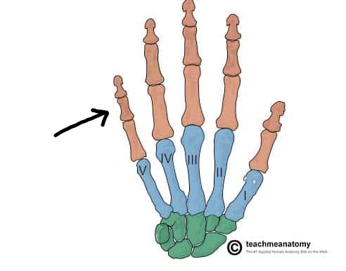
Phalanges of Hand
59
New cards
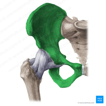
Hip Bones
60
New cards
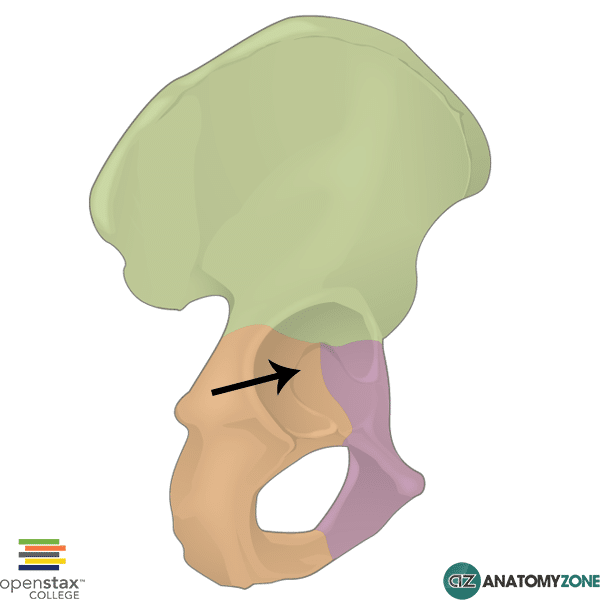
Acetabulum (Socket)
The socket where the head of the femur fits
61
New cards
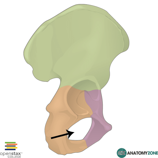
Obturator Foramen
Large opening in the hipbone between pubic and ischium
62
New cards
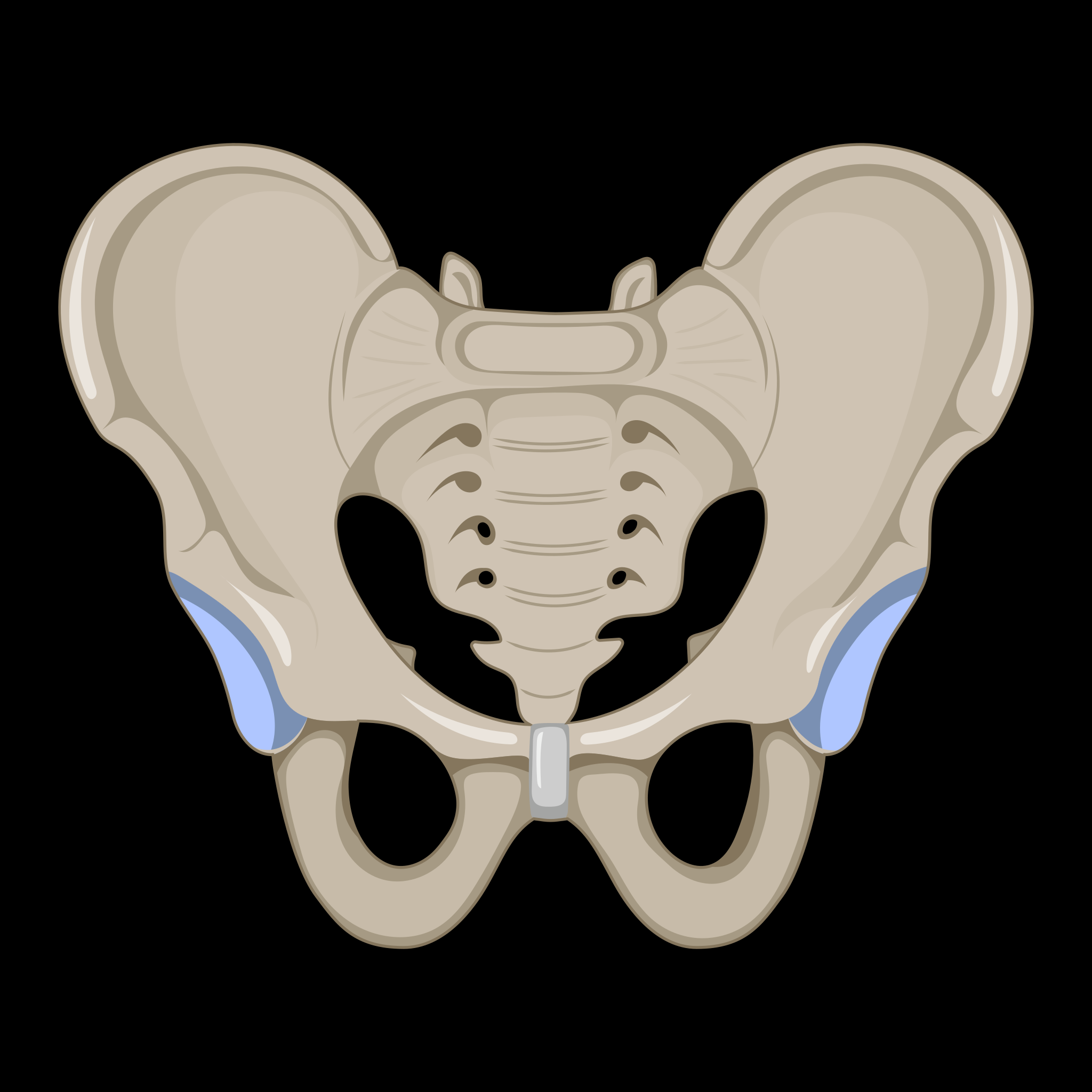
Male Pelvis
Which Pelvis is this?
*
*
63
New cards
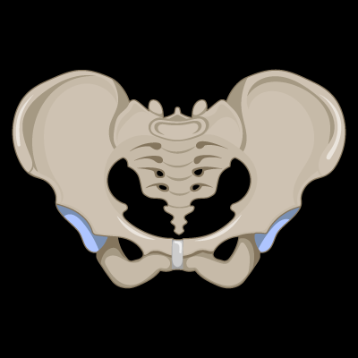
Female Pelvis
Which Pelvis is this?
* >90° Pubic Arch (Angle at the Bottom of the Pelvis)
* Shorter and Wider
* Shallow Pelvic Cavity
* >90° Pubic Arch (Angle at the Bottom of the Pelvis)
* Shorter and Wider
* Shallow Pelvic Cavity
64
New cards
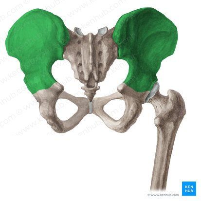
Ilium
The large broad bone forming the upper part of each half of the pelvis
65
New cards
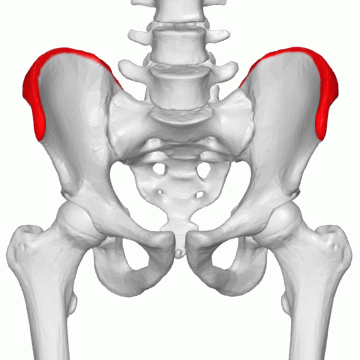
Iliac Crest
Commonly known as the hip bone
66
New cards
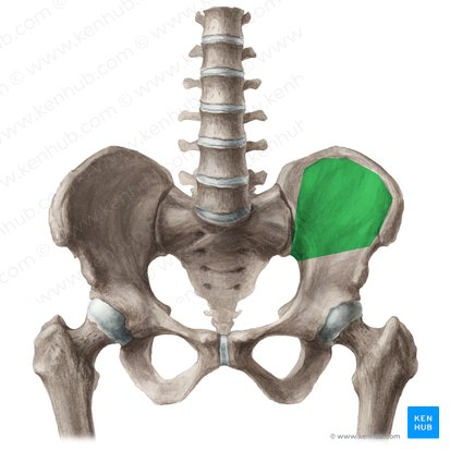
Iliac Fossa
Large, smooth, concave surface on the internal surface of the ilium
67
New cards
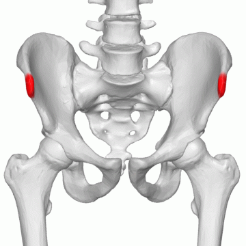
Anterior Superior Iliac Spine
68
New cards
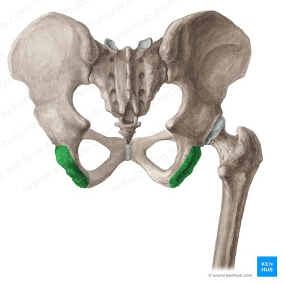
Ischium
69
New cards
Greater Sciatic Notch
\
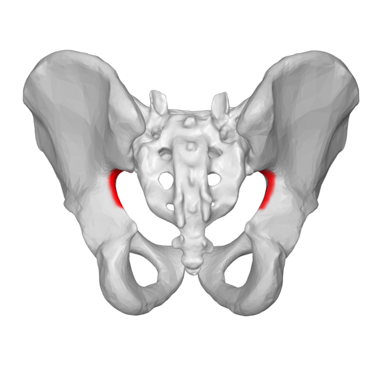
70
New cards
Ischial Tuberosity
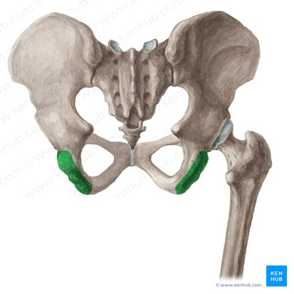
71
New cards
Ischial Spine
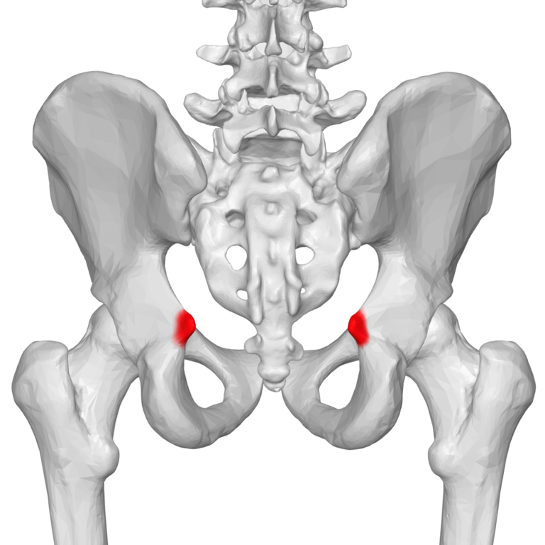
72
New cards
Pubis
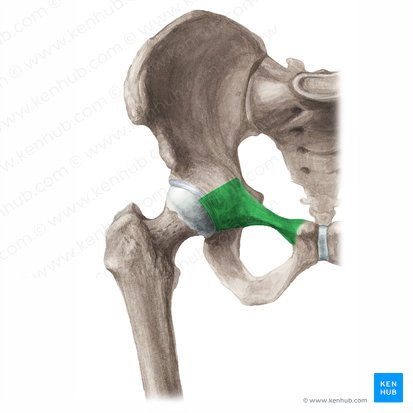
73
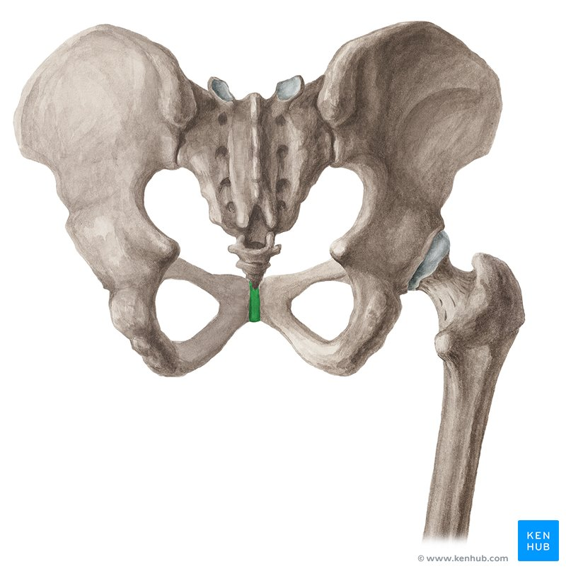
New cards
Pubic Symphysis
A joint sandwiched between the left and right pelvic bone
74
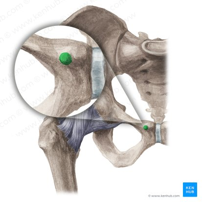
New cards
Pubic Tubercle
75
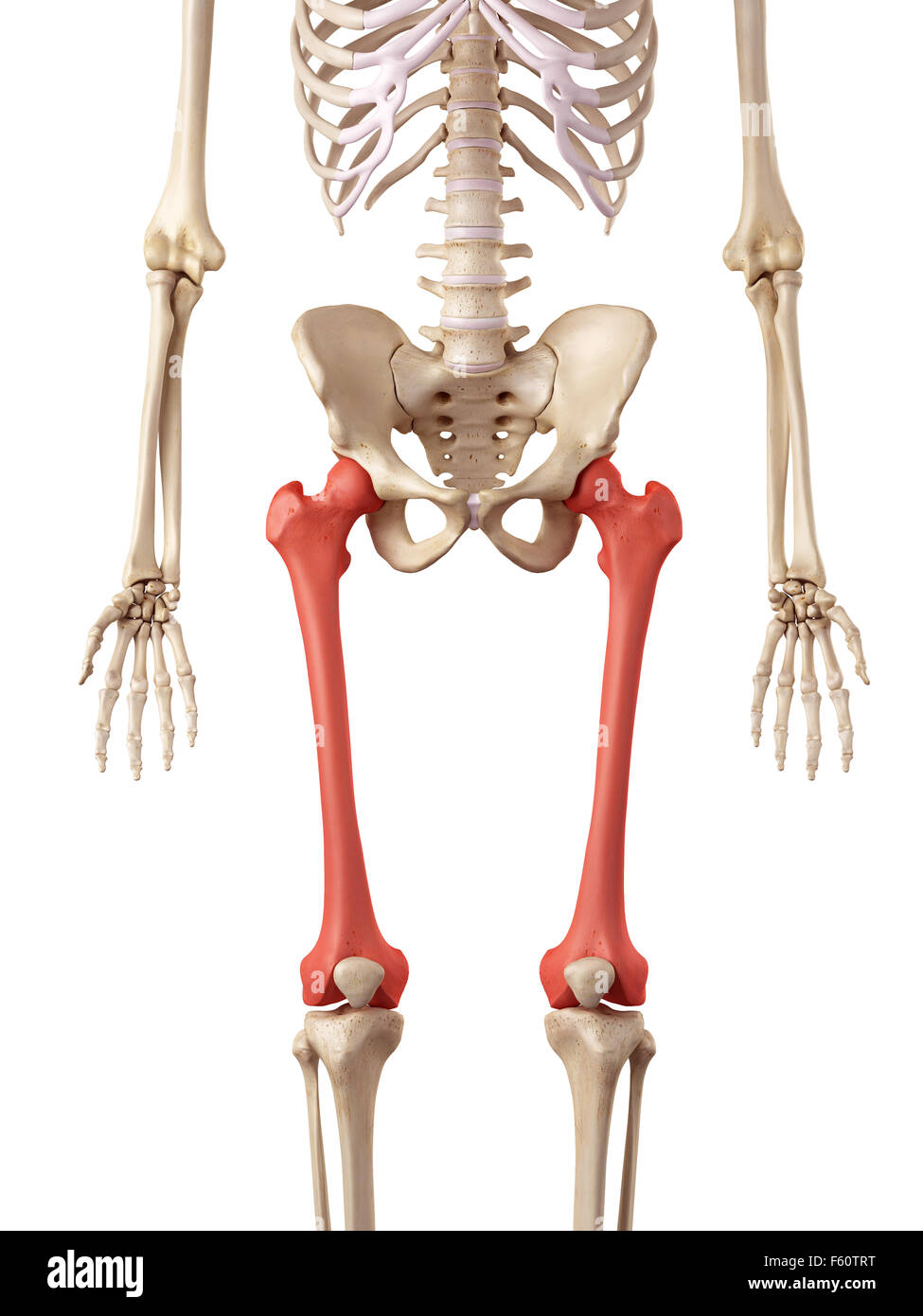
New cards
Femur
\
76
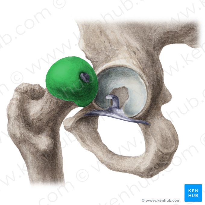
New cards
Head of Femur
77
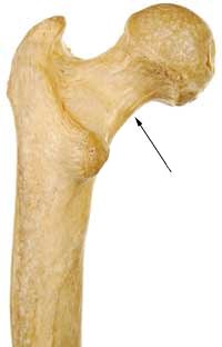
New cards
Neck of Femur
78
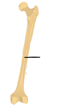
New cards
Linea Aspera
79
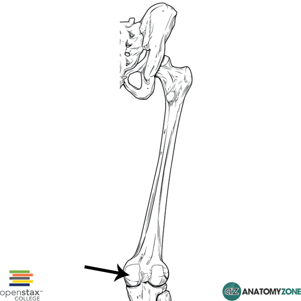
New cards
Medial Condyle
80
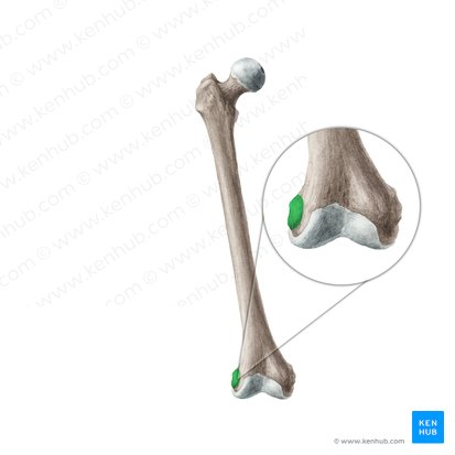
New cards
Lateral Condyle
\
81
New cards
Tibia
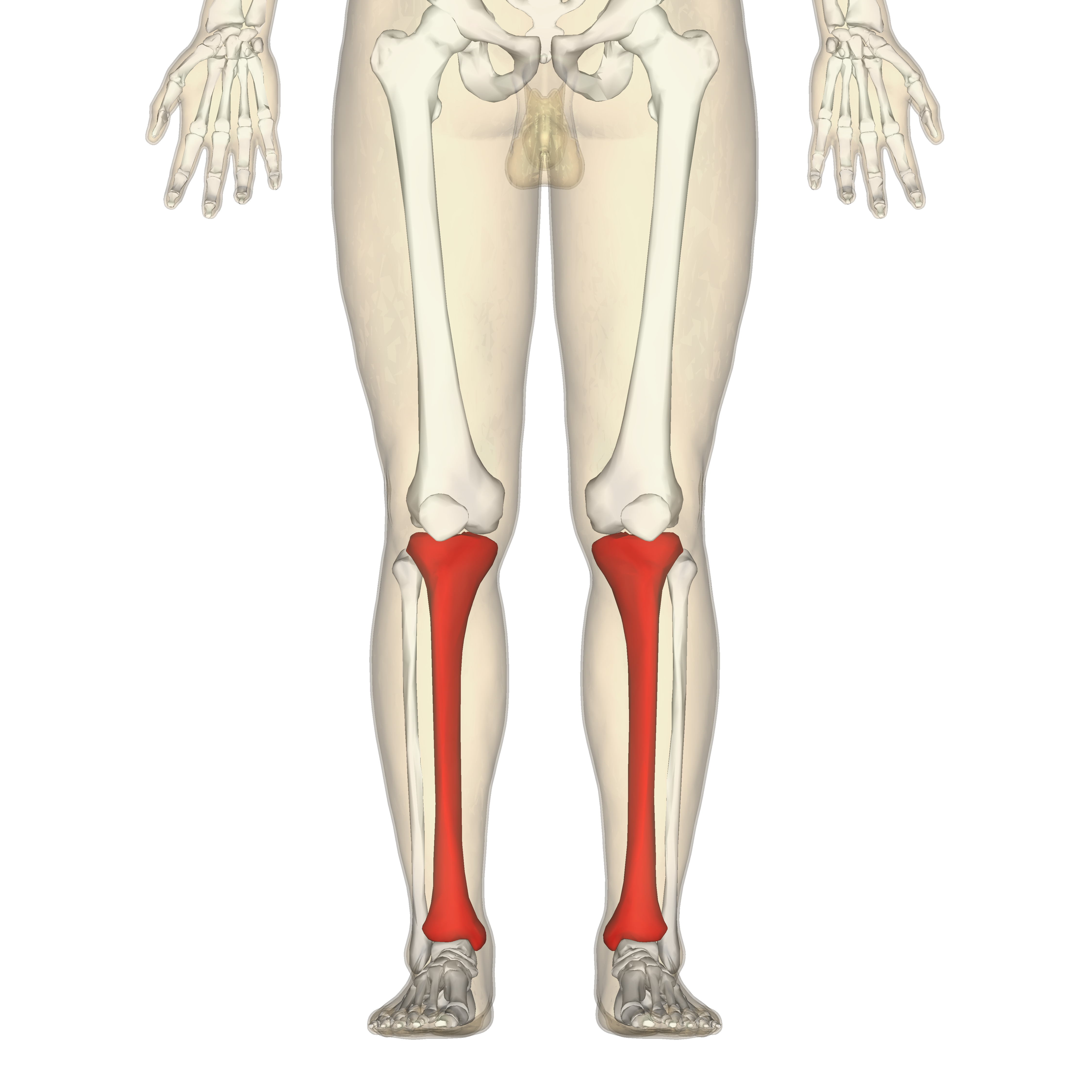
82
New cards
Medial Condyle of the Tibia
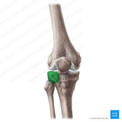
83
New cards
Lateral Condyle of the Tibia
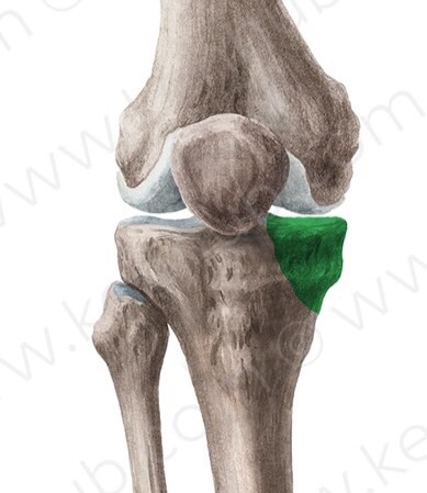
84
New cards
Medial Malleolus of Tibia
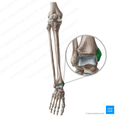
85
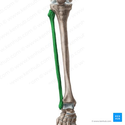
New cards
Fibula
86
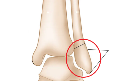
New cards
Lateral Malleolus
87
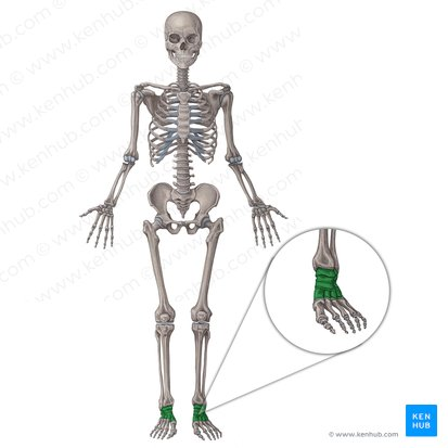
New cards
Tarsal Bones
88
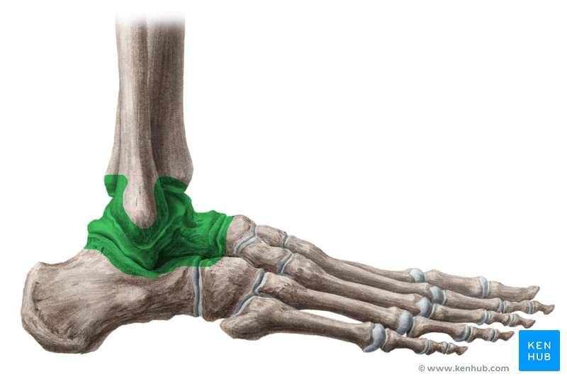
New cards
Talus
89
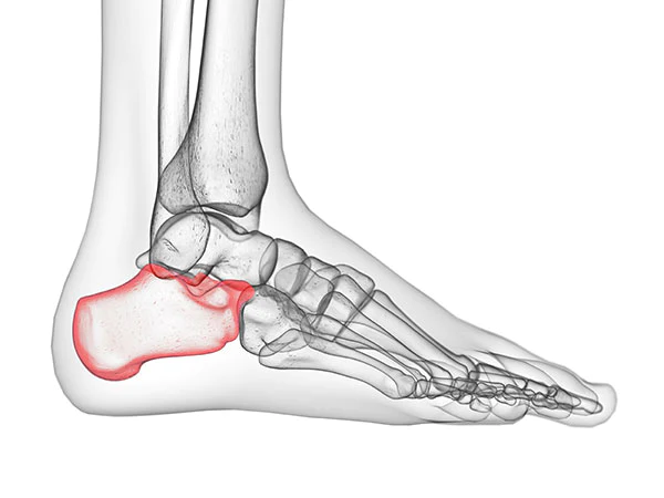
New cards
Calcaneus
90
New cards
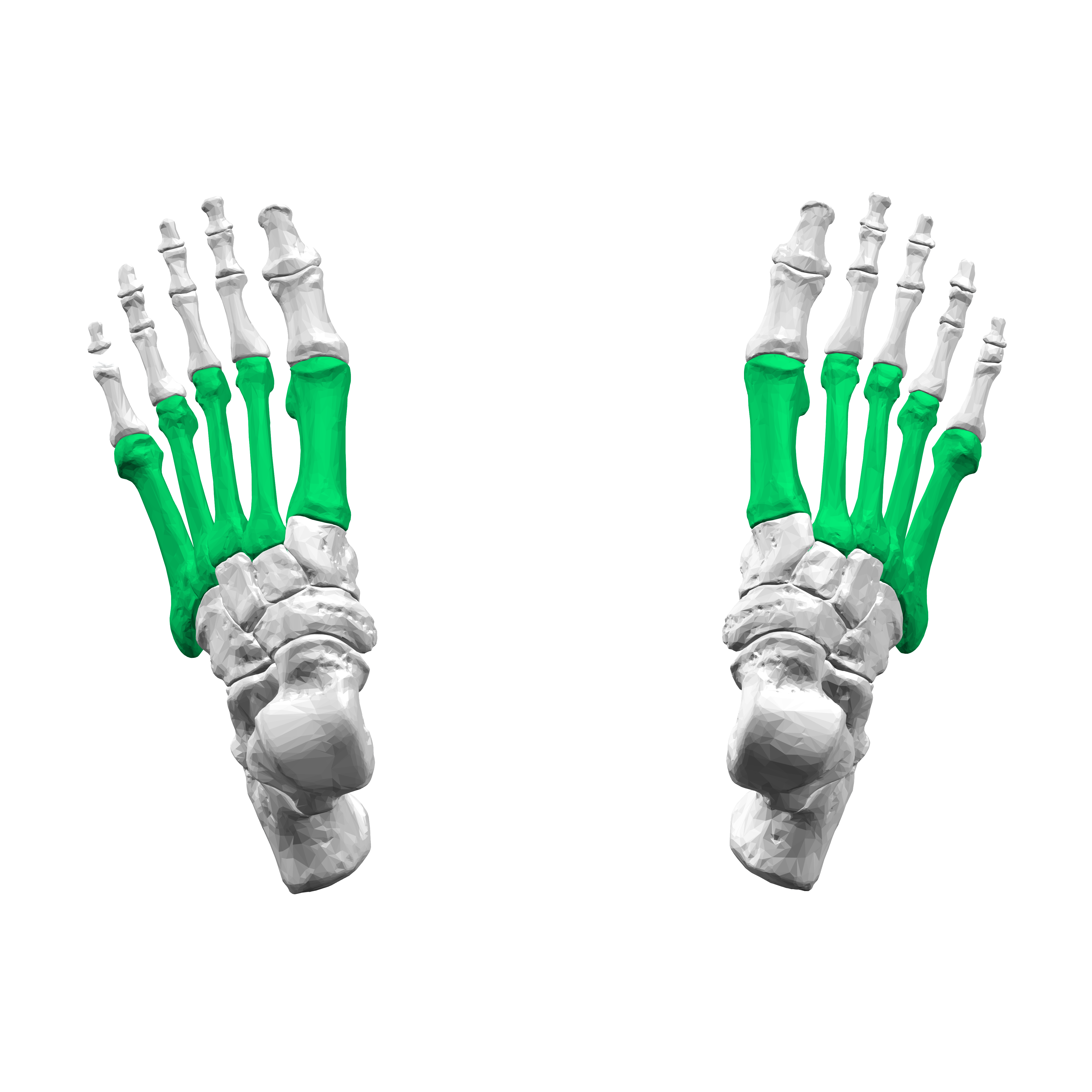
Metatarsals
I-V from inside out
91
New cards
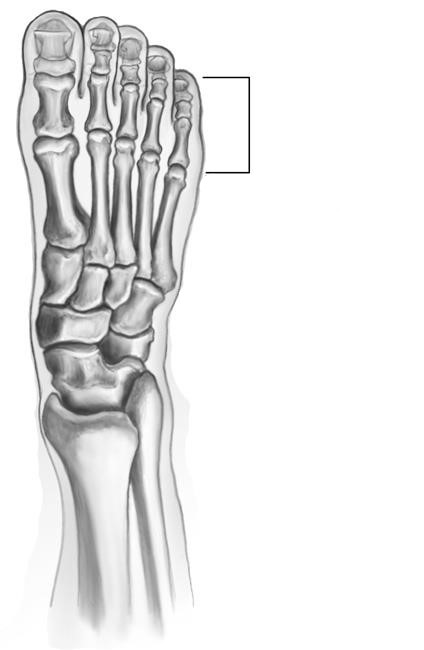
Phalanges of Foot
I-V from inside out
92
New cards
Synarthrosis Joint
Immovable Joints
Ex: Sutures of Skull
Ex: Sutures of Skull
93
New cards
Amphiarthrosis Joint
Slightly Moveable Joints
Ex: Intervertebral Discs
Ex: Intervertebral Discs
94
New cards
Diarthrosis Joint
Freely Moveable Joints
Ex: Shoulder
Ex: Shoulder
95
New cards
Fibrous Joint
Joints where adjacent bones are strongly united by fibrous connective tissue
Ex: Sutures of Skull
Ex: Sutures of Skull
96
New cards
Cartilaginous Joint
Type of joint where the bones are entirely joined by cartilage
Ex: Intervertebral Discs
Ex: Intervertebral Discs
97
New cards
Synovial Joints
Joints where the ends of bones are encased in smooth cartilage
Ex: Shoulder Joints in the arm
Ex: Shoulder Joints in the arm
98
New cards
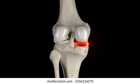
Medial Meniscus
99
New cards
Lateral Meniscus
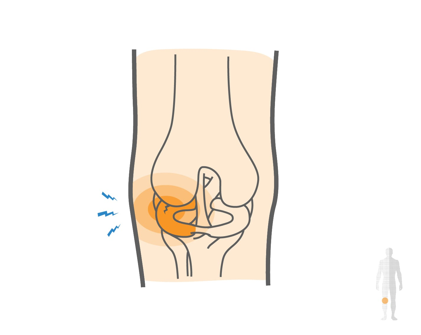
100
New cards
Articular Cartilage
