Anatomy Exam #4: Respiratory System
1/163
There's no tags or description
Looks like no tags are added yet.
Name | Mastery | Learn | Test | Matching | Spaced | Call with Kai |
|---|
No analytics yet
Send a link to your students to track their progress
164 Terms
parts of the upper respiratory system
nose, nasal cavity, paranasal sinuses, pharynx
parts of the lower respiratory system
larynx, trachea, bronchus, bronchioles, alveoli
function of the conducting respiratory system?
tubes to get oxygen in
function of the respiratory system?
- providing an extensive surface area for gas exchange
- moving air to and from (conducting)
- protecting respiratory surfaces from dehydration, temp changes, or other environmental variation
- producing sounds (phonation)
- facilitating the detection of olfactory stimuli (olfaction = smelling)
mucus escalator
The upward movement of mucus in the lungs caused by the coordinated movement of cilia.
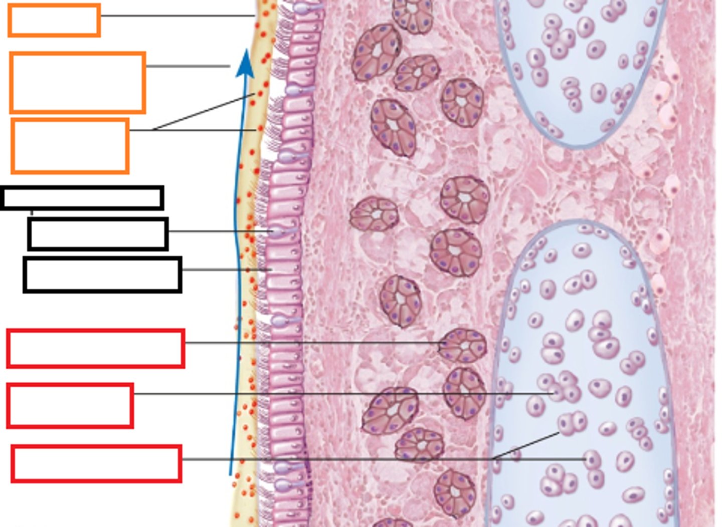
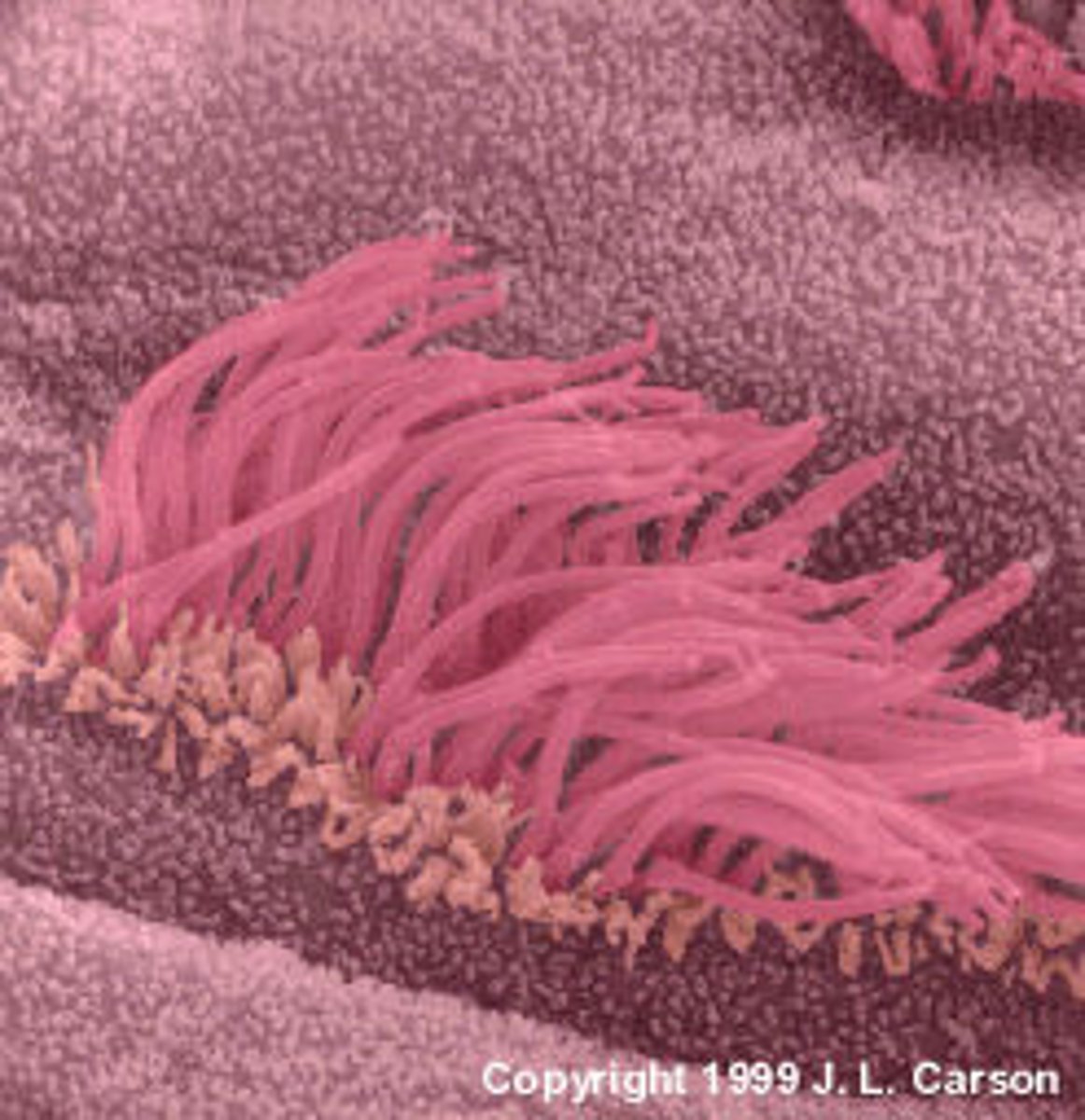
cilia
The hairlike projections on the outside of cells that move in a wavelike manner
goblet cells
pseudo-stratified columnar cell found in the respiratory and intestinal tracts, which secretes the main component of mucus through merocrine secretion
lamina propria
areolar connective tissue (loose connective tissue) underlying a mucous membrane
nasal
pseudo-stratified columnar epithelium (mucus)
pharynx
stratified squamous epithelium (protection)
trachea and primary bronchi
pseudo-stratified columnar epithelium (mucus)
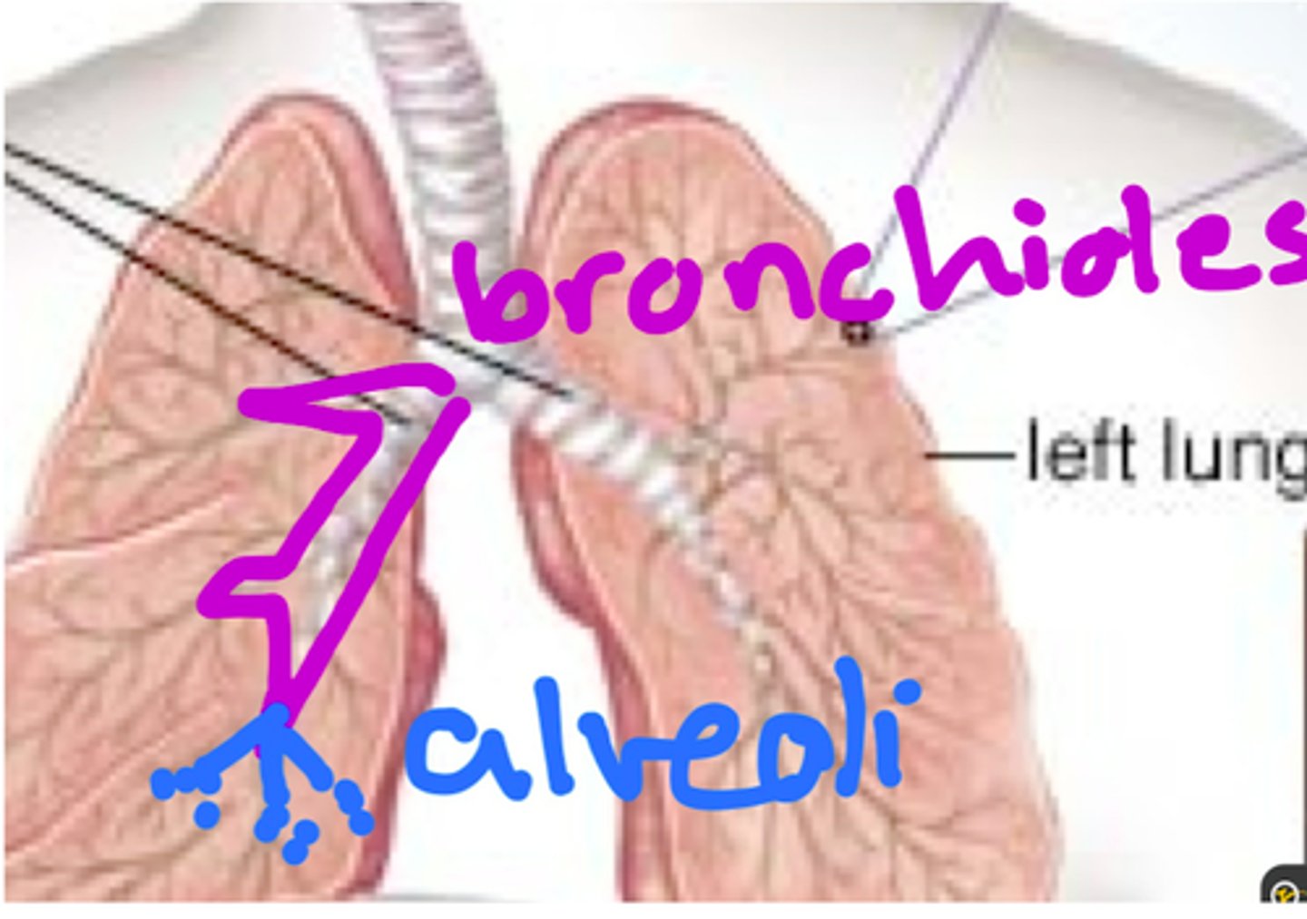
bronchioles
simple cuboidal epithelium (secretion/absorption)
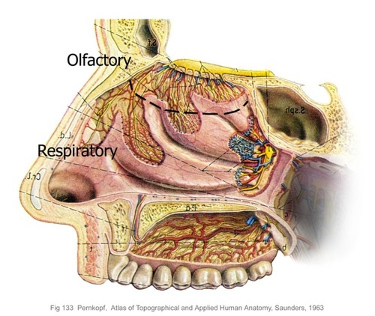
respiratory
simple squamous epithelium (communication/gas exchange)
location of nasal cavity
Inferior to the nasal bone and superior to the oral cavity
location of pharynx
behind the oral cavity, between the nasal cavity and larynx
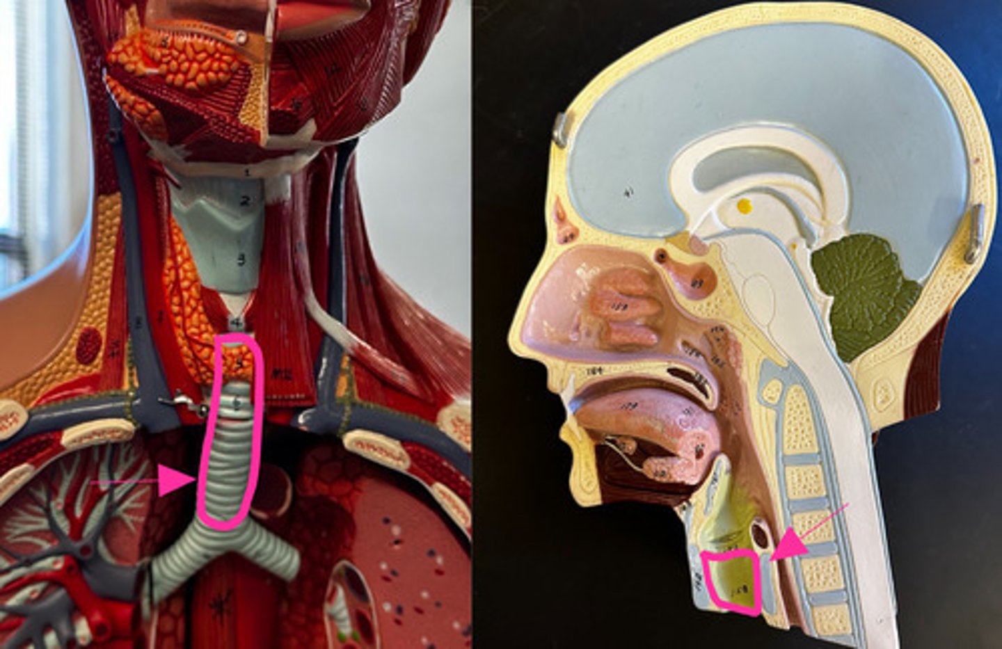
location of trachea
anterior to the esophagus
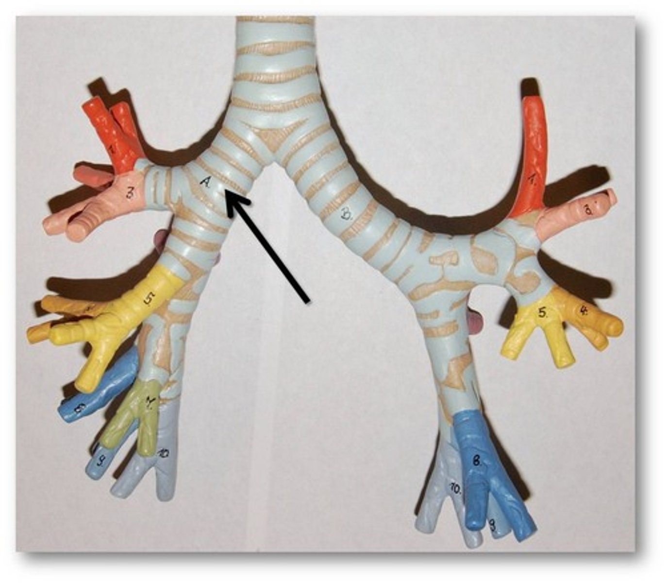
location of primary bronchi
runs obliquely in the mediastinum before plunging into the medial depression of the lungs
location of bronchioles
lungs
extrapulmonary
outside the lungs
intrapulmonary
inside the lungs
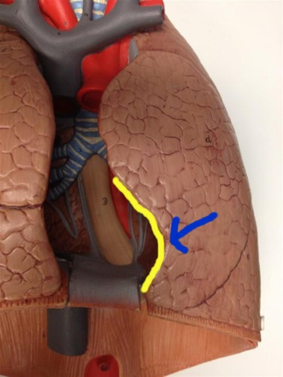
left lung
2 lobes: superior and inferior
1 fissure: oblique
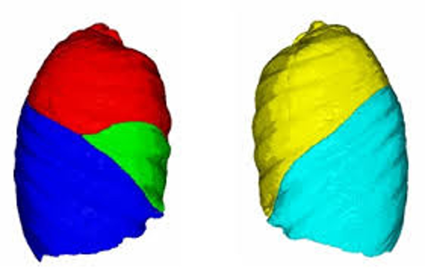
right lung
-3 lobes: Superior, Middle, and Inferior
-2 fissures: Horizontal and Oblique

cardiac notch
a concave space on the left lung in which the heart lies
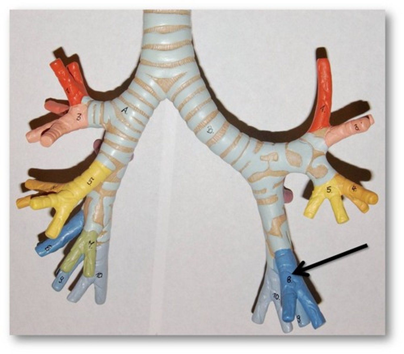
secondary bronchus
branches of the primary bronchi which supply each lobe of lung; there are 2 in the left lung and 3 in the right lung
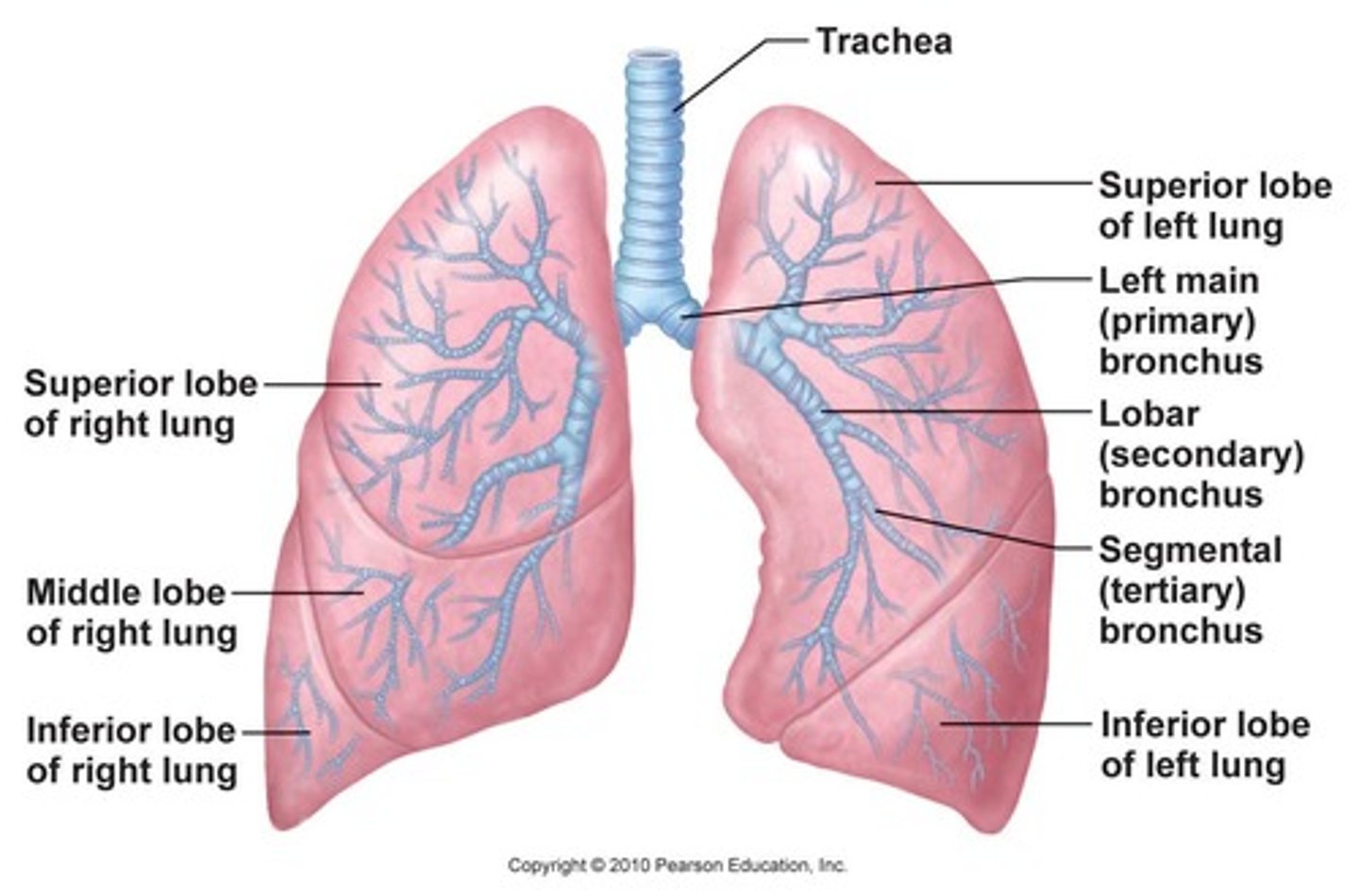
tertiary bronchus
Extends from the secondary bronchus and conducts air to each lobule of the lungs.
layers of the pleural cavity
- parietal
- pleural
- visceral
parietal pleura
outer layer of pleura lying closer to the ribs and chest wall
viseral pleura
the inner layer of pleura that surrounds each lung
air conducing passage
- nasal/oral cavity
- pharynx
- larynx
- trachea
- primary bronchi
- secondary bronchi
- tertiary bronchi
- bronchioles
- terminal bronchioles
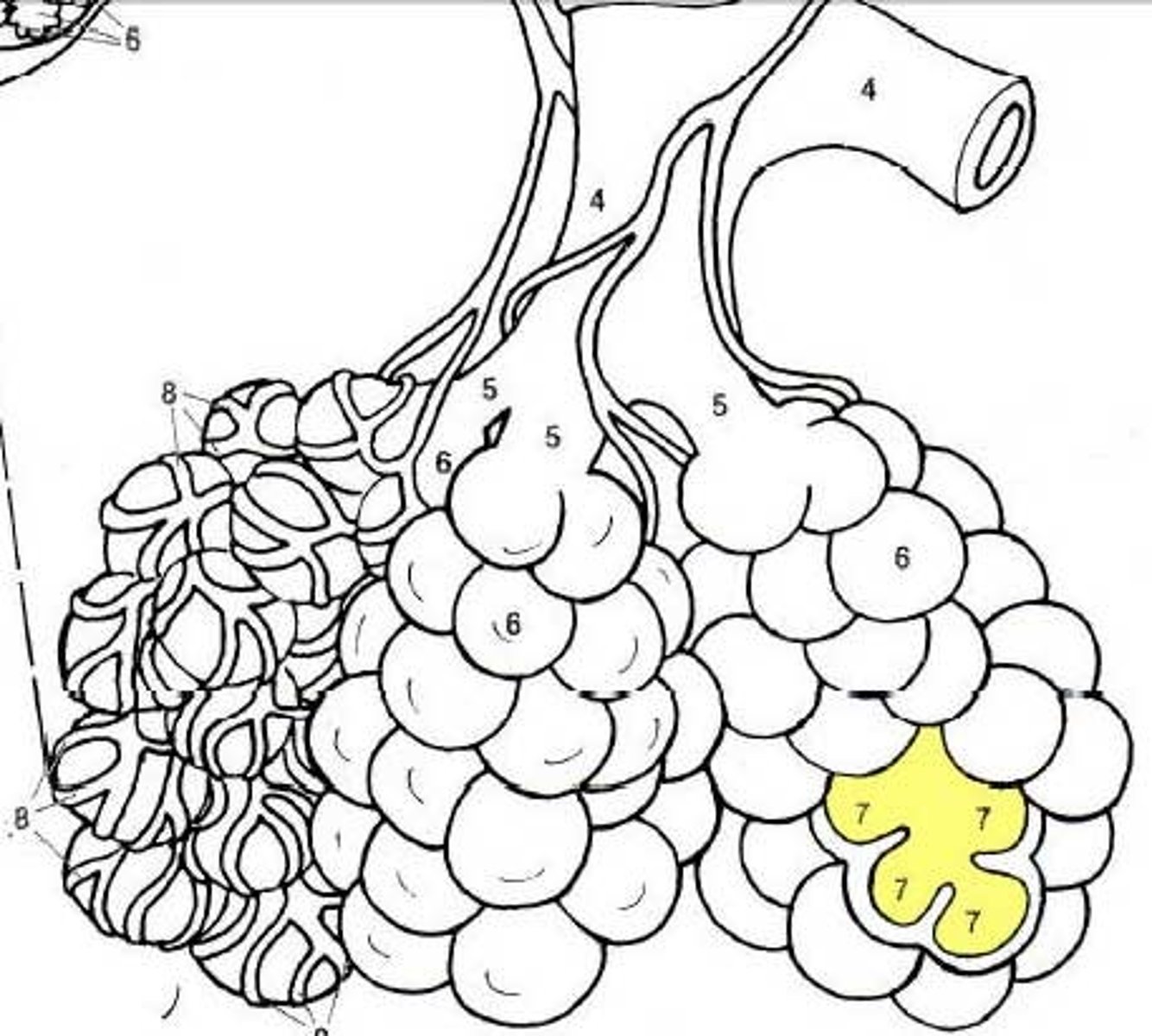
terminal bronchioles
smallest bronchioles
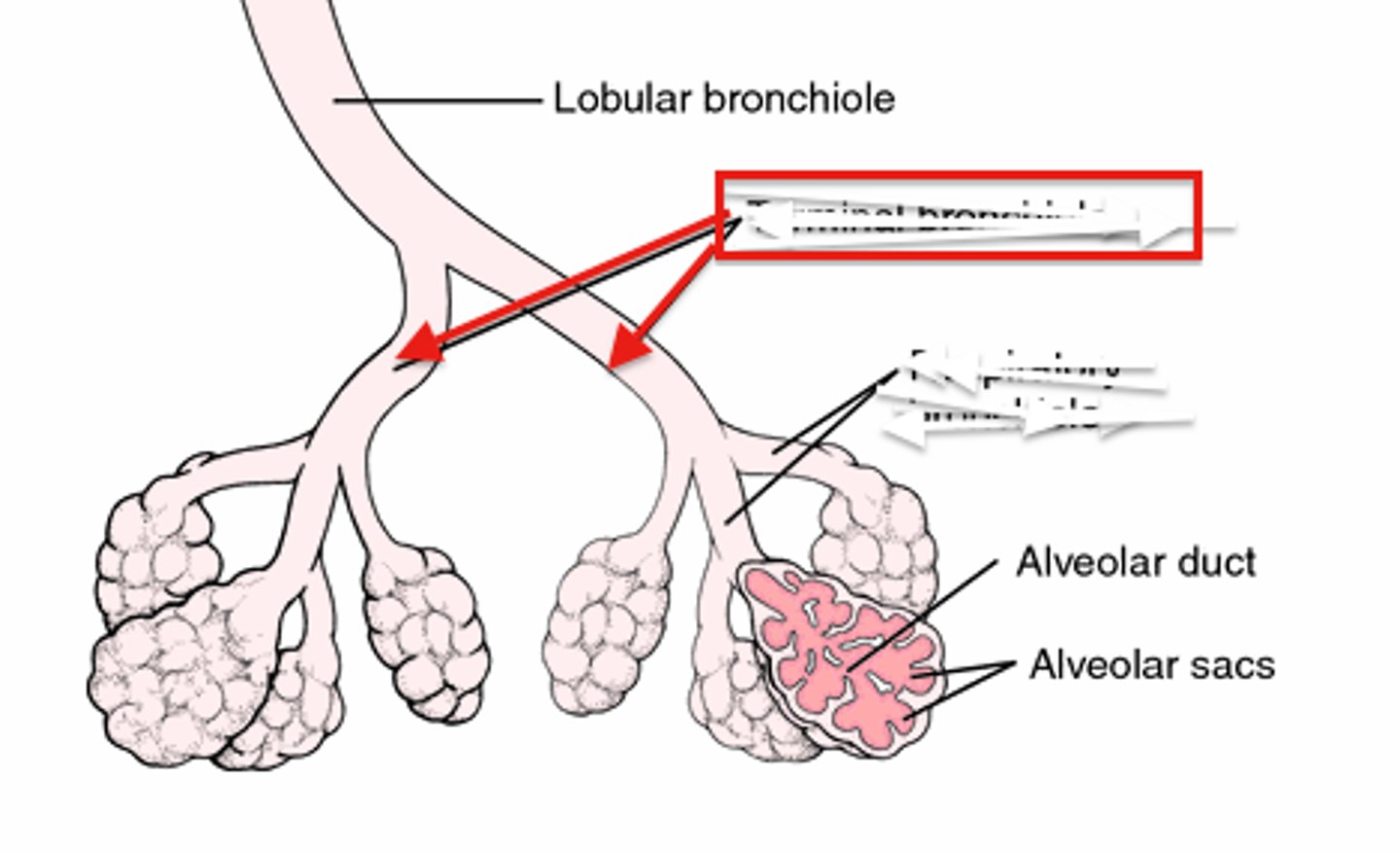
alveoli
tiny sacs of lung tissue specialized for the movement of gases between air and blood
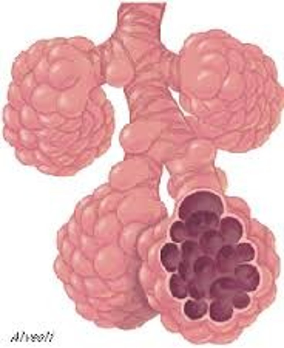
alveolar sac
2 or more alveoli sharing a common opening
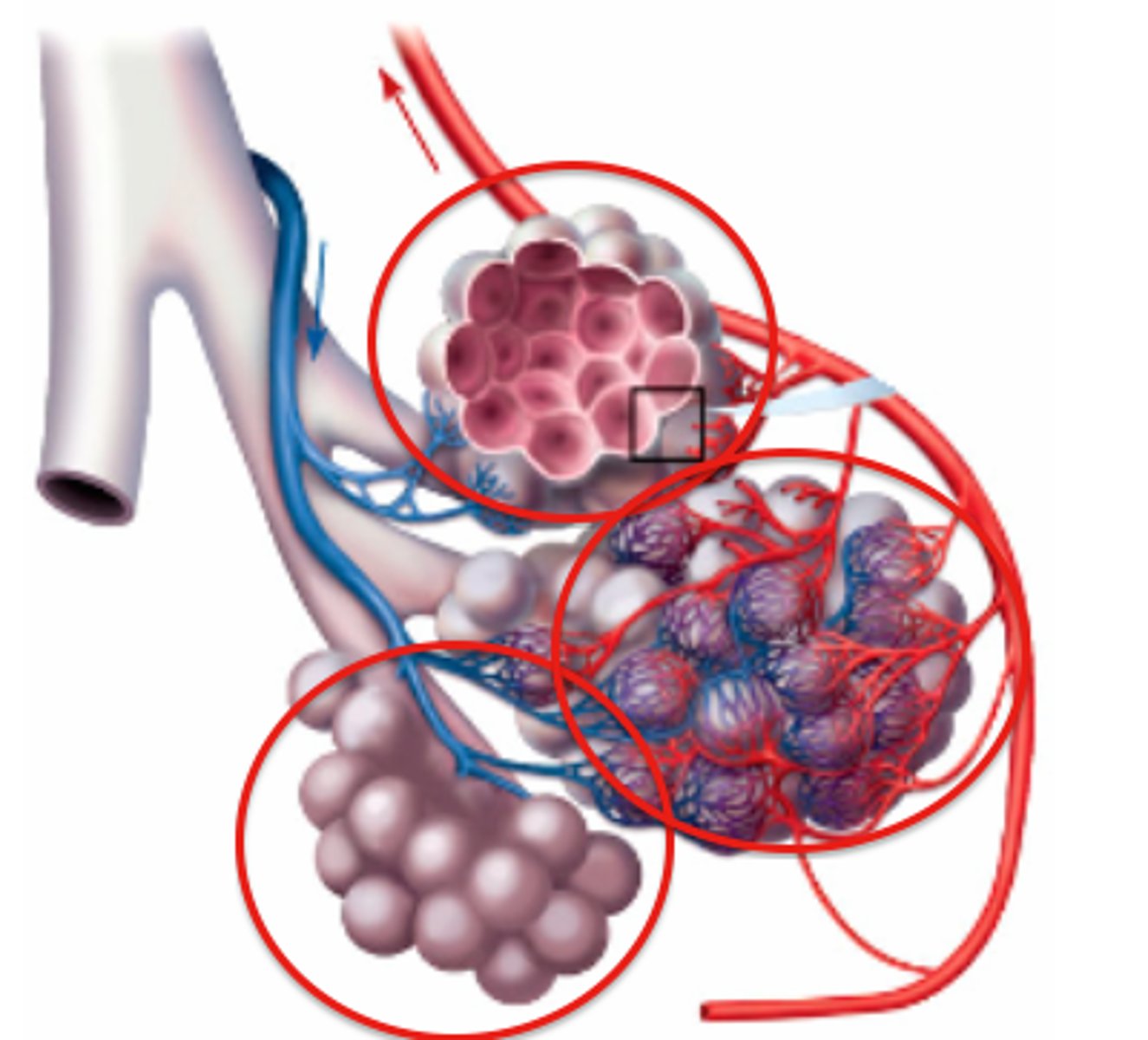
pulmonary artery
artery carrying oxygen-poor blood from the heart to the lungs
pulmonary vein
carries oxygenated blood from the lungs to the heart
capillary
A tiny blood vessel where substances are exchanged between the blood and the body cells.
alveolus
tiny air sac at the end of a bronchiole in the lungs that provides surface area for gas exchange to occur
nasal cavity
hollow space behind the nose
concha
used to mix oxygen to help catch particles and mix with H20 vapor (humidifying) - superior, middle, inferior
frontal sinus
cavity within the frontal bone
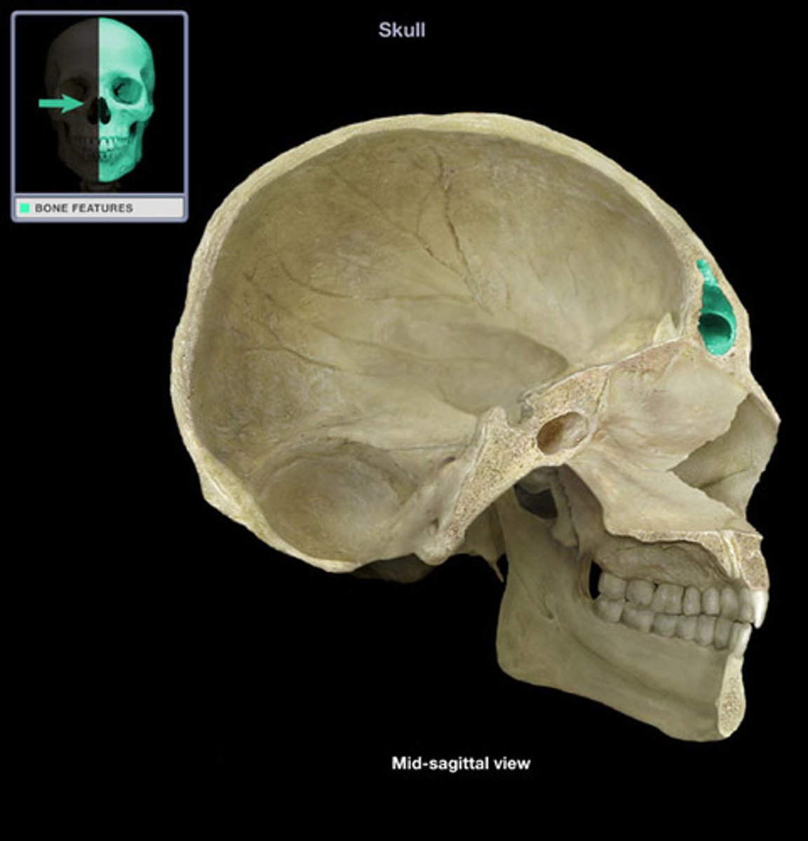
paransal sinuses
cavities found in the skull bones which empty into the nasal cavity, they lighten the skull, resonate with the voice, produce mucous, and warm air
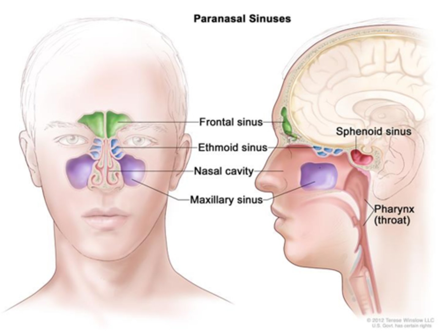
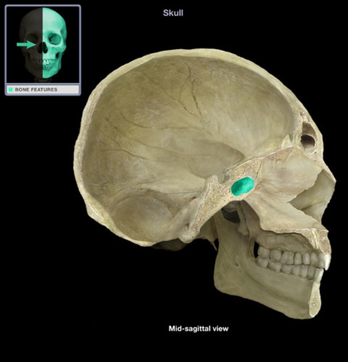
sphenoid sinus
air-filled space located within the sphenoid bone; most posterior of the paranasal sinuses
function of sinuses
reduce weight of head, strengthen skull, modify voice during phonation
vestibule
entrance (your nose hole)
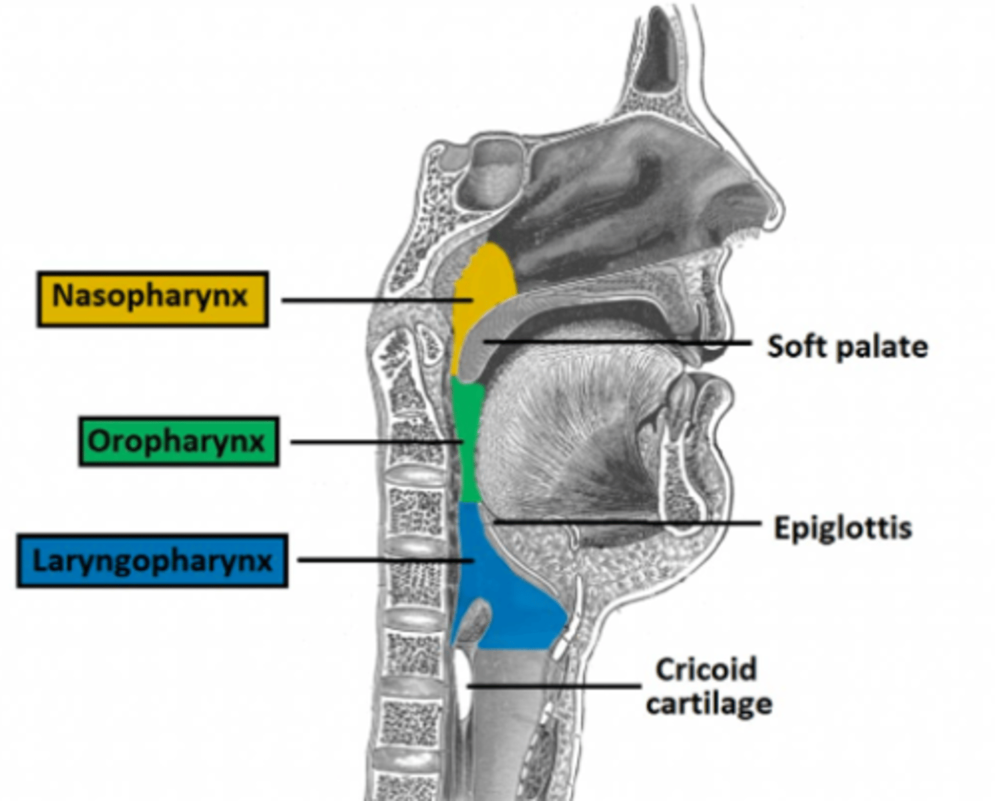
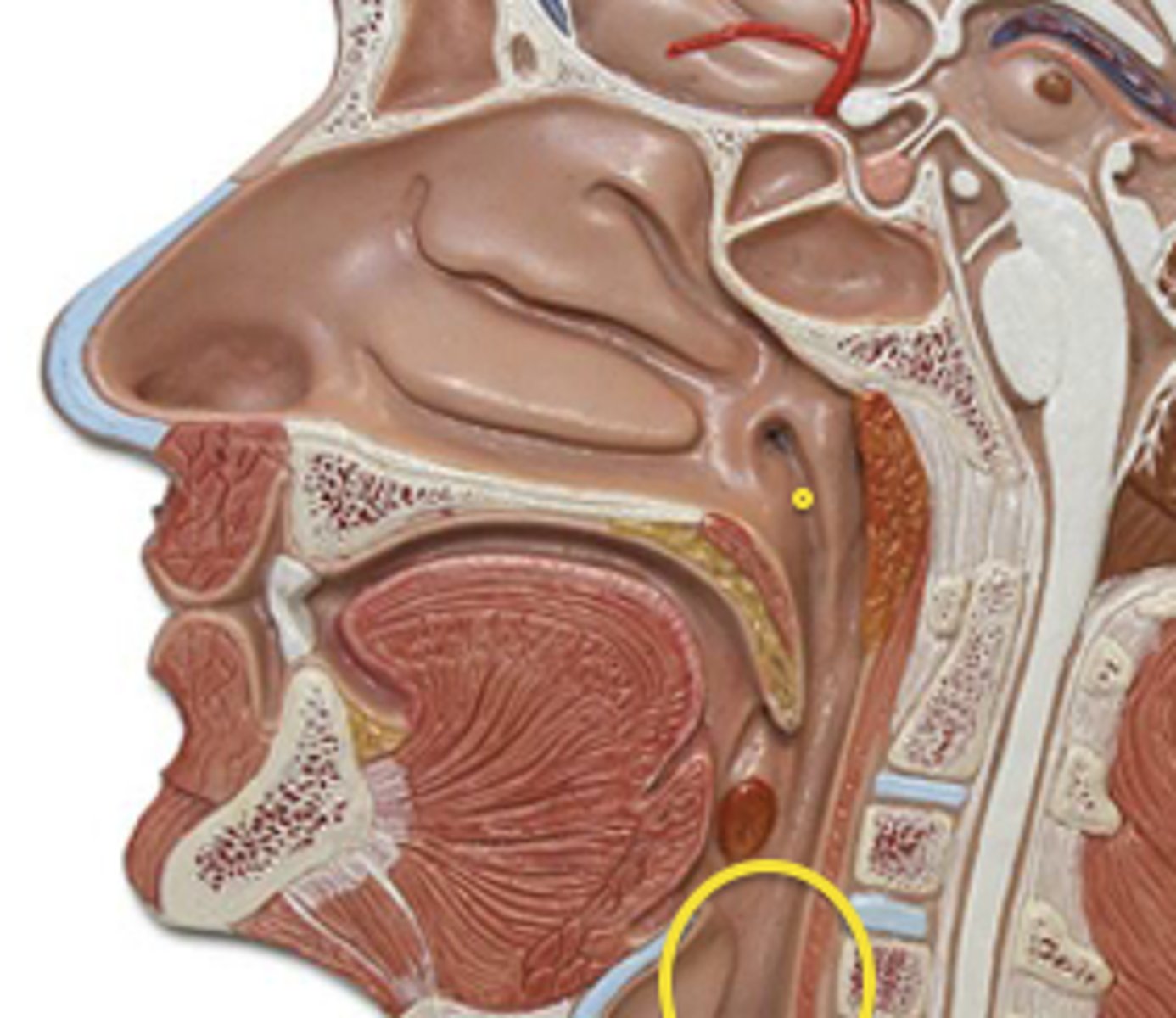
nasopharynx
part of the pharynx directly behind the nasal passages
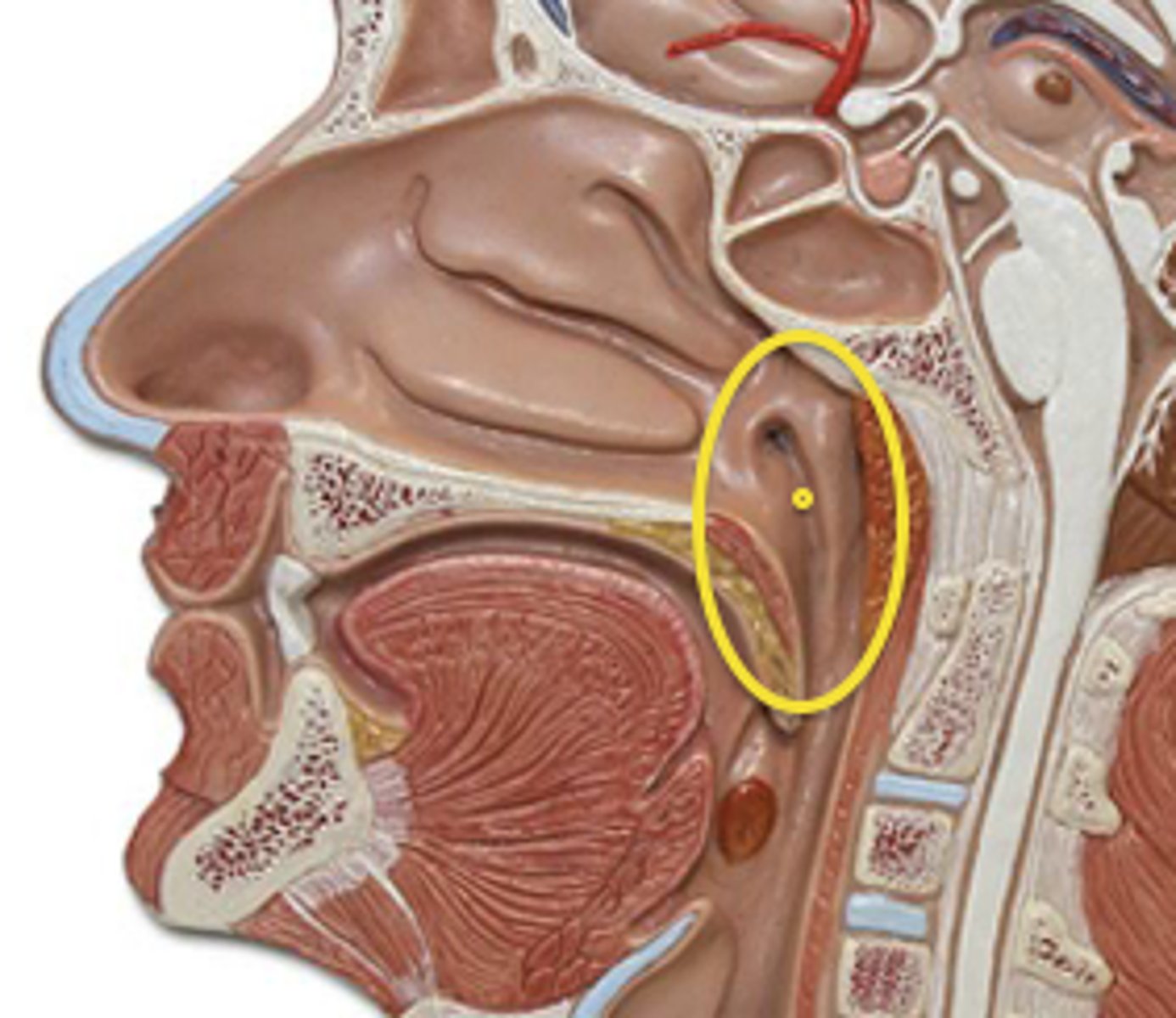
oropharynx
central portion of the pharynx between the roof of the mouth and the upper edge of the epiglottis
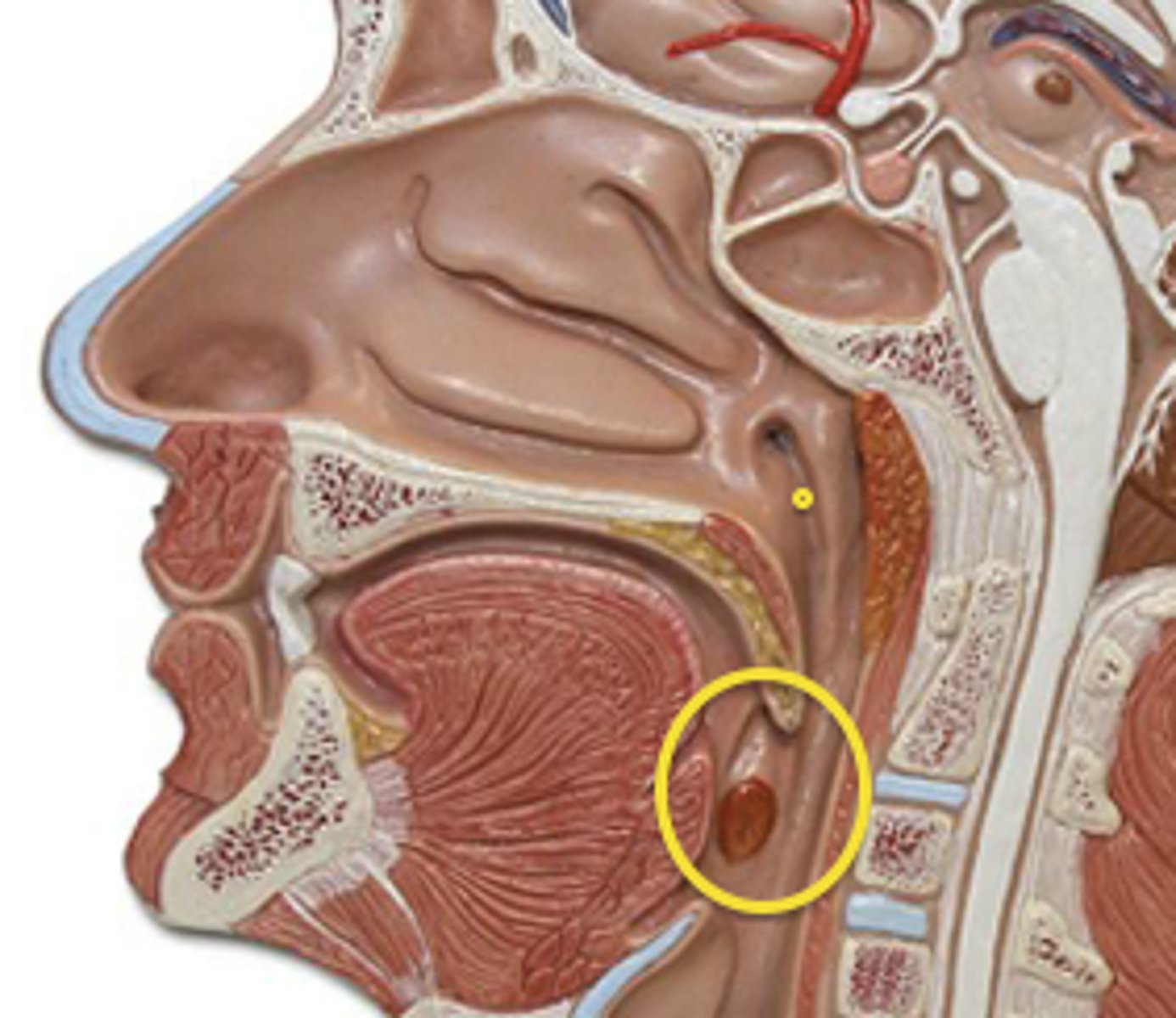
laryngopharynx
lower part of the pharynx, just below the oropharyngeal opening into the larynx and esophagus
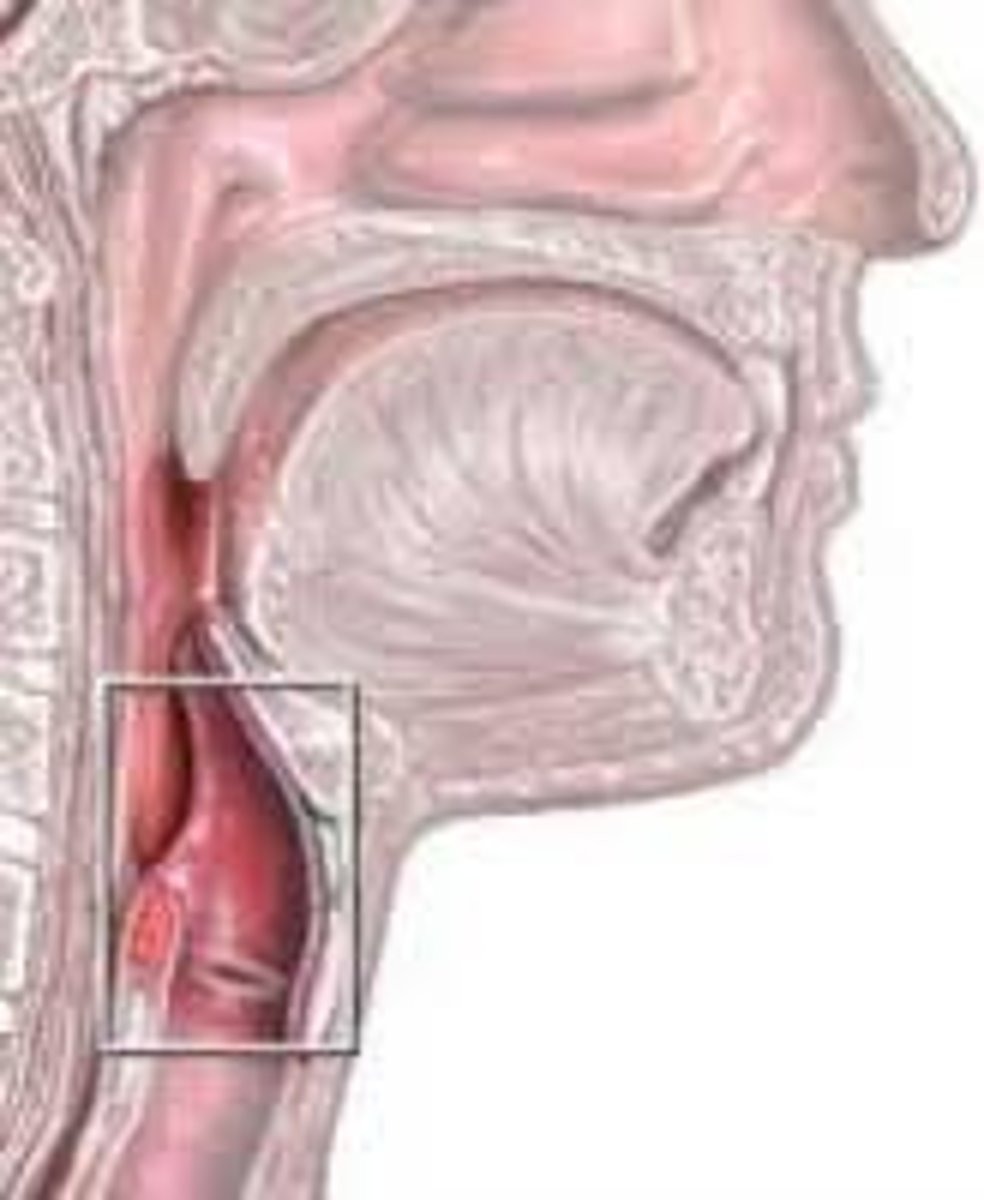
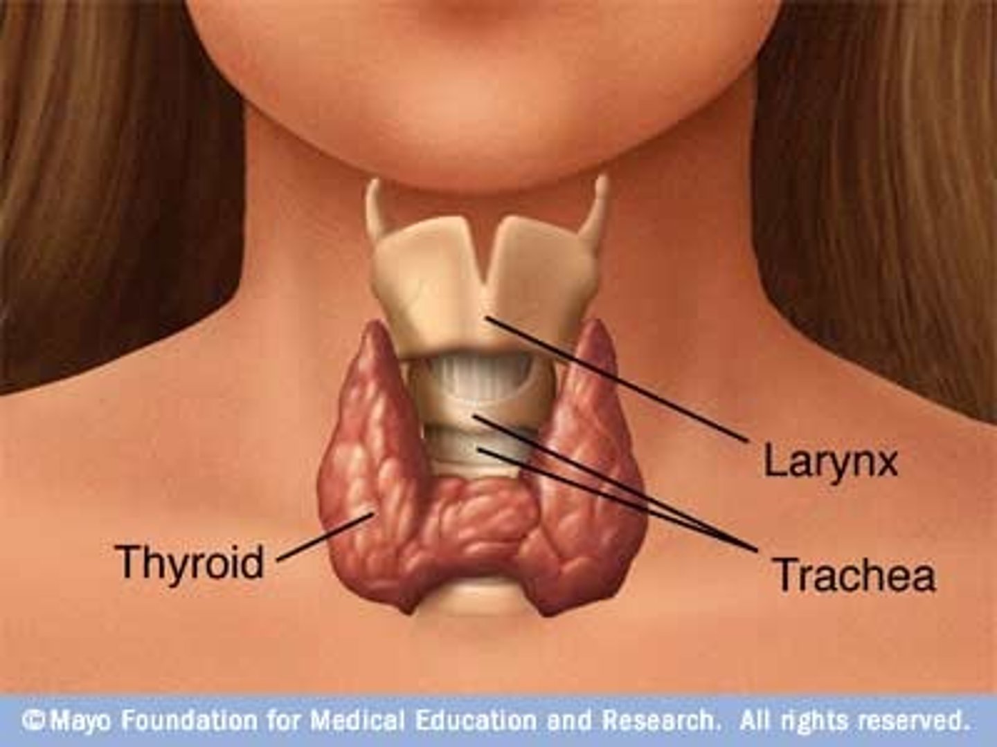
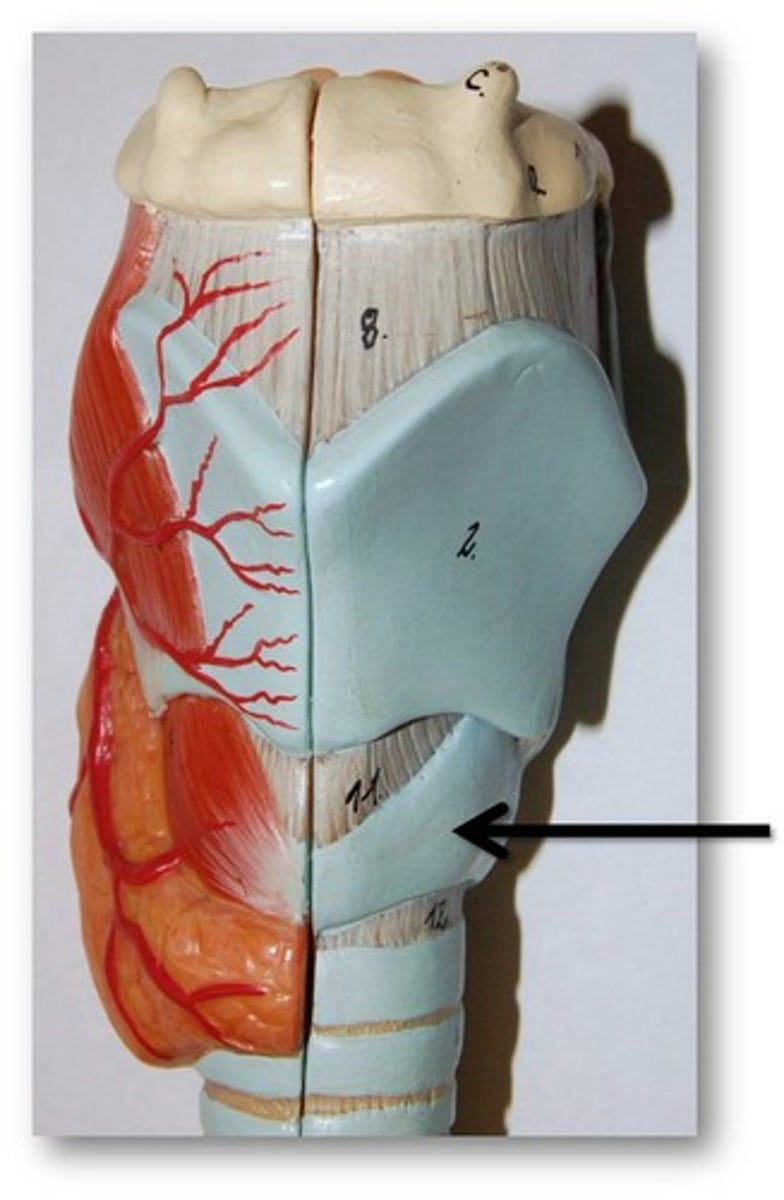
larynx
voice box; passageway for air moving from pharynx to trachea; contains vocal cords - made of hyaline cartilage
thyroid
secretes hormones that regulate growth, metabolism, and appetite
thyroid cartilage
A firm prominence of cartilage that forms the upper part of the larynx; the Adam's apple.
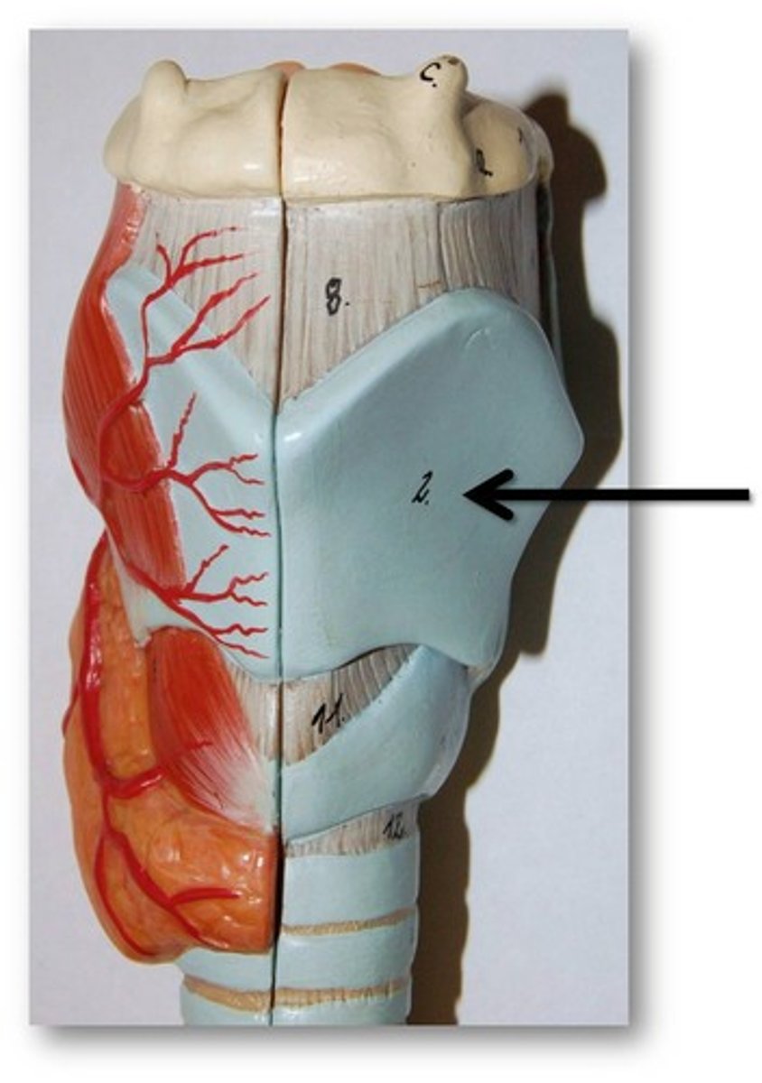
cricoid cartilage
the ring-shaped structure that forms the lower portion of the larynx
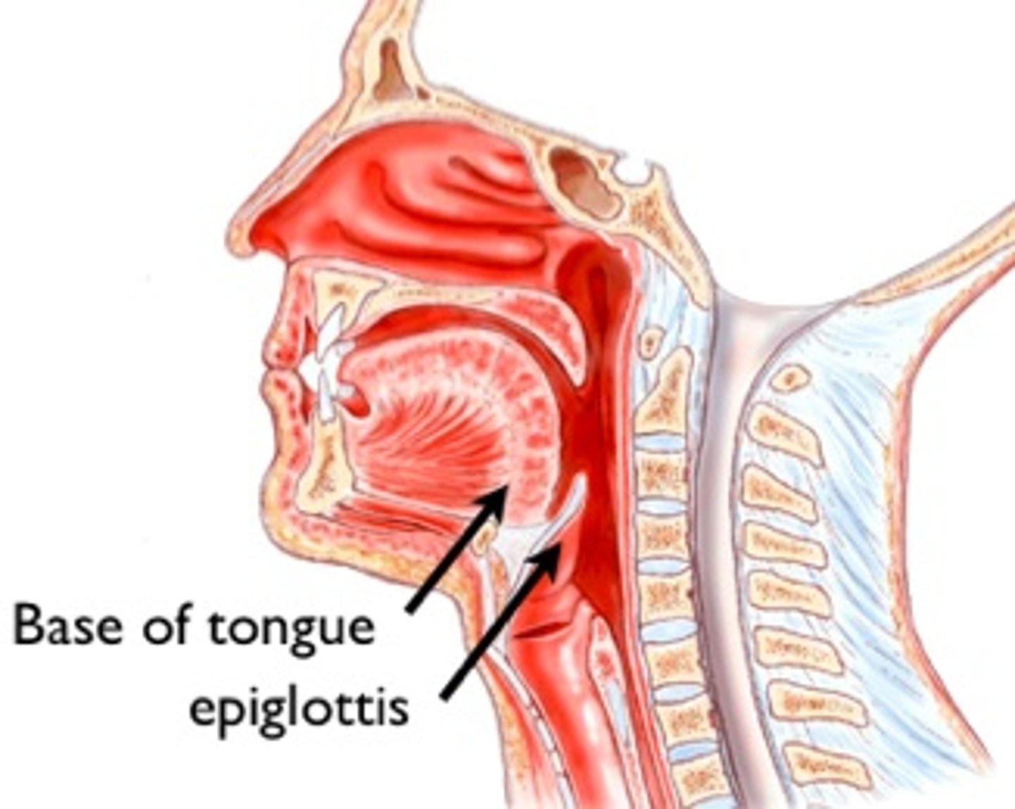
epigottis
A flap of tissue that seals off the windpipe and prevents food from entering.
laryngeal prominence
Adam's apple
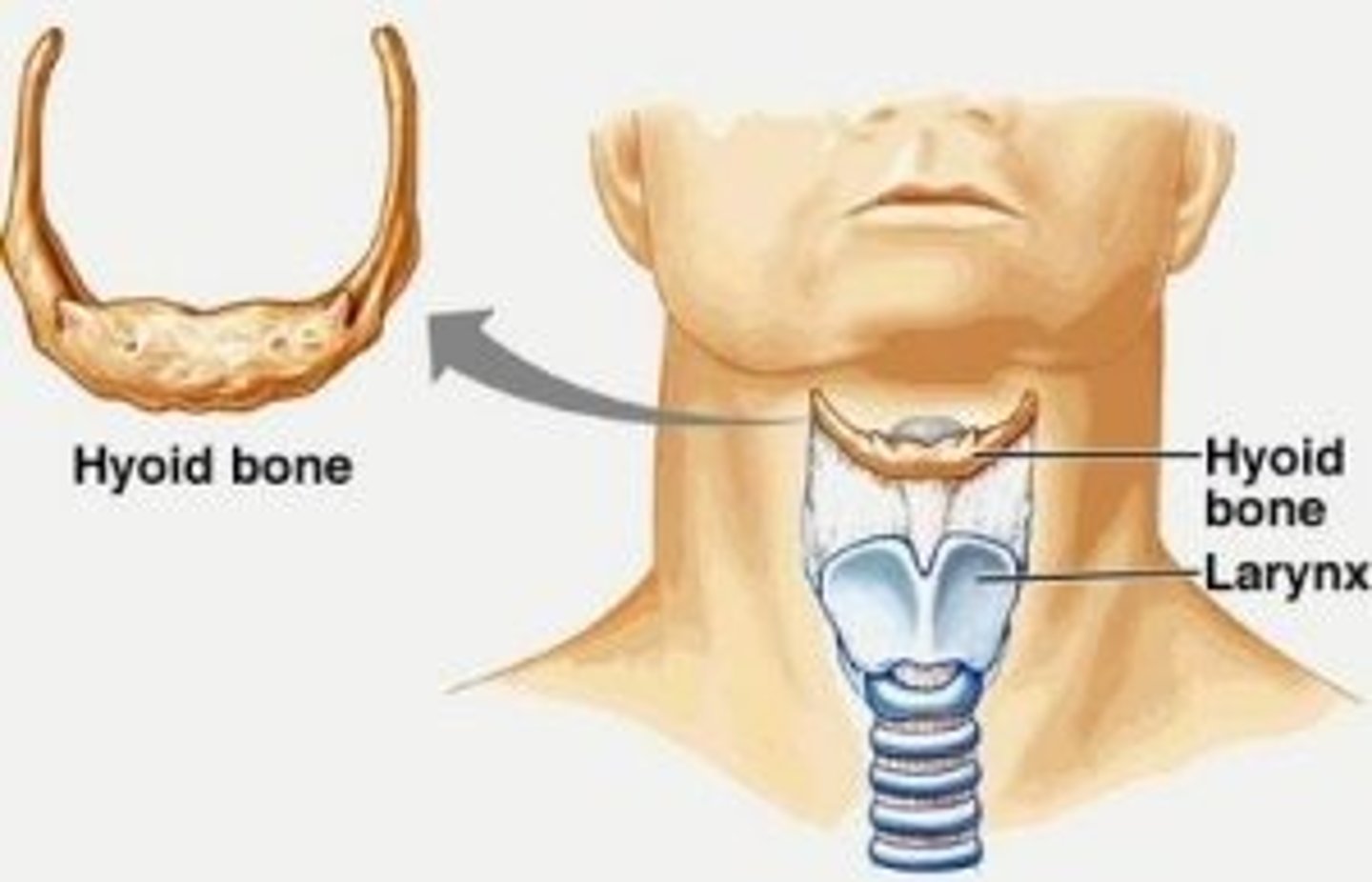
hyoid bone
U-shaped bone at the base of the tongue that supports the tongue and its muscles. (floating)
cuneiform cartilage
a pair of club- or wedge-shaped elastic cartilages anterior to the corniculate cartilages that support the vocal folds and lateral aspects of the epiglottis
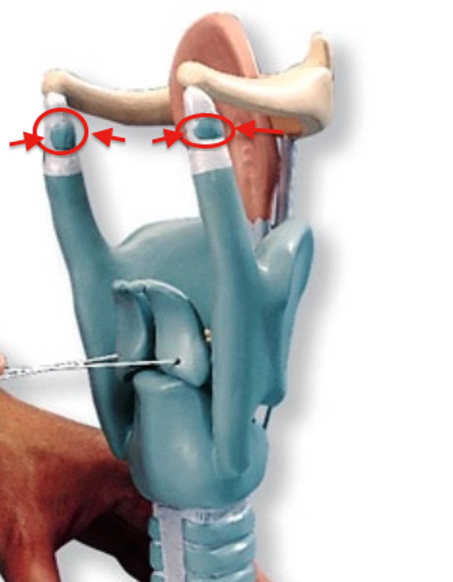
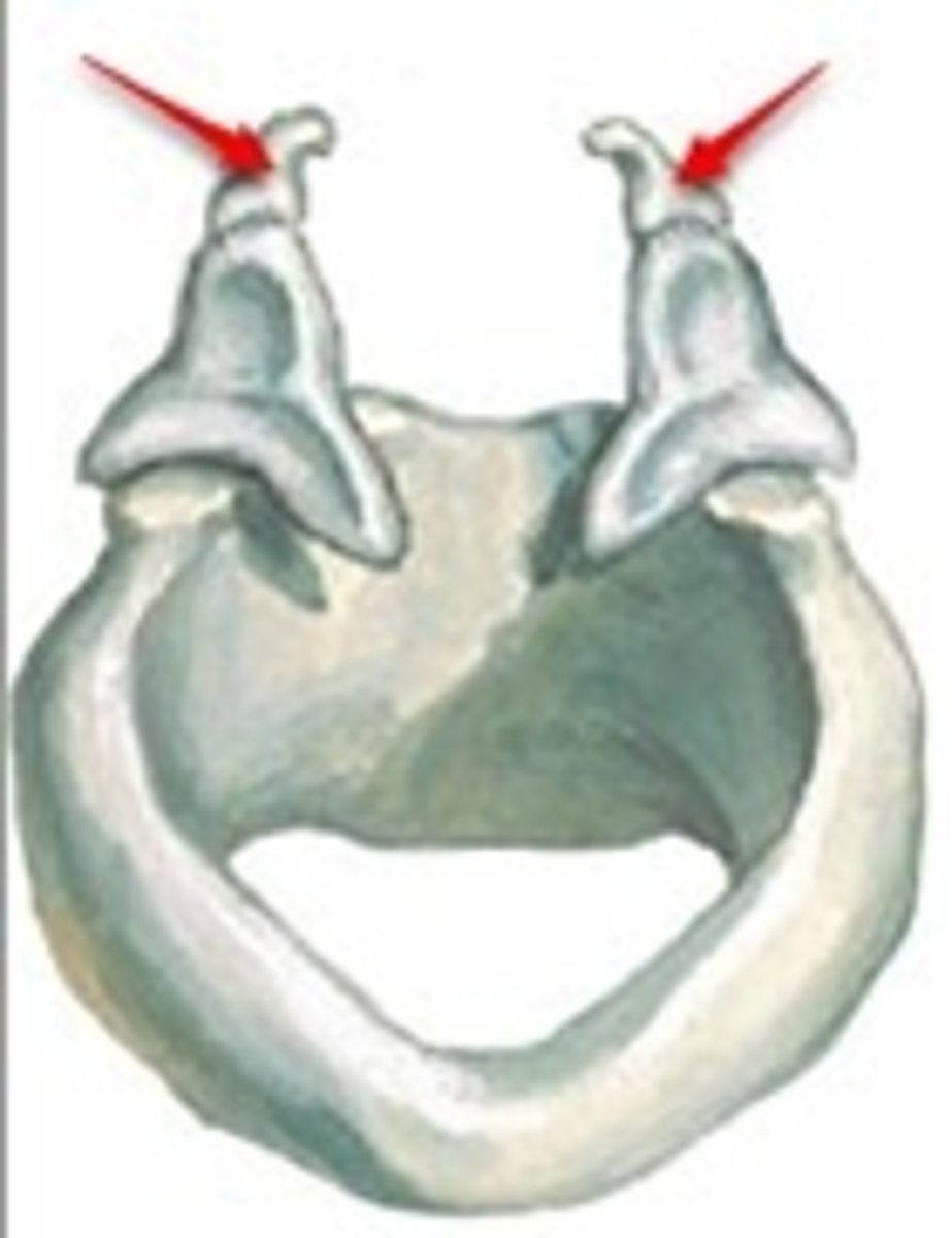
corniculate cartilage
a pair of horn-like pieces of elastic cartilage located at the apex of each arytenoid cartilage
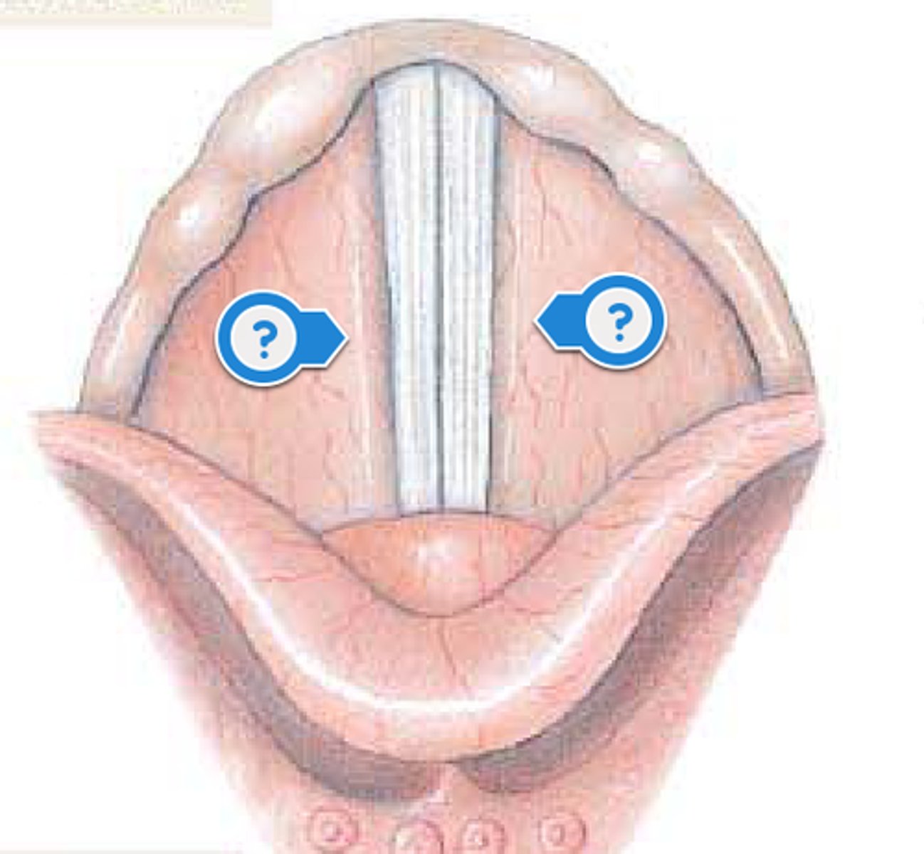
vocal ligament
elastic ligament within the vocal fold (true vocal cords) that vibrates and produces phonation (sound production at the larynx)
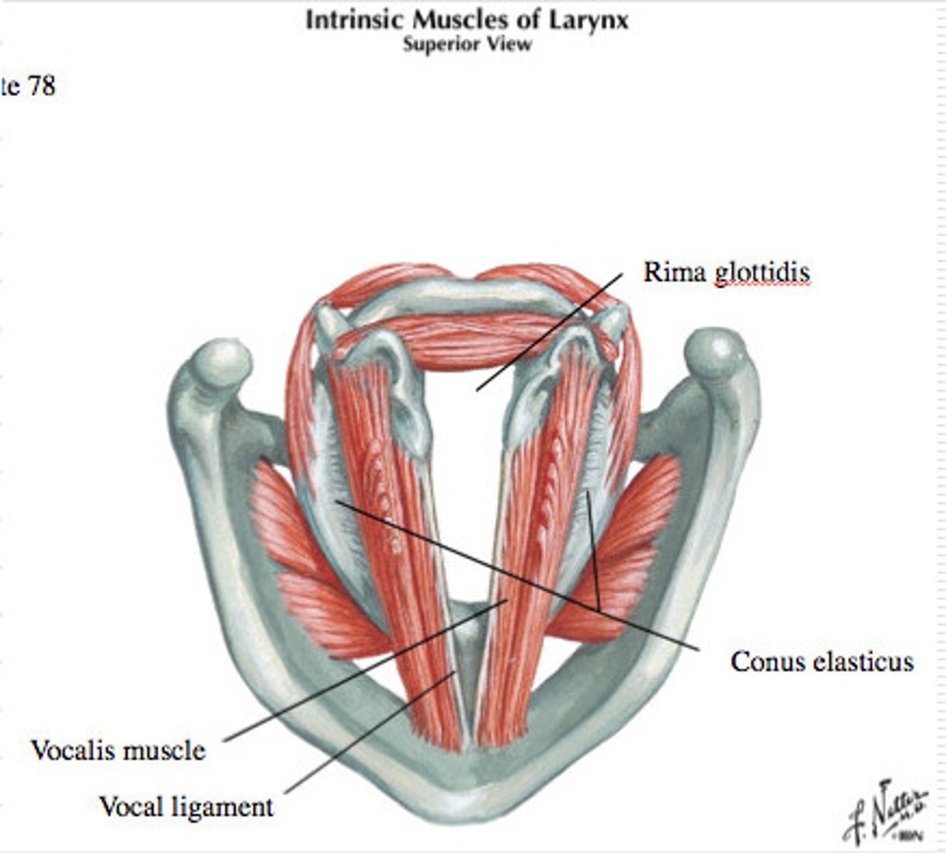
glottis
Opening between vocal cords
vestibular folds
false vocal cords
why are the rings important on the trachea
help keep the trachea open
ventilation
movement of air in and out of the lungs - breathing
always want to move
high to low pressure
boyle's law
A principle that describes the relationship between the pressure and volume of a gas at constant temperature - P1V1=P2V2 (with same number of particles and same temperature)
pressure and volume are...
inversely proportional
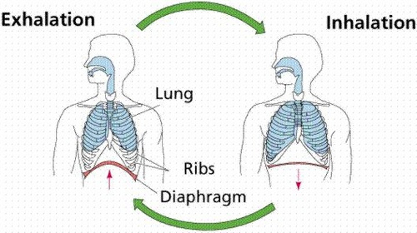
during an inhale
increase volume
decrease pressure
- inhale pressure < atmospheric pressure
during an exhale
decrease volume
increase pressure
- exhale pressure > atmospheric pressure
function of the diaphragm
separates the digestive cavity from the respiratory cavity, contracts and relaxes to help the lungs inflate and deflate
Primary muscles of inspiration
diaphragm and external intercostals
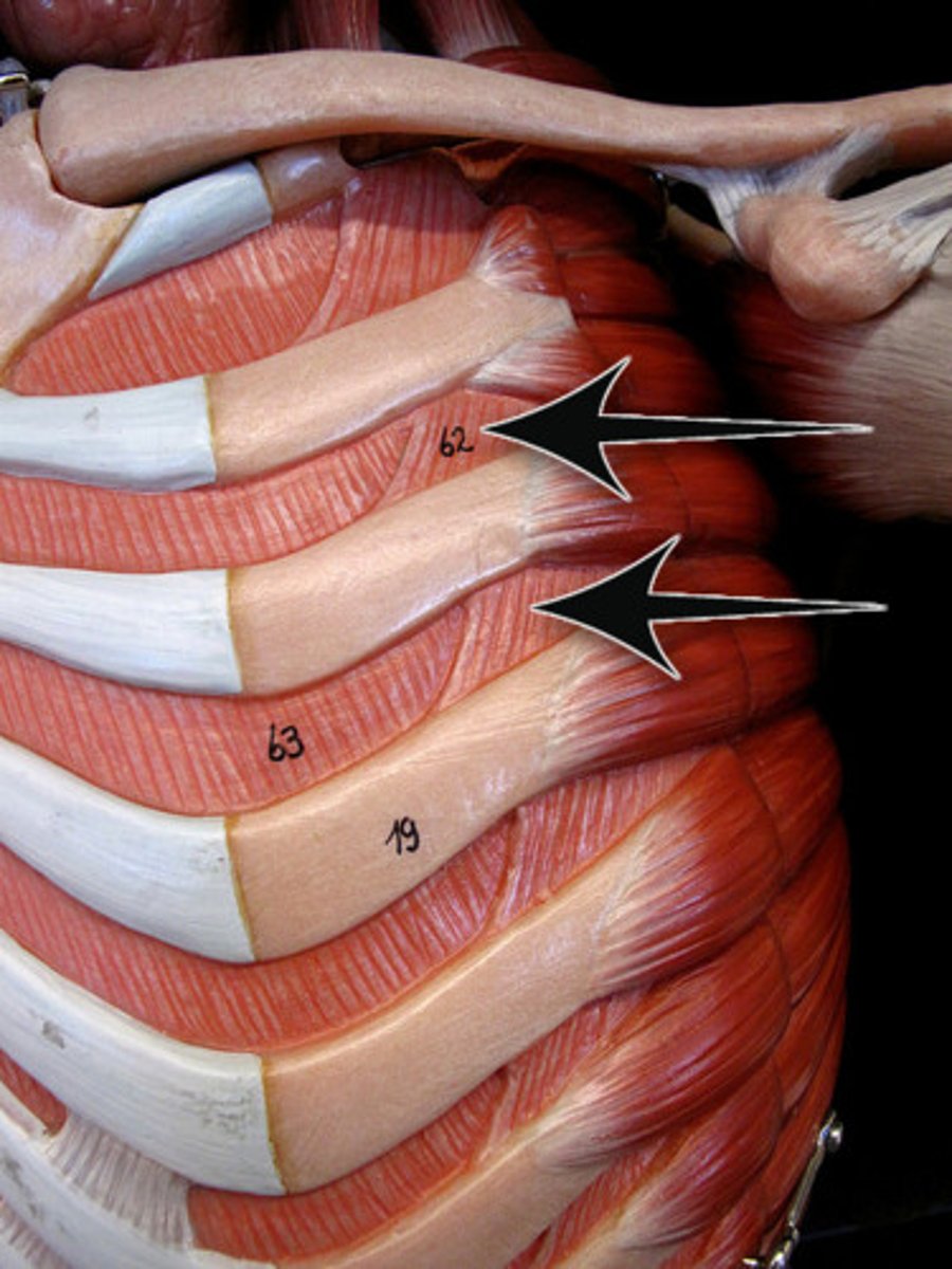
external intercostals
elevates ribs during inspiration
secondary muscles of inspiration
Sternocleidomastoid (elevates sternum)
Scalene muscles (elevate ribs)
Pectoralis minor (elevate ribs)
internal intercostals (makes space smaller)
oblique
function of secondary muscles
assists in the external intercostals muscles in elevating the ribs
function of primar muscles
contraction of external intercostals muscles elevates the ribs and the contraction of diaphragm flattens the floor of the thoracic cavity, increasing its volume and drawing in air into the lungs
intrapleural pressure
pressure within the pleural cavity
intrapulmonary pressure
pressure in the alveoli
transpulmonary pressure
difference between intrapulmonary and intrapleural pressure
atmospheric pressure (Patm)
Pressure exerted by air surrounding the body
760 mm Hg at sea level = 1 atmosphere
normal lungs at rest
P = -3 mm Hg
process of ventilation
- just prior to inspiration, atmospheric pressure = intrapulmonary pressure
- lungs start to expand, so intrapulmonary pressure decreases
- air moves from atmosphere to lungs
- pressure return to equilibrium
- during expiration, thoracic volume declines and intrapulmonary pressure > atmospheric pressure
- air moves out of lungs until pressure equilibrates again
compliance
how much air flow
if compliance increases
the lower the tension in the walls of the lungs
resistance
conducting portion that resists air flowing through them (always want a low resistance so you have greater flow)
if resistance increases
the harder it is to force air along the conducting passages
if resistance increases, what happens to the radius
decreases
diseases of compliance increase
COPD
Emphysema
disease of resistance
asthma
diseases of compliance decrease
fibrosis
pneumonia
neonates lungs
the surfactant is produced late
surfactant
chemical produced in the lungs to maintain the surface tension of the alveoli and keep them from collapsing - reduces surface tension and produced in epithelium
if surface tension is reduced then...
- attraction of H20 molecules
- increase in compliance (good amount)
large alveolus
low collapsing pressure
smal alveolus
high collapsing pressure
small alveolus with surfactant
low collapsing pressure
equation for respiratory minute volume
Ve= f x Vt (volume of air moved out each minute = respiratory rate x tidal volume)
relationship of respiratory minute volume
Ve increases, f increases, Vt increases
IRV
Inspiratory reserve volume: the maximal volume that can be inhaled from the end-inspiratory level
TV
tidal volume - amount at normal breath
ERV
Expiratory reserve volume: the maximal volume of air that can be exhaled from the end-expiratory position
RV
residual volume - air that cannot get out
IC
inspiratory capacity
TV + IRV
FRC
functional residual capacity
ERV + RV
VC
vital capacity - everything moving out