Biology 2.1.1- Cell Structure
1/19
Earn XP
Description and Tags
Name | Mastery | Learn | Test | Matching | Spaced | Call with Kai |
|---|
No analytics yet
Send a link to your students to track their progress
20 Terms
Outline the structure and function of the: cytoplasm, nucleus, nucleolus and nuclear envelope
Cytoplasm: contains all cell organelles
Nucleus: contains chromatin, transcription of DNA
Nucleolus: inside the nucleus, makes RNA and ribosomes
Nuclear envelope: a double membrane, contains the nucleus, has pores to allow substances like RNA to leave the nucleus
Outline the structure and function of the: rough and smooth ER, Golgi apparatus and cell membrane
Rough ER: membrane-bound sacs called cisternae, connected to nuclear envelope, contains ribosomes, folds and processes proteins
Smooth ER: produces and packages lipids
Golgi apparatus: made up of flat membrane-bound sacs, processes, packages and can modify proteins and lipids, makes lysosomes
Cell membrane: made of lipids and proteins, regulates transport of substances
Outline the structure and function of the: ribosomes, lysosomes and mitochondria
Ribosomes: attached to rough ER or independent, not membrane-bound, site of protein synthesis
Lysosomes: round membrane-bound organelles, contain enzymes that break down materials
Mitochondria: releases energy via respiration, producing ATP, consists of the matrix within a membrane folded into cristae, surrounded by an outer membrane
Outline the structure and function of the: vesicles, cilia and centrioles
Vesicles: small fluid-filled sac, transports substances between cells and between organelles
Cilia: hair-like structures on the plasma membrane in some animal cells, contain protein microtubules in a 9+2 formation which let the cilia move, used to move substances in multicellular eukaryotes
Centrioles: hollow cylinders made from protein microtubules, form spindle fibres in mitosis
Outline the structure and function of the: cell wall, vacuole, amyloplasts and chloroplasts
Cell wall: made of cellulose, cell structure and rigidity, pores called plasmodesmata connect cells via cytoplasm for the exchange of substances
Vacuole: membrane called the tonoplast, filled with cell sap, stores substances and maintains cell turgidity
Amyloplast- contains starch grains for energy storage
Chloroplasts: release energy via photosynthesis, contains thylakoid membranes stacked to form grana, and the stroma (like cytoplasm), surrounded by a double membrane
(plant cells only)
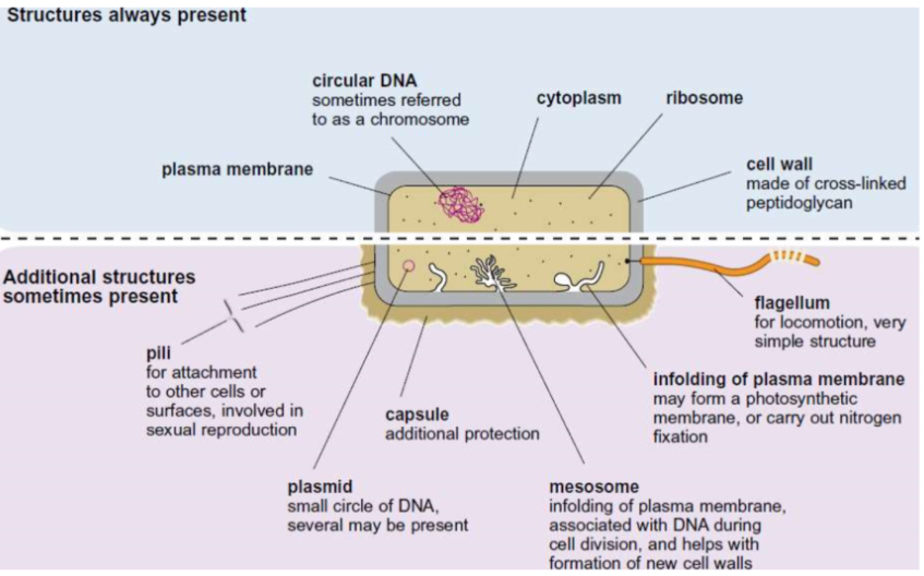
Name the organelles found in prokaryotes and outline their functions
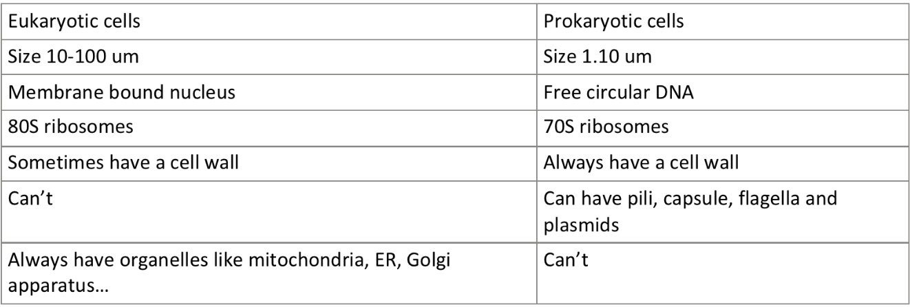
What are the differences between prokaryotic and eukaryotic cells?
Process of the excretion of proteins
The protein is produced in the ribosomes of the rough ER
The protein is pinched off in a vesicle, which carries it to the Golgi apparatus
The vesicle fuses with the Golgi apparatus, which processes, packages, and may modify the protein
The protein is pinched off in another vesicle which travels to and fuses with the cell membrane
The cell membrane opens to secrete the protein
What are the 4 main functions of the cytoskeleton?
Support the cell organelles and keep them in place
Provide strength and structure to the cell
Movement of materials eg. for mitosis
Cell movement, as cytoskeleton filaments run through flagella and cilia
Name the three types of cytoskeleton structures, and outline their structure + function
Microfilaments: dynamic, contractile fibres made of actin, positioned near cell membrane, cause cell contraction during cytokinesis
Microtubules: dynamic, hollow cylinders made of tubulin, can contract and polymerase to change their length, involved in anaphase in mitosis as contracting spindle fibres, help move organelles like vesicles around the cell
Intermediate filaments: fixed within the cell, maintain the structure and keep organelles in place
What is the difference between magnification and resolution?
Magnification is how much bigger the image is than the specimen
Resolution is how close together two objects can be before they appear as one under a microscope, ie. how detailed the microscope can see
How do laser scanning confocal microscopes work?
The specimen is tagged with a fluorescent dye
Laser beams are fired at the specimen, which gives off light
This can be detected by a computer to generate an image
How do the two electron microscopes work?
Transmission electron microscope:
Use electromagnets to focus a beam of electrons at a thin specimen in a vacuum
The electrons are transmitted but are absorbed by denser parts of the specimen or areas stained using heavy metals
A computer generates a black and white image, which is darker corresponding to where electrons have been absorbed
Scanning electron microscope:
Electrons are fired at a specimen in a vacuum and bounce off
These can be detected at an angle and an image is produced by a computer
What are the pros and cons of light microscopes?
Pros:
Cheap
Small size, don’t need installation
Easy to use and prepare slides for
Specimens can be living
Images are coloured, or can be stained
Doesn’t require a vacuum
Cons:
Low magnification- max 1500 x
Low resolution- 0.2 micrometers (because light has a longer wavelength than electrons)
Can only create 2D images
Can’t see viruses or full cell structure
What are the pros and cons of SEMs?
Pros:
High magnification - max 500,000 x (not as high as TEMs)
High resolution- 0.002 micrometers (as electrons have a shorter wavelength than light)
Images produced are 3D
Can see viruses and shows more detail into cell structure and ultra structure
Cons:
Expensive
Large, need to be installed
Hard to use and prepare specimens for
Requires a vacuum, so samples have to be dead
Images are black and white (but can be digitally coloured)
What are the pros and cons of TEMs?
Pros:
High magnification - can be greater than 1,000,000 x
High resolution- 0.0002 micrometers (as electrons have a shorter wavelength than light)
Can see viruses and shows more detail into cell structure and ultra structure
Cons:
Expensive
Large, need to be installed
Hard to use and prepare slides for
Specimens have to be very thinly sliced and can get distorted in preparation
Images produced are 2D
Requires a vacuum, so samples have to be dead
Images are black and white (but can be digitally coloured)
What are the pros and cons of laser scanning confocal microscopes?
Pros:
Small (but bigger than light microscopes)
High resolution
Images produced are 3D
Cons:
Images are black and white (but colours can be added afterwards)
Low magnification
How can you use an eyepiece graticule to measure cells?
You first have to calibrate it to find out the actual length of each division on the ruler:
Place a stage micrometer slide on the stage
Focus the microscope on the lowest power objective lens
Move the stage micrometer around to line up the two rulers
Count the number of small divisions on the graticule equivalent to 100 micrometers on the stage micrometer
Divide 100 by the number counted to find the length that one division represents, and record the result
Repeat for the medium and highest power objective lenses
You can then replace the stage micrometer with a slide and measure the length of a cell using the calculated values for the divisions
What is differential staining?
Different stains can be applied to one slide, which are absorbed by different parts of the cell to identify them
What stains can be used in differential staining?
Acetic orcein stains chromosomes dark red
Eosin stains cytoplasm dark red/pink
Iodine stains starch blue-black
Iodine in potassium iodide solution stains cellulose yellow
Hematoxylin stains RNA and DNA purple/blue
Methylene blue is an all-purpose stain, used often to stain DNA blue