A2 - THE HUMAN BRAIN
1/60
There's no tags or description
Looks like no tags are added yet.
Name | Mastery | Learn | Test | Matching | Spaced | Call with Kai |
|---|
No analytics yet
Send a link to your students to track their progress
61 Terms
how will the neural tube enlarge and develop into different components of the nervous system during embryonic development?
The anterior (front) part of the neural tube will expand to form the brain during cephalisation (development of the head)
The remainder of the neural tube will develop into the spinal cord
Cells that comprised the neural crest will differentiate to form most of the peripheral nervous system
Formation of the Human Brain

What does the human brain act as and process
The human brain acts as an integration and coordination system for the control of body systems
It processes sensory information received from the body and relays motor responses to effector organ
How is the human brain organized
The human brain is organised into clearly identifiable sections that have specific roles
Internal structures include the hypothalamus, pituitary gland and corpus callosum
major external structures of the brain
The major external structures include the cerebral cortex, cerebellum and brainstem
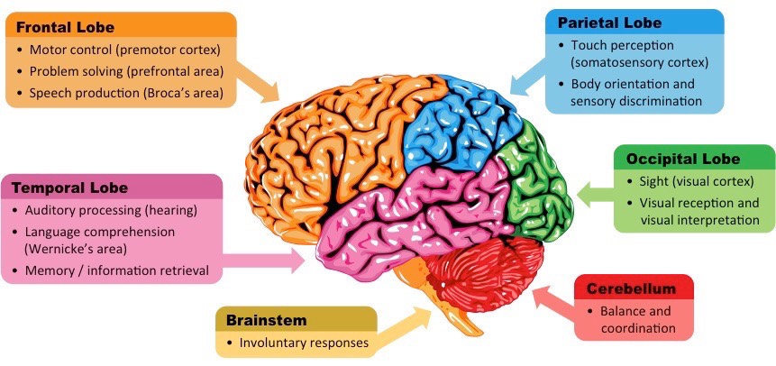
cerebral cortex
The cerebral cortex is an outer layer of tissue organised into two cerebral hemispheres and composed of four distinct lobes:
frontal lobe
parietal lobe
temporal lobe
occipital lobe
frontal lobe
controls motor activity and tasks associated with the dopamine system (memory, attention, etc.)
parietal lobe
responsible for touch sensation (tactility) as well as spatial navigation (proprioception)
temporal lobe
involved in auditory processing and language comprehension
occipital lobe
the visual processing centre of the brain and is responsible for sight perception
cerebellum - what does it appear as and what is it responsible for
The cerebellum appears as a separate structure at the base of the brain, underneath the cerebral hemispheres
It is responsible for coordinating unconscious motor functions – such as balance and movement coordination
brainstem
The brainstem is the posterior (rear) part of the brain that connects to the spinal cord (which relays signals to and from the body)
The brainstem includes the pons, medulla oblongata (often referred to as the medulla) and the midbrain
The brainstem (via the medulla) controls automatic and involuntary activities (breathing, swallowing, heart rate, etc.)
major internal structures of the brain
Hypothalamus
Pituitary Gland
Corpus Callosum
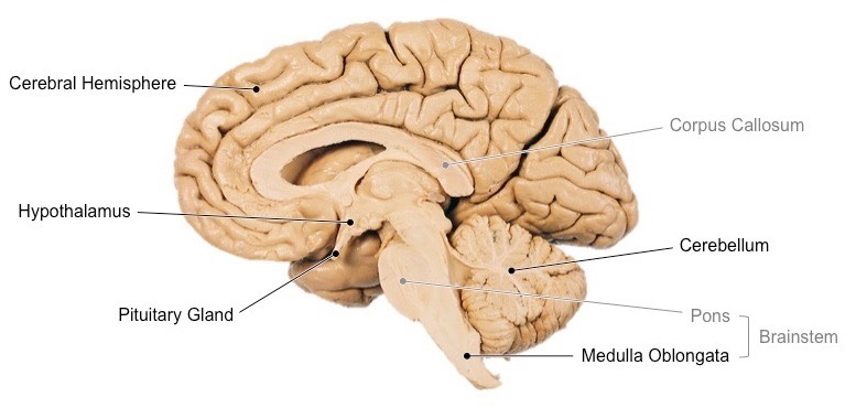
Hypothalamus
The hypothalamus is the region of the brain that functions as the interface with the pituitary gland
As such, the hypothalamus functions to maintain homeostasis via the coordination of the nervous and endocrine systems
The hypothalamus also produces some hormones directly, which are secreted via the posterior pituitary (neurohypophysis)
Pituitary gland
The pituitary gland is considered the ‘master’ gland – it produces hormones that regulate other glands and target organs
The anterior lobe is called the adenohypophysis and secretes hormones such as FSH, LH, growth hormone and prolactin
The posterior lobe is called the neurohypophysis and secretes hormones such as ADH and oxytocin
Corpus Callosum
The corpus callosum is a bundle of nerve fibres that connects the two cerebral hemispheres
It is the largest white matter structure in the brain, consisting of roughly 250 million axon projections
Damage to the corpus callosum can prevent information exchange between left and right hemispheres (split brain disorders)
how can the role of a specific brain part can be identified by either stimulating or removing the region to assess its effect
by either stimulating or removing the region to assess its effect
Identification of brain roles can be made via the use of animal experiments, autopsy, lesions and fMRI
animal experiments
Animal experimentation can be used to identify function by stimulating regions with electrodes or removing via lobotomy
Because such methods are highly invasive and potentially damaging, animal models are frequently used
Experimentation on animals involves less ethical restrictions than human studies (although ethical standards do exist)
Animal studies are limited by the differences between animal and human brains, making valid comparisons difficult
Example: Animal studies using mice and rats have been used to develop drug treatments for diseases such as MS
lesions
Lesions are abnormalareas of braintissue which can indicate the effect of the loss of a brain area
Lesions can be identified via post-mortem analysis (autopsy) or via scans of the brain (CT scans or MRI)
The effects of lesions can be difficult to identify, as many functions may involvemultiple brain areas
Additionally, the brain has the capacity to re-learn certain skills by re-routing instructions to other areas (plasticity)
Example: Split brain patients have been used to identify specific roles of the left and right cerebral hemisphere
autopsy
An autopsy is a post-mortem examination of a corpse via dissection in order to evaluate causes of death
Comparisons can be made between the brains of healthy and diseased corpses to identify affected brain areas
Example: Cadavers who suffered from aphasia (language impairment) in life demonstrate damage to specific areas
fMRI
Functional magnetic resonance imaging (fMRI) records changes in blood flow within the brain to identify activated areas
Oxygenated haemoglobin responds differently to a magnetic field than deoxygenated haemoglobin
These differences in oxygenation can be represented visually and reflect differences in the level of brain activity
fMRI is non-invasive and can be used to identify multiple brain regions involved in complex, integrated brain
Example: fMRI studies have been used to diagnose ADHD and dyslexia, as well as monitor recovery from strokes
examples of brain areas with clearly defined functions
visual cortex, Broca’s area and the nucleus accumbens
Visual Cortex
Located within the occipital lobe of the cerebrum and receives neural impulses from light-sensitive cells in the eyes
The visual cortex is the region of the brain responsible for visual perception (sight)
Broca’s Area
Located within the frontal lobe of the left cerebral hemisphere (not present in the right hemisphere)
Is responsible for speech production (if damaged, the individual cannot produce meaningful speech despite intending to)
Nucleus Accumbens
The nucleus accumbens is involved in the pleasure reward pathway and is found within each cerebral hemisphere
It secretes neurotransmitters responsible for feelings of pleasure (dopamine) and satiety (serotonin)
It communicates with other centres involved in the mechanisms of pleasure, such as the ventral tegmental area (VTA)
What is the cerebral cortex
the outer layer of neural tissue found in the cerebrum of humans and other mammals
It is composed of grey matter and is involved in complex actions, such as memory, perception, consciousness and thought
is much more highly developed in humans than other animals and forms a larger proportion of the brain
result of evolution of human cerebral cortex and what is this responsible for
Through evolution, the human cerebral cortex has been greatly enlarged in comparison to other brain structures
The disproportional enlargement of the cerebral cortex in humans is responsible for our capacity for cognitive thought
what is the increase in human cerebral cortex mediated by and what does this allow for
The increase in total area is mediated by extensive folding (gyrification) to form wrinkled peaks (gyrus) and troughs (sulcus)
This greatly increases surface area without increasing volume – allowing the brain to fit within the cranium
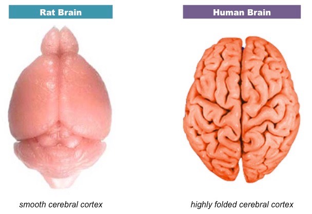
what does the extent of gyrification of the cerebral cortex indicate
The extent of gyrification of the cerebral cortex is a reliable indicator of potential cognitive capacity
Primates and humans have a greater degree of folding compared to lower mammals (e.g. rats have a smooth cortex)
Brain Comparison – Human versus Rat
what is the cerebrum organised into
two hemispheres that are responsible for higher order functions and complex skills
functions of hemispheres of cerebrum (6)
memory,
speech,
cognitive thought,
problem solving,
attention
and emotions
are some activities localised to a single side of the cerebral hemispheres? example
yes - Not all complex tasks are equally represented by both cerebral hemispheres – some activities are localised to a single side
Speech production is coordinated by Broca’s area, which is situated in the left frontal lobe of the brain
what is the left/ right cerebral hemisphere responsible for
The left cerebral hemisphere is responsible for processing sensory information from the right side of the body
processed on the left side of the visual cortex
The right cerebral hemisphere is responsible for processing sensory information from the left side of the body
processed on the right side of the visual cortex
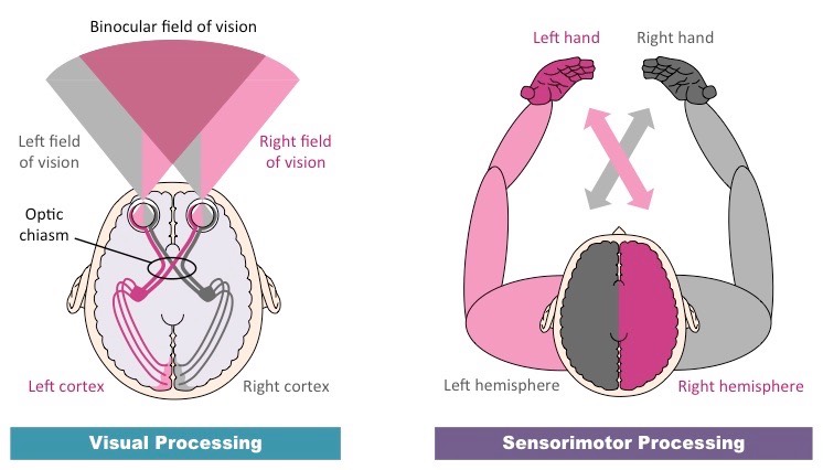
contralateral processing
The processing of information on the opposite side of the body
how does contralateral processing work
Tactile information from the left side of the body is transferred to the right side in the spinal cord or brainstem and vice versa
Visual information from the left visual field is transferred to the right cerebral hemisphere at the optic chiasma and vice versa
how is motor information processesed on the diff sides of the cerebral hemispheres
The left cerebral hemisphere is also responsible for processing motor information for the right side of the body (and vice versa)
Muscular contractions are coordinated by the motor cortex (premotor cortex = preparation ; primary motor cortex = execution)
Contralateral Processing diagram
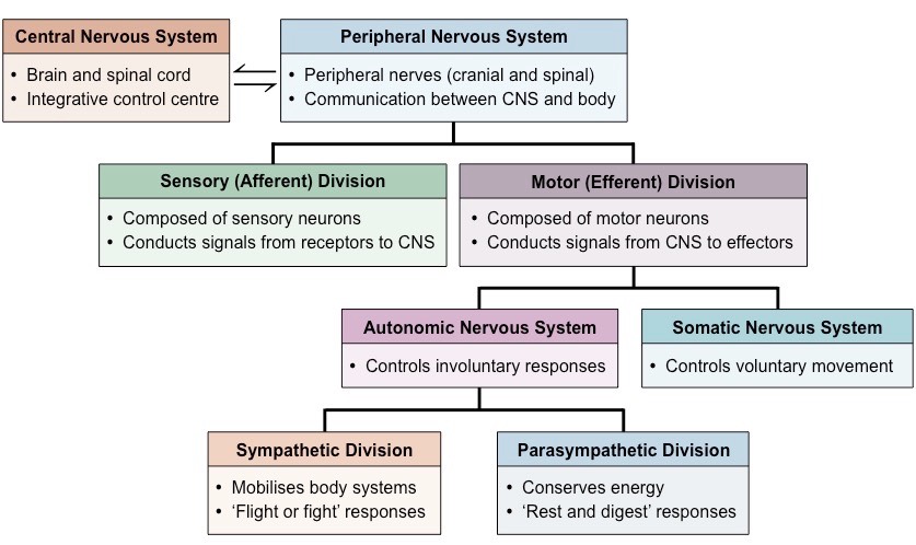
what sub-system is the nervous system divided into
the nervous system can be divided into the
central nervous system (brain and spine) and
peripheral nervous system
what sub-system is the peripheral nervous system divided into
The peripheral nervous system (PNS) can be divided into the
sensory (afferent) pathway or
the motor (efferent) pathway
how can the motor pathway of the peripheral nervous system be subdivided
The motor pathway can be subdivided according to whether the response is
voluntary (somatic) or
involuntary (autonomic)
what does the autonomic nervous system control - using what two things
The autonomic nervous system controls involuntary processes in the body using sympathetic and parasympathetic nerves
Sympathetic nerves release noradrenaline (adrenergic) to mobilise body systems (‘fight or flight’ responses)
Parasympathetic nerves release acetylcholine (cholinergic) to relax body systems and conserve energy (‘rest and digest’)
Divisions of the Nervous System diagram
what is the medulla oblangata
The medulla oblongata is a part of the brainstem responsible for coordinating many autonomic (involuntary) activities
This includes the regulation of body activities such as swallowing, breathing and heart rate
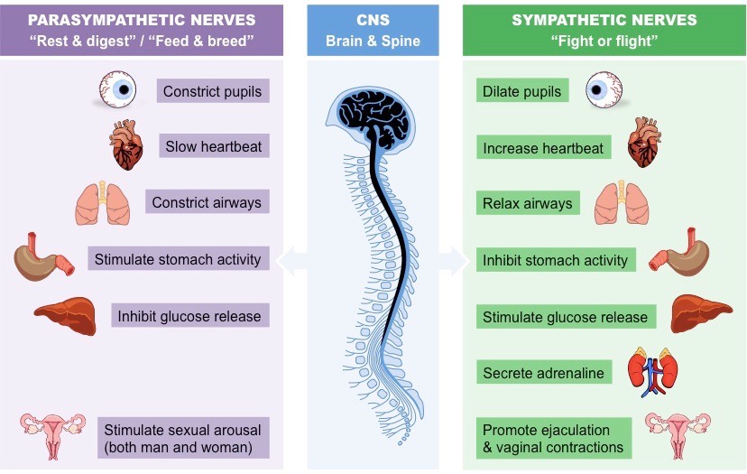
what occurs during sympathetic Responses
‘Fight or flight’ mode
Decreases salivary release and blood flow to the gut in response to swallowing
Increasesventilationrate and dilates airways in response to a reduction in bloodpH (caused by increased levels of CO2)
Increasesheartrate by raising the normal sinus rhythm of the pacemaker of the heart
what occurs during parasympathetic responses
‘Rest & Digest’ mode
Increases salivary release and blood flow to the gut in response to swallowing
Lowers ventilation rate and constrictsairways in response to an increase in blood pH (caused by lower levels of CO2)
Reduces heart rate (via vagus nerve) by lowering the normal sinus rhythm of the pacemaker of the heart
Autonomic Control of Body Systems diagram
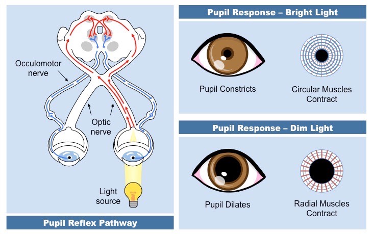
what is the pupil reflex
The pupil reflex is an involuntary response originating at the brainstem and under the control of the autonomic nervous system
It involves the resizing of the iris to regulate the amount of light that reaches the retina (excess light can damage the retina)
how do pupils respond to diff. levels of light
Pupils constrict in bright light (to prevent overstimulation of photoreceptors) and dilate in dim light (to maximise light exposure)
how do the para and sympathetic nerve control pupil constriction
In bright light, parasympathetic nerves trigger circular muscles to contract and cause the pupils to constrict
In dim light, sympathetic nerves trigger radial muscles to contract and cause the pupils to dilate
Overview of the Pupil Reflex diagram
What is brain death
Brain death is defined as the permanent absence of measurable activity in both the cerebrum and brainstem
when will a vegatative state occur and how
The brainstem is responsible for involuntary autonomic responses and may function alone to maintain homeostasis
Hence, individuals with a non-functioning cerebrum but a functioning brainstem may be kept alive in a vegetative state
how can brain death be determined
Brain death can be determined by medical professionals by testing the function of specific autonomic responses
The pupil reflex is one autonomic test used to assess brain death – brain dead individuals will not exhibit a pupil reflex
The Glasgow Coma Scale uses multiple tests to determine the neurological health of someone with suspected brain injury
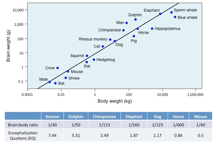
correlation between body and brain size in diff. animals
There is a positive correlation between body size and brain size in different animals – larger animals have larger brains
This correlation follows a linear pattern of progression but is not directly proportional
brain:body ratio in larger animals explanation
While an increase in body size results in an increase in brain size, the brain:body ratio decreases in larger animals
Body mass increases disproportionately to an increase in brain mass as most tasks only require a fixed brain capacity
what is Encephalization
the amount of brain mass relative to an animal's body mass
Encephalization graph
brain energy consumption
The human brain consumes ~20% of the body’s energy levels, despite making up only ~2% of the body’s mass
why are large amounts of energy needed/ what is it used for in the brain
The large amounts of energy required by the brain are used to sustain neurons and their processes
Energy is needed to maintain a resting potential when neurons are not firing (Na+/K+ pump uses ATP)
Energy is used to synthesise large numbers of neurotransmitters to facilitate neuronal communication