GRISTO Module 4 Anatomy: Blood Supply of the CNS
1/117
There's no tags or description
Looks like no tags are added yet.
Name | Mastery | Learn | Test | Matching | Spaced | Call with Kai |
|---|
No analytics yet
Send a link to your students to track their progress
118 Terms
center, furthest
Arterial supply is delivered from the outside, so the ____ of the CNS is ______ from the largest vessels
segmental, regional
The arterial supply to the spinal cord is largely ________, while the blood supply of the brain is ______
arterioles and capillary beds
Anastomoses between larger arteries are limited and there are almost no anastomoses between
The internal carotid system and the vertebral system
There are two pathways of blood into the brain:
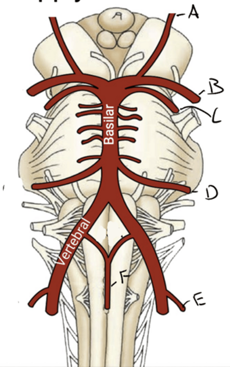
BLOOD SUPPLY OF THE BRAIN AND BRAINSTEM
BLOOD SUPPLY OF THE BRAIN AND BRAINSTEM
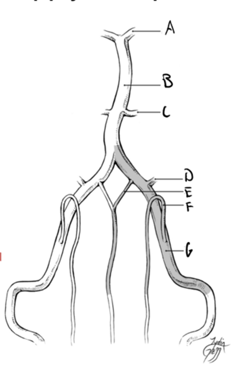
A. Basilar
B. Vertebral artery
C. Internal carotid artery
D. External carotid artery
E. Common carotid
F. Brachiocephalic trunk
G. Subclavian artery
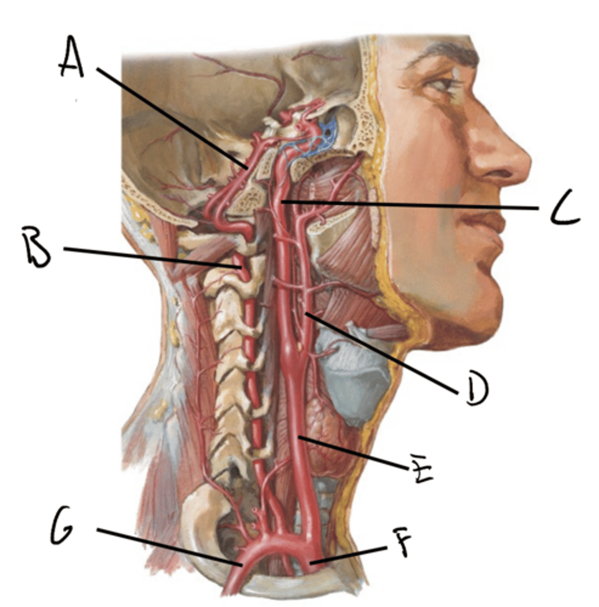
Enters the skull at the opening to the carotid canal, passes through the petrous temporal bone before emerging into the middle cranial fossa, where it passes anteriorly through the cavernous sinus
The artery loops superiorly and posteriorly before dividing into its terminal branched
The internal carotid artery is a branch of the common carotid artery. The artery travels:
Anterior cerebral artery and middle cerebral artery
Terminal branches of the internal carotid artery
ascend the neck through the transverse foramina of cervical vertebrae before entering the skull at the foramen magnum
These two vessels then come together to form the basilar artery at the base of the medulla
The vertebral arteries are branches of the subclavian artery. These vessels travel:
brainstem
The basilar artery is the main blood supply to the
circle of willis
The basiler arter is connected to the carotid system via the
Circle of Willis Isolated
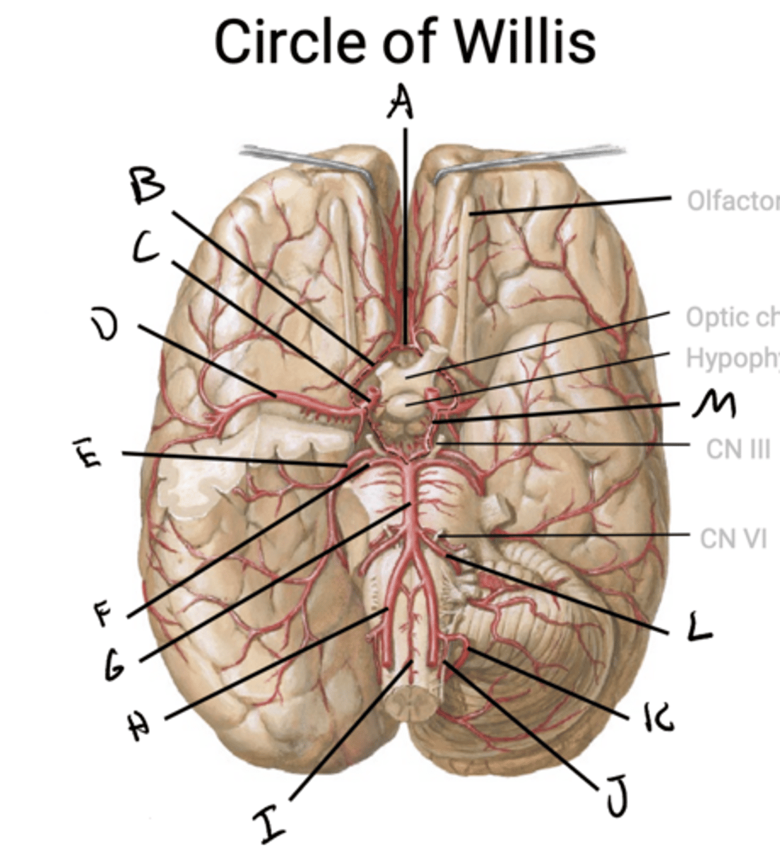
A. Middle cerebral artery (MCA)
B. Posterior cerebral artery
C. Superior cerebellar artery (SCA)
D. Anterior inferior cerebellar artery
E. Posterior inferior cerebellar artery (PICA)
F. Anterior spinal artery (ASA)
G. Posterior spinal artery (PSA)
H. Vertebral artery
I. Basilar artery
J. Posterior communicating artery (PCOM)
K. Internal carotid artery
L. Anterior communicating artery (ACOM)
M. Anterior cerebral artery (ACA)
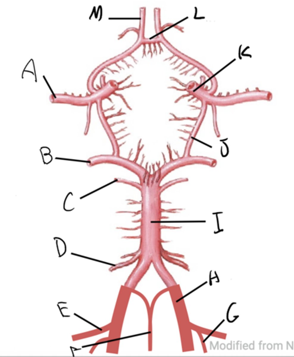
A. Anterior communicating branch (ACOM)
B. Anterior cerebral artery
C. Internal carotid artery
D. Middle cerebral artery
E. posterior cerebral artery
F. superior cerebellar artery
G. Basilar artery
H. Vertebral artery
I. Anterior spinal artery
J. Posterior spinal artery
K. Posterior inferior cerebellar artery (PICA)
L. Anterior inferior cerebellar artery (AICA)
M. Posterior communicating artery (PCOM)
cerebral cortex and deep telencephalic structures like basal ganglia and internal capsule
The internal carotid system irrigates most of the
middle cerebral artery (MCA) and anterior cerebral artery (ACA)
The main branches of the internal carotid artery are the
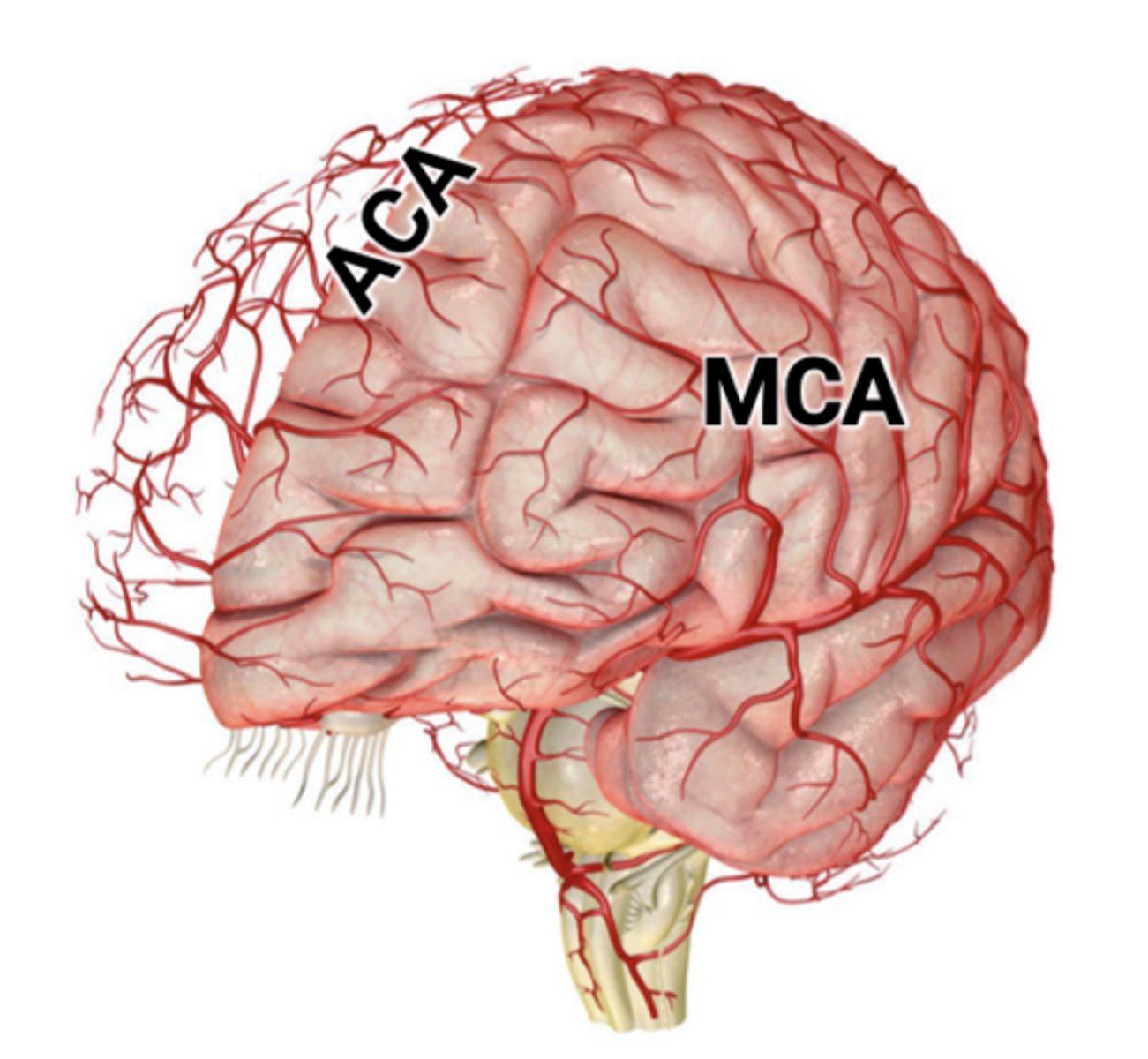
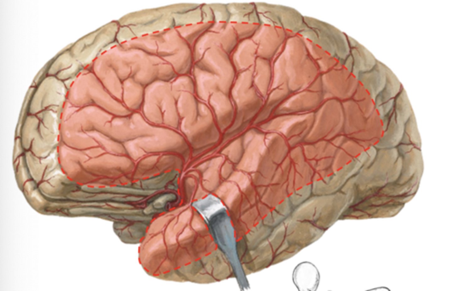
Covers the largest territory in the brain, supplying most of the lateral frontal, parietal, and temporal lobes and insula
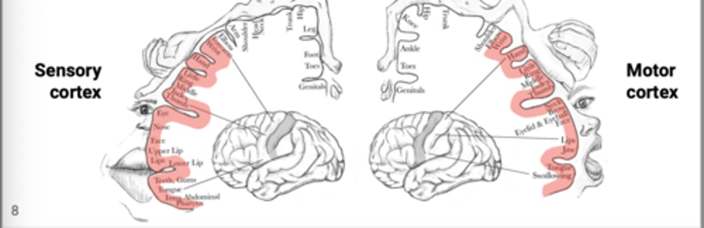
Branches of the MCA supply the portions of the primary motor and sensory cortices dedicated to the arm and face.
Middle cerebral artery:
will generally have paresis of the contralateral face and upper limb with sensory loss from the corresponding area
Patients with MCA strokes
ACA and PCA can NOT take over it does not anastomoses with anything
If we lose MCA
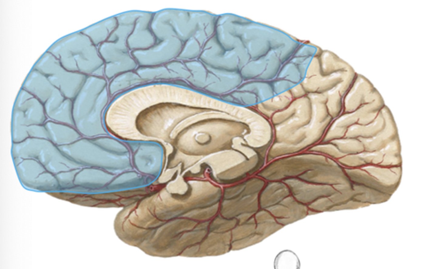
Supplies the medial surfaces of the frontal and parietal lobes. The paired anterior cerebral arteries are joined anterior to the optic chiasm by the anterior communicating artery
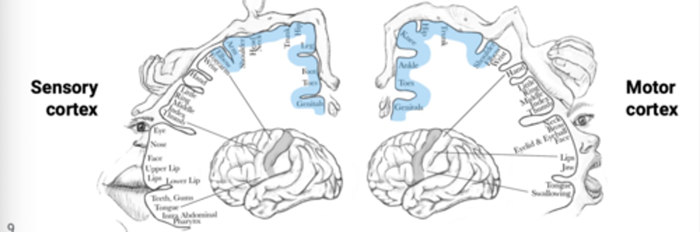
Branches of the ACA supply the portions of the primary motor and sensory cortices dedicated to the lower limb and perineum.
Anterior cerebellar artery:
have paresis of the contralateral lower limb and foot with sensory loss from the corresponding areas
Patients with ACA strokes will generally
the entire spinal cord, brainstem, cerebellum, thalamus, occipital lobes, and inferior aspect of temporal lobes
The vertebrobasilar system irrigates
Basilar artery terminates in two posterior cerebral arteries
The PCA supplies the medial surface of the occipital lobe (visual cortex) and the inferior and lower lateral surfaces of the temporal lobe
Posterior cerebral artery (PCA)
prominent visual deficits
Strokes to PCA results in
A. PCOM
B. PCA
C. SCA
D. AICA
E. PICA
F. ASA
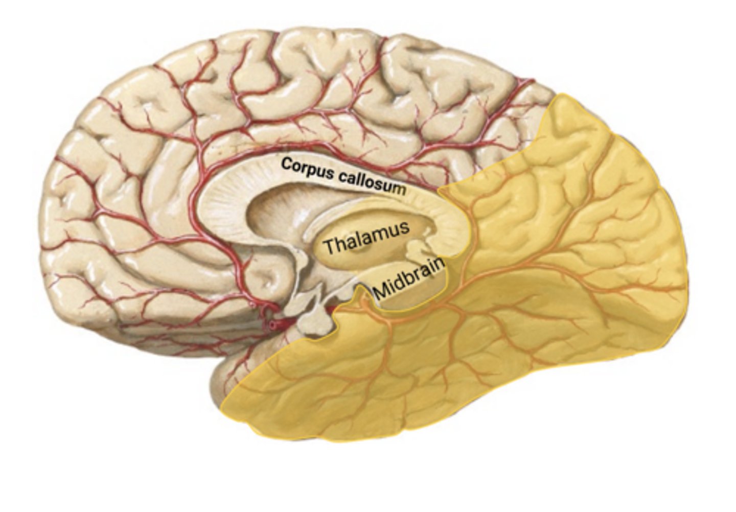
PCA aa
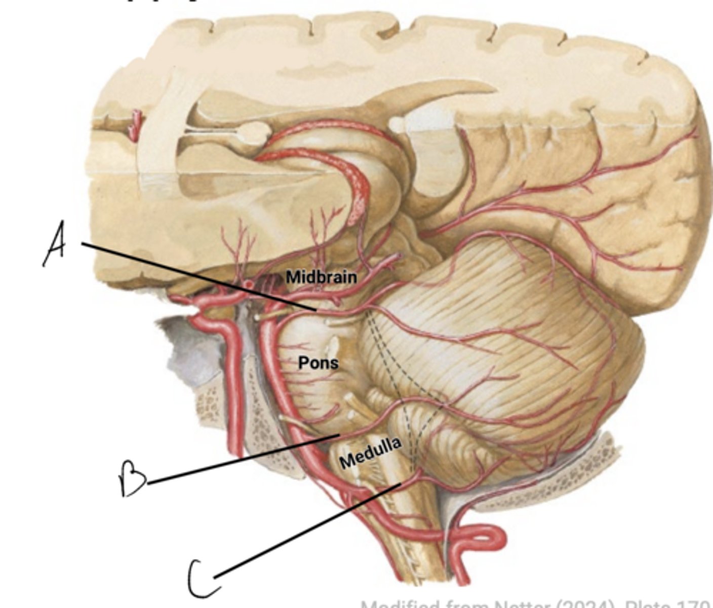
Blood supply to the Midbrain
Basilar a.
Blood supply to the pons
vertebral aa
Blood supply to the medulla
PCA
Blood supply to occipital lobe
PCA
Blood supply to inferior temporal lobe
PCA
Blood supply to thalamus
PICA, AICA, and SCA
Blood supply to cerebellum
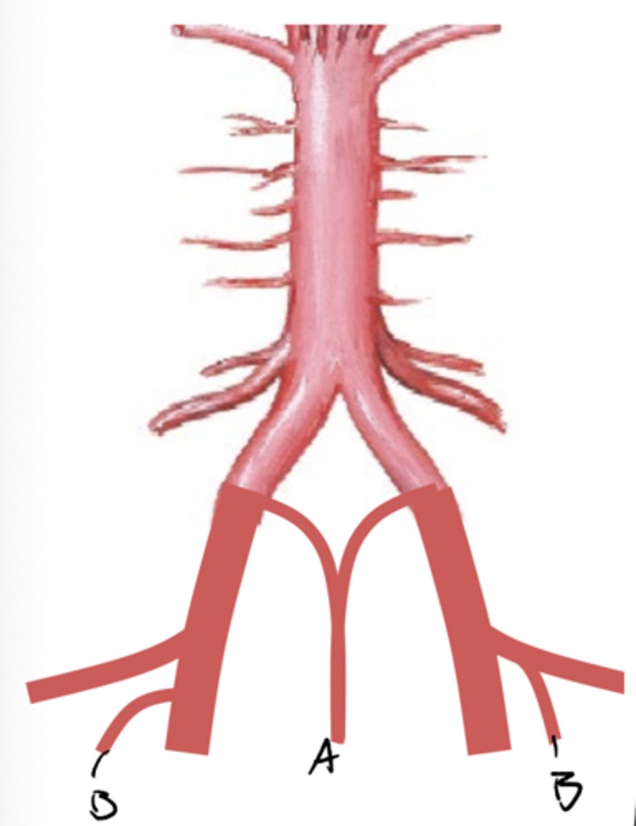
vertebral artery system --> anterior and posterior spinal arteries
Blood supply to spinal cord
A. Superior cerebellar artery (SCA)
B. Anterior inferior cerebellar artery (AICA)
C. Posterior inferior cerebellar artery (PICA)
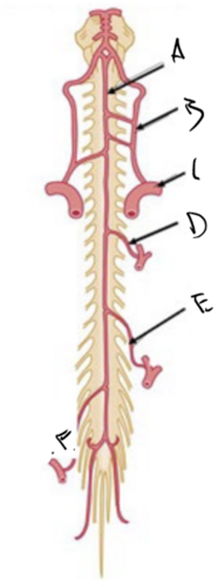
BLOOD SUPPLY OF THE SPINAL CORD
BLOOD SUPPLY OF THE SPINAL CORD
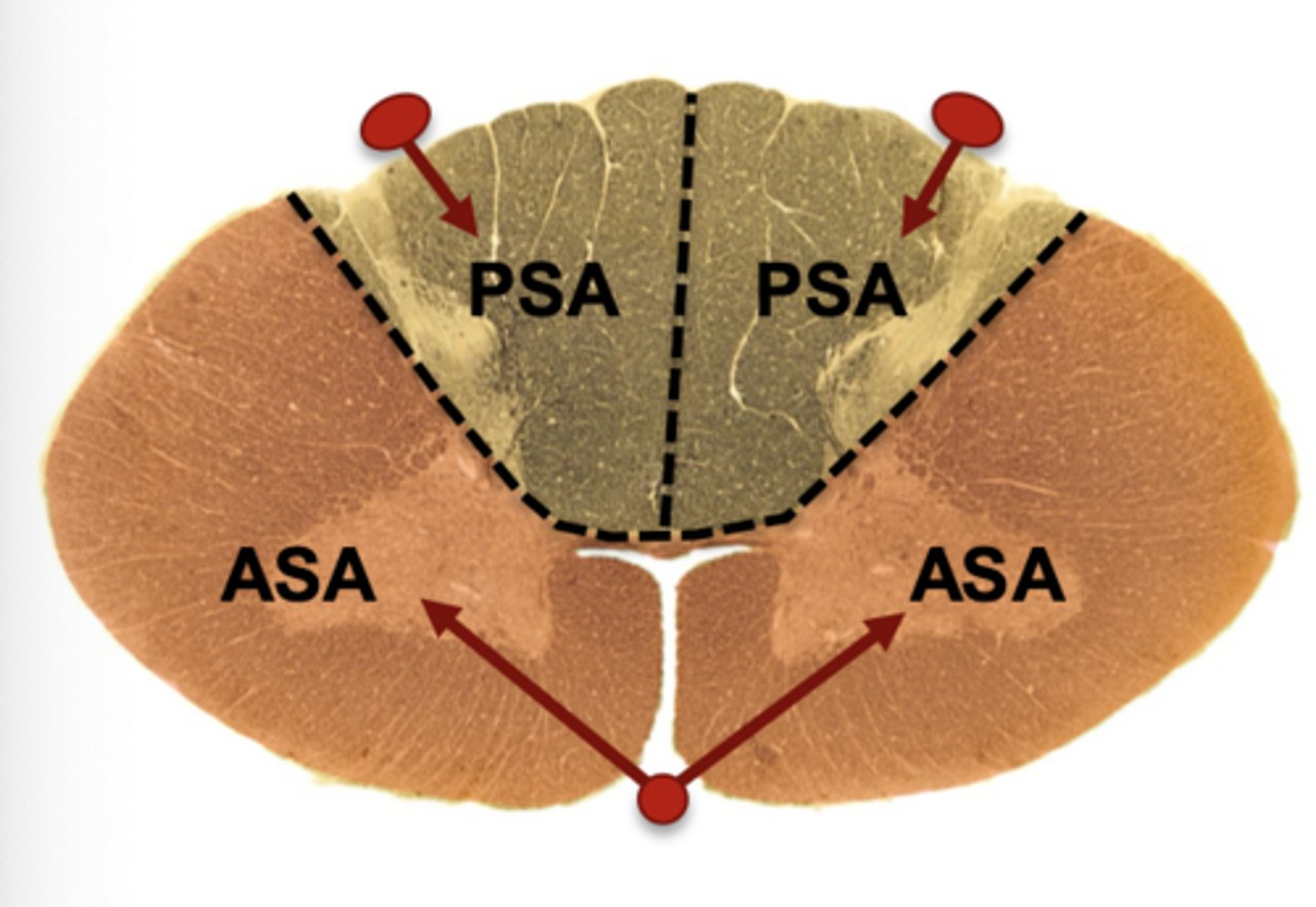
A. ASA
B. PSA
also B: PSA
A. SCA
B. Basilar artery
C. AICA
D. PICA (gives off posterior spinal a in 75%)
E. Anterior spinal a
F. Posterior spinal a. (25% of cases)
G. vertebral artery
entire spinal cord
The vertebrobasilar system supplies the
the vertebral artery
The anterior spinal artery branches directly off
PICA but can also branch off the vertebral arteries as well
The posterior spinal artery most often branches off
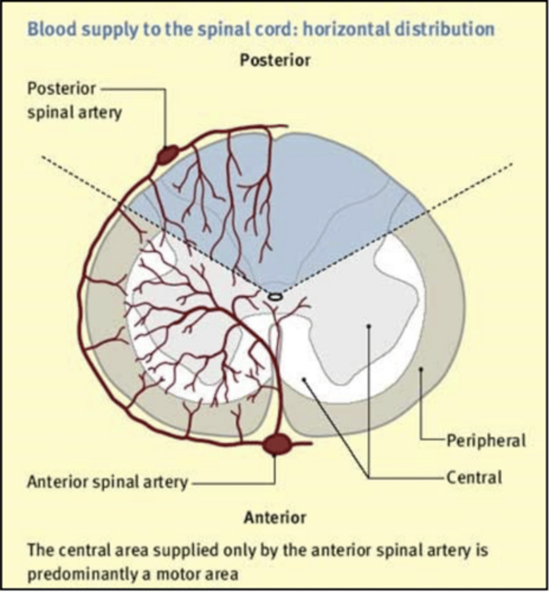
Supplies the ventral horn and intermediate gray areas, plus the ventral and lateral funiculi bilaterally, as well as the ventral white commissure
Along the length of the spinal cord:
The Anterior spinal artery
is bilateral. Together they supply most of the dorsal horn and dorsal column unilaterally
Along the length of the spinal cord:
The Posterior spinal artery
weakly anastomose, but this is inconsistent
ASA and PSA territories
have some serious consequences
A lesion to either the ASA or PSA can
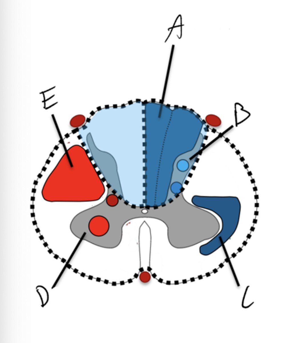
A. Dorsal column system
B. Dorsal horn
C. Spinothalamic tract
D. Ventral horn
E. Corticospinal tract
descend
Although the ASA and PSA arteries run the length of the cord their robusticity decreases as they
medullary, great radicular artery (of adamkiewicz)
Occasionally, _______ arteries will extend to anastomose with the ASA. The largest and most discernable of these is the __________
A. Anterior spinal a.
B. Vertebral a.
C. Subclavian
D. Anterior medullary a.
E. Great radicular a.
F. Lumbar anterior medullary a.
roots
The ASA and PSA supply the cord itself but rarely reach out to supply the
segmental arteries, Radicular arteries
The roots are supplies by ____________, which branch from larger local arteries. they in turn give off ____
ventral and dorsal roots
- Very small and terminate in the roots
The radicular arteries supply the
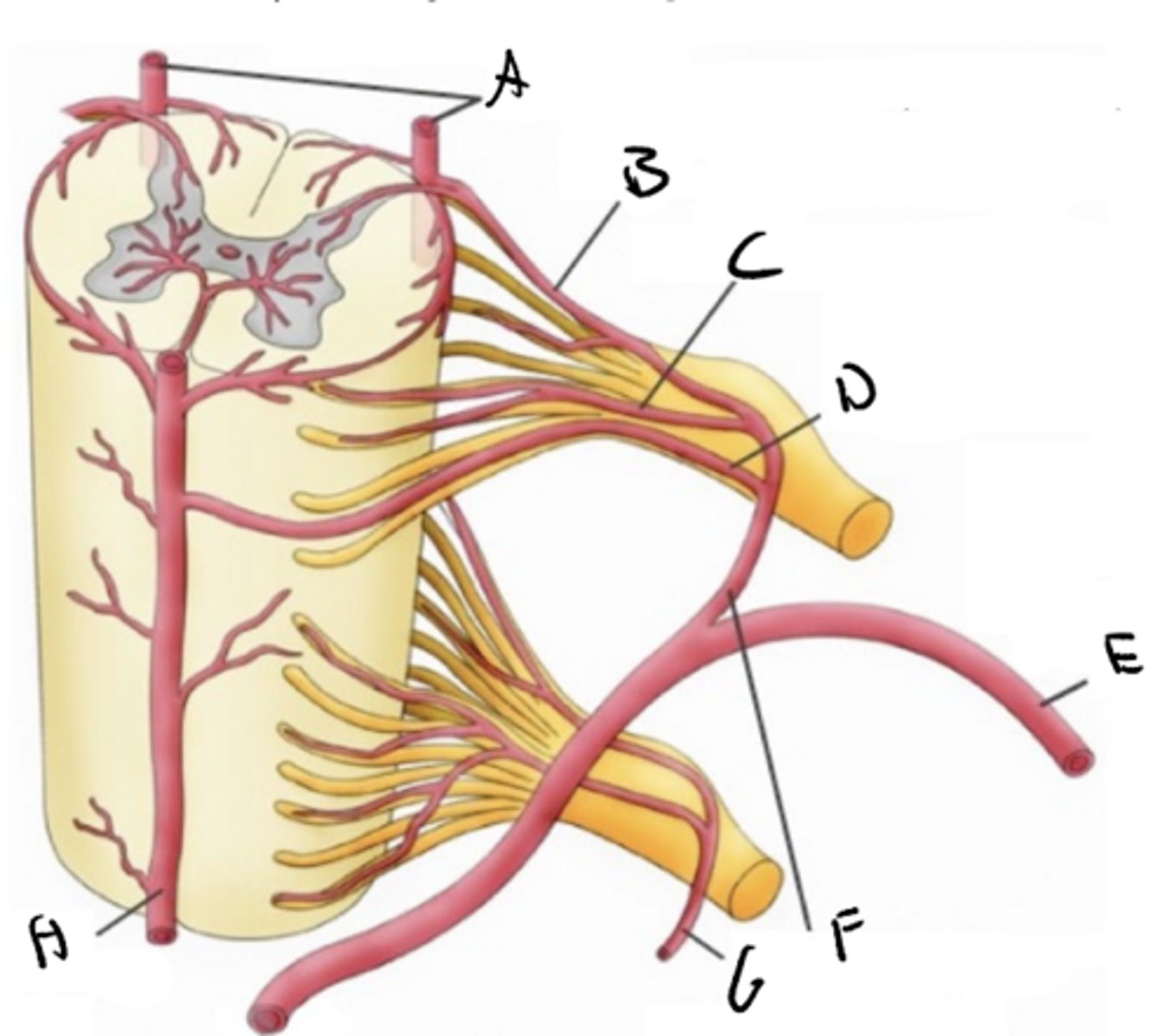
A. Posterior spinal aa (from PICA)
B. Posterior medullary a
C. Radicular artery
D. Anterior medullary a
E. Posterior intercostal a
F. Segmental aa.
G. Segmental aa
H. Anterior spinal a
VENOUS DRAINAGE
VENOUS DRAINAGE
Cerebral
______ veins drain the brain and associated structures
dural venous sinuses (openings in the layers of the dura mater)
Cerebral veins drain into
internal jugular vein, which leaves the neurocranium
Dural venous sinuses drain predictably through the skull and coalesce to form the
A. External cerebral veins
B. Superior sagittal sinus
C. Bridging veins from external cerebral veins
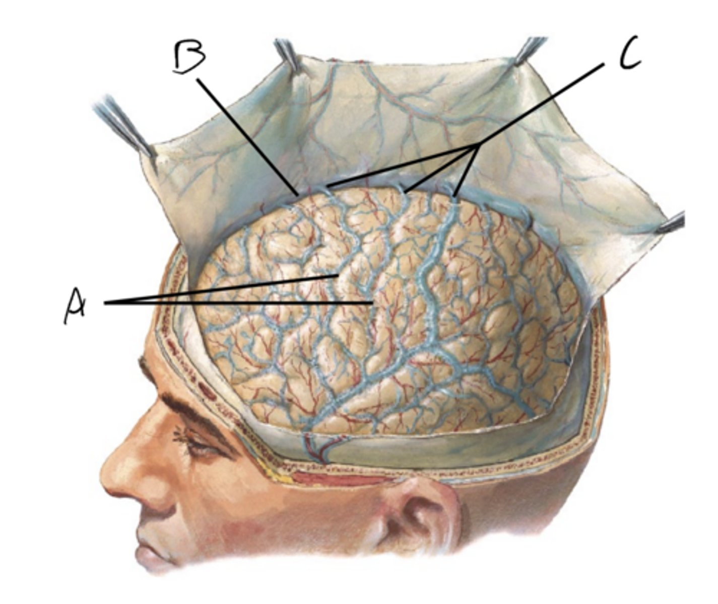
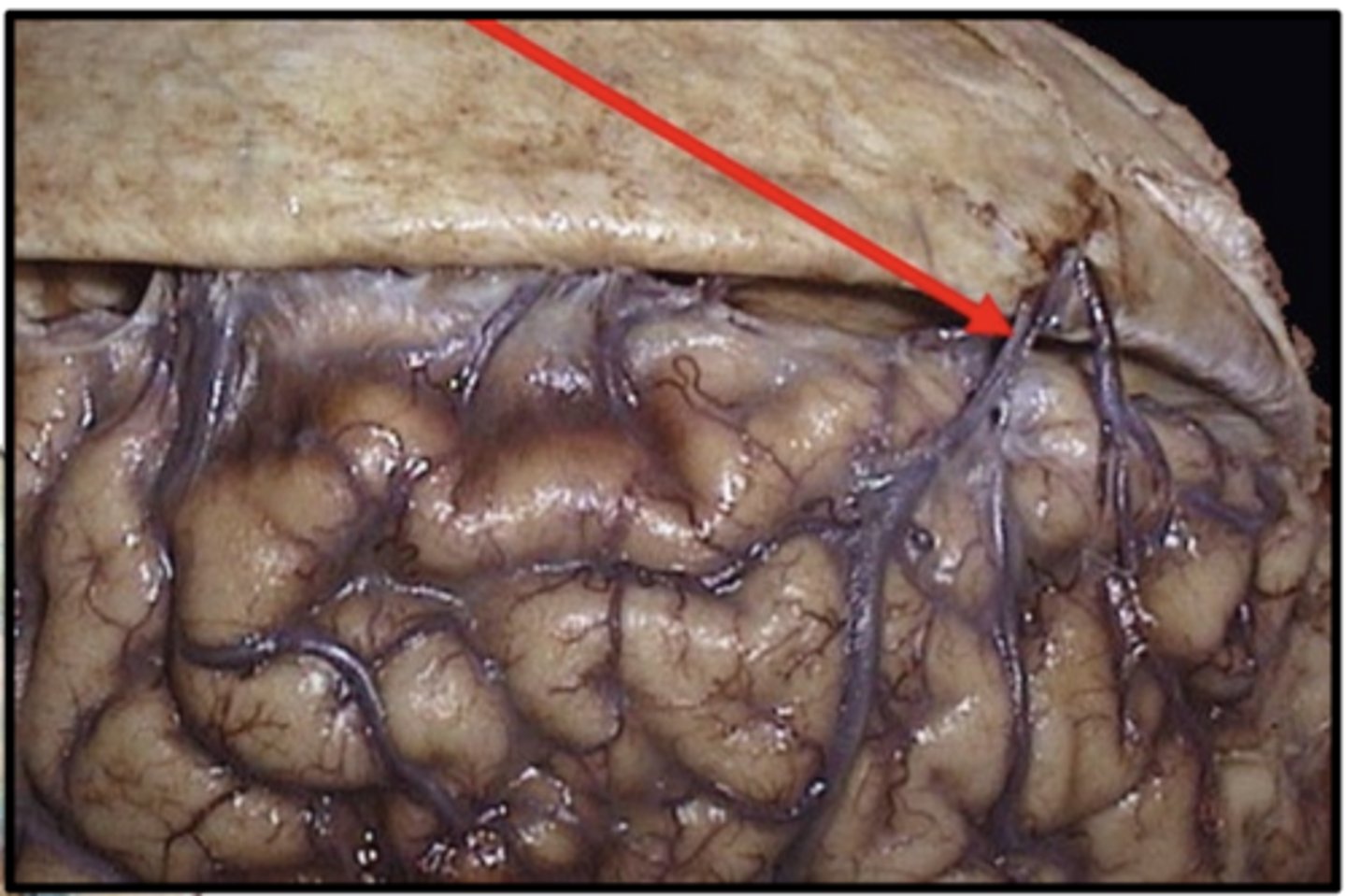
Bridging veins
- if broken can cause subdural hematoma
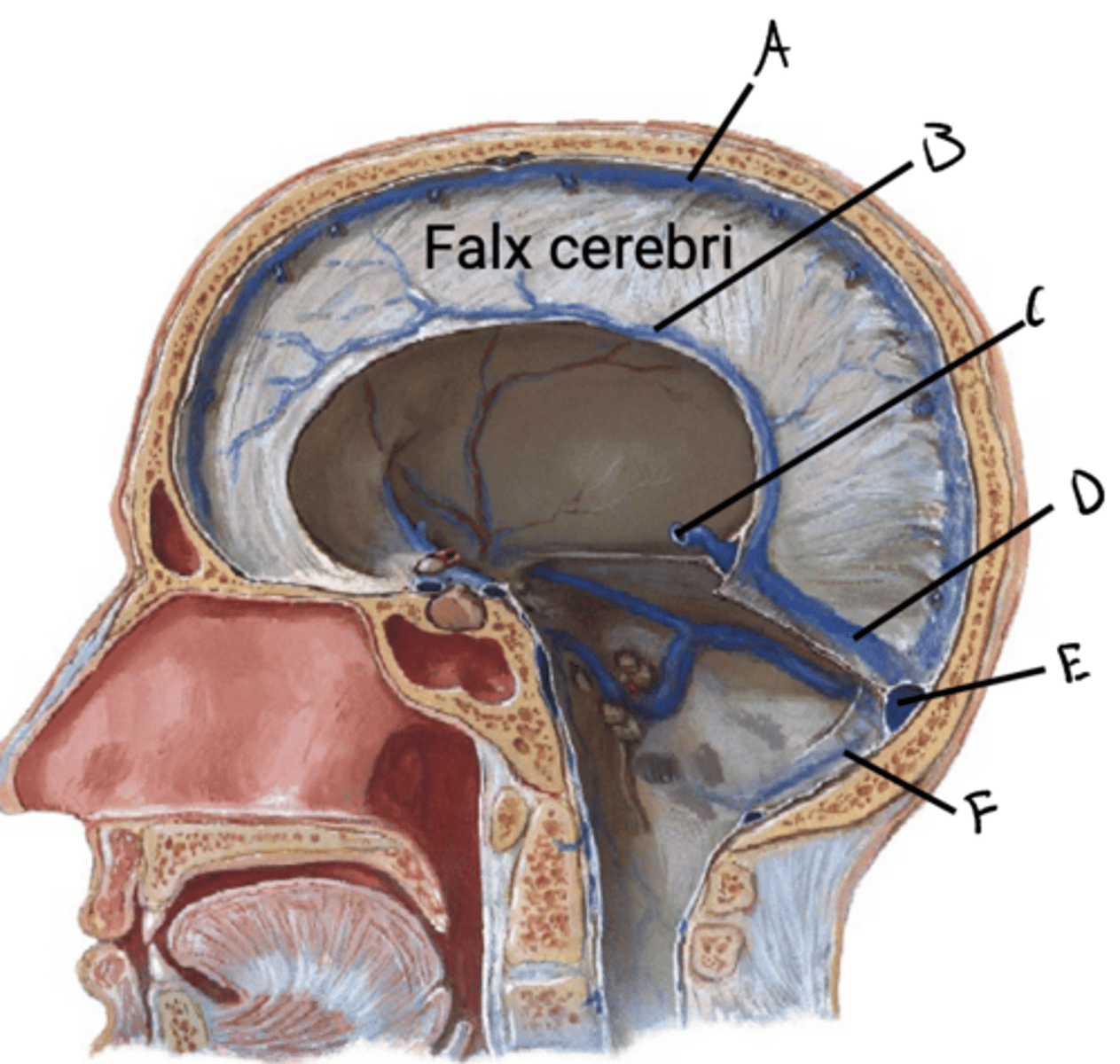
crescent-shaped dural fold that sits in the longitudinal fissure of the cerebrum
The falx cerebri is a
the crista galli to the occiput where it connects with the tentorium cerebelli
The falx cerebri runs from
A. Superior sagittal sinus
B. Inferior sagittal sinus
C. Great cerebral vein (galen)
D. Straight sinus
E. Confluence of sinuses
F. Occipital sinus
A. Superior sagittal sinus
B. Superior ophthalmic vein
C. Cavernous sinus
D. Tentorial notch
E. Inferior sagittal sinus
F. Straight sinus
G. Confluence of sinuses
H. Transverse sinus
I. Sigmoid sinus
J. Internal jugular vein
K. Superior petrosal sinus
L. Inferior petrosal sinus
M. Falx cerebri
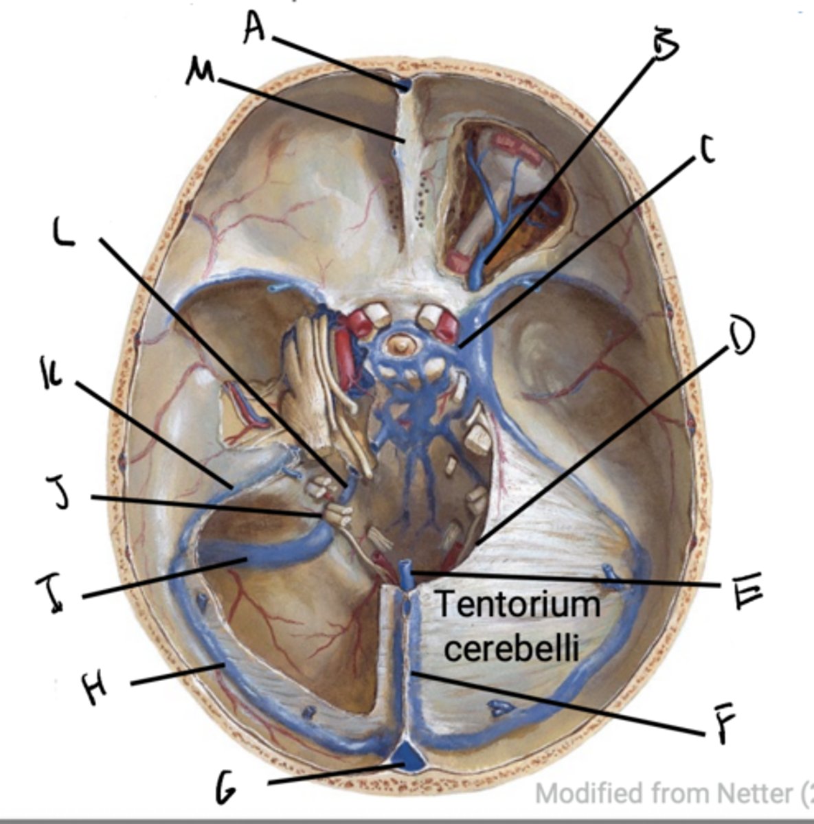
falx cerebri between the cerebral hemispheres
Tentorium cerebelli between occipital lobe of the cerebrum and cerebellum
Two specialized dural folds support and partition the major brain parts:
meningeal dura
The dural venous sinuses are venous channels found between layers of
internal and external veins of the brain
As well as CSF from the subarachnoid space, ultimately joining to empty into the internal jugular vein
The dural venous sinuses receive blood from the ......... as well as CSF from the ........
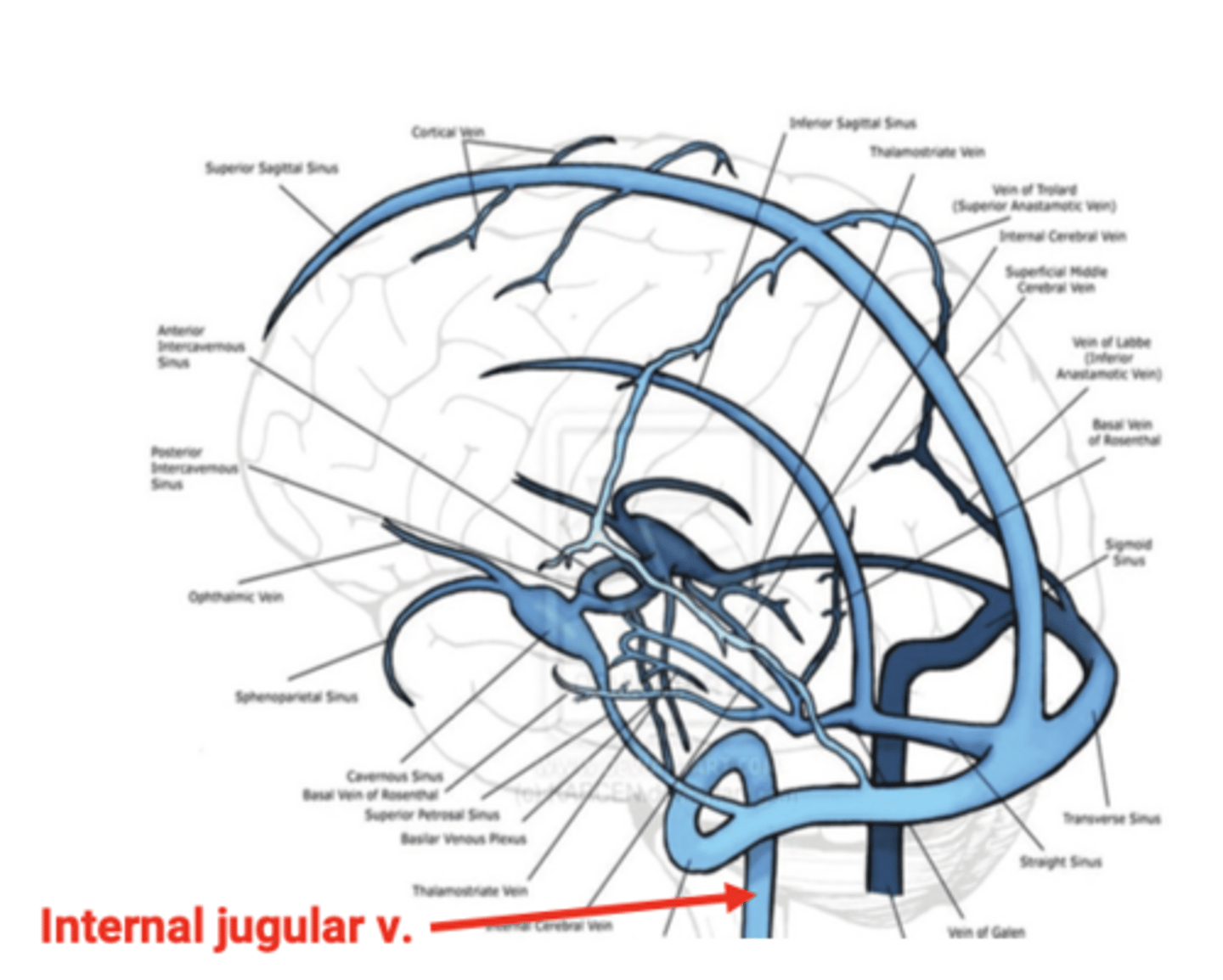
HOW BLOOD IS RETURNED TO THE INTERNAL JUGULAR
HOW BLOOD IS RETURNED TO THE INTERNAL JUGULAR
falx cerebri
The superior sagittal sinus arises in the roof of the
deeply placed great cerebral vein to form the straight sinus
The inferior sagittal sinus arises in the inferior free edge of the falx cerebri and joins the
superior sagittal sinus and the small occipital sinus which runs in the small falx cerebelli, at the confluence of sinuses
The straight sinus courses through the midline of the tentorium cerebelli before uniting with the
two transverse sinuses which run lateralward and forward in the attached margin of the tentorium cerebelli
The confluence of sinuses drains into the
Common superior ophtalmic vein, which in turn drains into the centrally located cavernous sinus
Meanwhile:
Blood from the orbit and face (mainly via the supraorbital and supratrochlear veins and nasal veins) drains into the
superior ophthalmic vein, which in turn drains into the centrally located cavernous sinus
The cavernous sinus is emptied by the
transverse sinus to form the S-shaped sigmoid sinus
The superior petrosal sinus units with teh
the internal jugular vein
The inferior petrosal sinus joins with the sigmoid sinus to form
A. Optic Chiasm
B. Internal carotid artery
C. CN III
D. CN IV
E. CN VI
F. CN V1
G. CN V2
H. Cavernous sinus
I. Pituitary
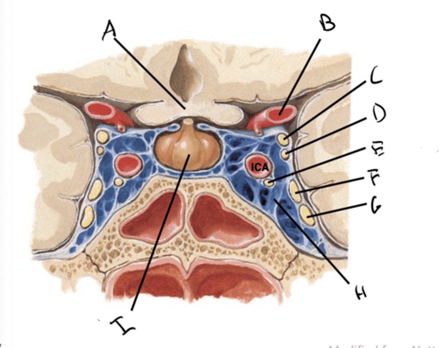
cool arterial blood entering the cranium
The cavernous sinus helps
nasal cavity
Across many heat adapted mammalian species, heat is dissipated through the vessels in the
cavernous sinus
The cool venous blood returns to the skull and drains into the
cools the arterial blood as it enters the cranium, thus aiding in maintaining brain thermodynamics
The internal carotid artery runs through the cavernous sinus. This cool venous blood of the cavernous sinus surrounds the carotid artery and
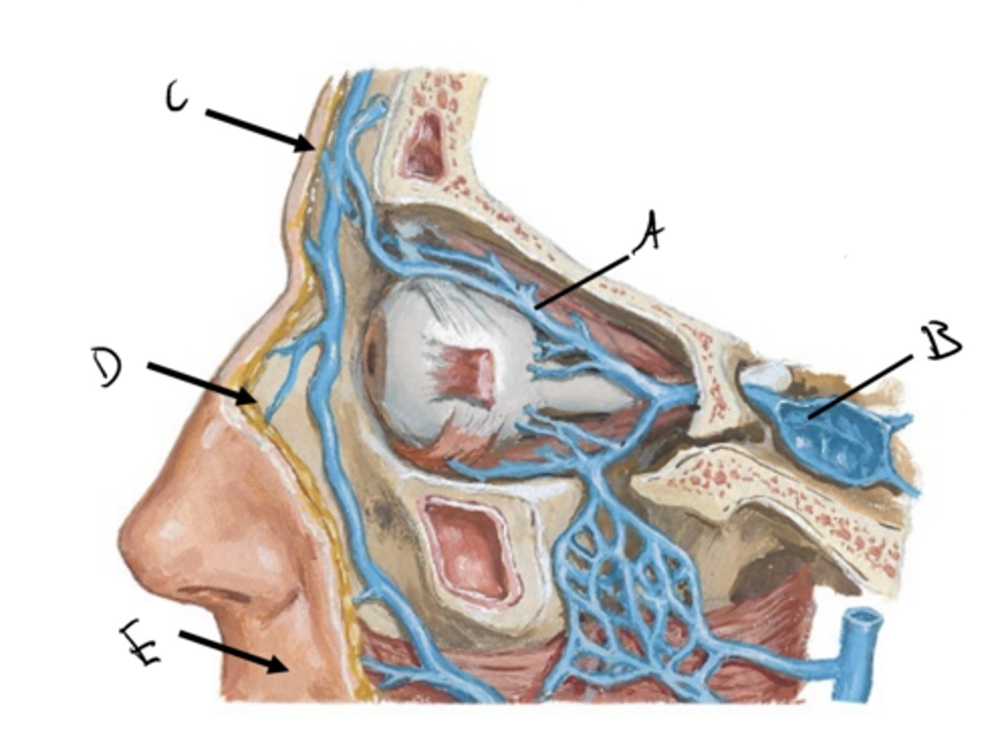
A. Ophthalmic vein
B. Cavernous sinus
C. Drains the forehead
D. Drains the nose
E. Drains the lips
Bacterial infection that spreads from the face such as sinusitis, infection of the nasal sinuses
Or
Facial acne which can become infected or introduce infections into the bloodstream if punctured
Cavernous sinus thrombosis is often caused by
region from which blood drains from the face via the superior ophthalmic vein into the cavernous sinuse
This is the way these infections can spread to the dural sinuses
The Danger Triangle of the face is the
QUIZ QUESTIONS
QUIZ QUESTIONS
False
True or false? There are many anastomotic connections between the arteries of the brain
Vertebral
Indicate the systemic origin (carotid versus vertebral)
Basilar artery
Vertebral
Indicate the systemic origin (carotid versus vertebral)
AICA
Vertebral
Indicate the systemic origin (carotid versus vertebral)
Posterior spinal artery
Carotid
Indicate the systemic origin (carotid versus vertebral)
Anterior cerebral artery
Carotid
Indicate the systemic origin (carotid versus vertebral)
Middle cerebral artery
Vertebral
Indicate the systemic origin (carotid versus vertebral)
Posterior cerebral artery
Vertebral
Indicate the systemic origin (carotid versus vertebral)
PICA
Vertebral
Indicate the systemic origin (carotid versus vertebral)
Anterior spinal artery
Vertebral
Indicate the systemic origin (carotid versus vertebral)
Superior cerebellar artery
Posterior communicating artery
What is the name of the artery that connects the vertebral and carotid systems
Lateral cerebrum (UL & head motor and sensory cortex and insula)
Match the artery with its general territory:
MCA