Sensory modalities
0.0(0)
0.0(0)
Card Sorting
1/12
Earn XP
Description and Tags
Colour vision, hearing, and multimodal integration
Study Analytics
Name | Mastery | Learn | Test | Matching | Spaced |
|---|
No study sessions yet.
13 Terms
1
New cards
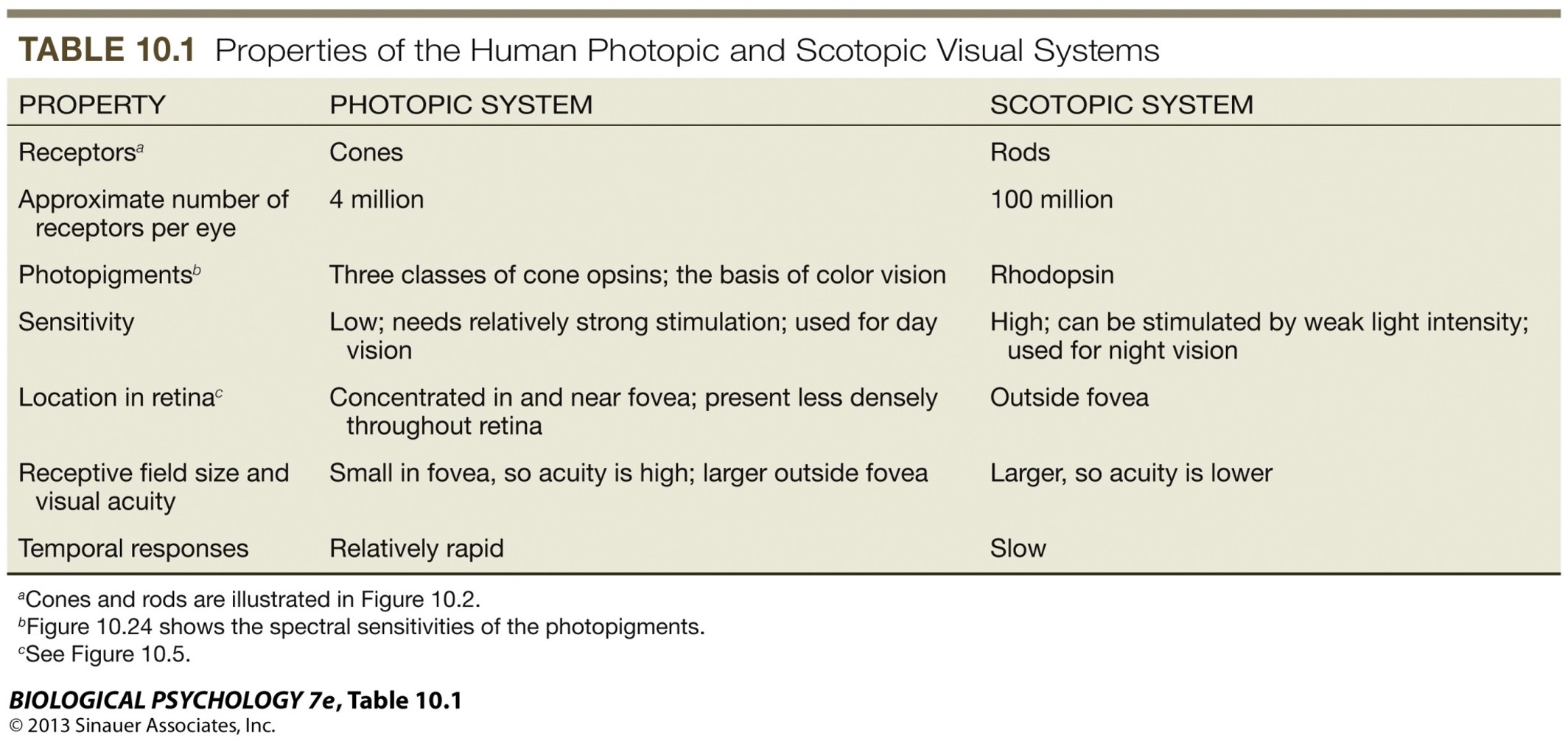
Cones vs rods
Cones:
* Photopic system
* 4 million receptors per eye
* 3 classes of opsins - **basis of colour vision**
* S-cones - short-wavelength sensitive receptor
* M-cones - medium-wavelength sensitive receptor
* L-cones - long-wavelength sensitive receptor
* Low sensitivity - needs strong stimulation, used for **day vision**
* Location - concentrated in and near fovea, less dense throughout retina
* Small in fovea, so higher acuity
* Relatively rapid temporal responses
Rods:
* Scotopic system
* 100 million receptors per eye
* Rhodopsin photopigments
* High sensitivity - stimulated by weak light intensity, used for **night vision. No colour vision.**
* Located outside fovea
* Larger, so lower acuity
* Slow temporal responses
2
New cards
Types of colour vision
* Dichromatic colour vision - only 2 cones, most mammals are dichromats and lack M-cone
* Trichomatic colour vision - have 3 cones therefore can discriminate more colours than dichromats, e.g. humans, few old-world primates
* Monochromatic vision - marine mammals do not see colour as they only have L-cone (e.g. whales, seals)
* Trichomatic colour vision - have 3 cones therefore can discriminate more colours than dichromats, e.g. humans, few old-world primates
* Monochromatic vision - marine mammals do not see colour as they only have L-cone (e.g. whales, seals)
3
New cards
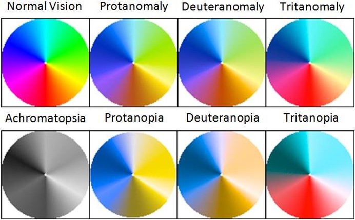
Colour vision deficiencies
* Prevalent in 5-8% of males and 0.5% of females
* X-chromosome recombination causes colour vision defects
* S gene on chromosome 7
* M and L gene on X
* Colour deficiency (mild) - trichromats
* Colour blindness (severe deficiency) - dichromats and truly colour blind
* More men affected than women because X chromosome defects are frequent and men only have an X chromosome
* See diagram for different variations (Melillo et al., 2017)
4
New cards
Colour pathway
P-ganglion cells:
* Project to parvocellular layer in lateral geniculate nucleus (LGN)
* Small receptive fields (RFs)
* Slower conduction speed
* High acuity
* Poor response to transient stimuli
* **Colour sensitive**
M-ganglion cells:
* Project to magnocellular layer in lateral geniculate nucleus (LGN)
* Large receptive fields
* Higher conduction speed
* Sensitive to motion
* Low acuity
* **No colour discrimination**
Parvocellular (P-pathways, small soma) and magnocellular (M-pathways, large soma) pathways:
* Only P-cells in the LGN show spectrally opponent responses to monochromatic light stimuli of different wavelength, i.e. excited by some wavelengths and inhibited by others
* M-cells project to layer 4ca, P-cells project to layer 4c and 2,3
5
New cards
Hearing functions
* Hearing to detect and discriminate locations and movement of sound sources
* Spatial orientation
* Echolocation - bats, whales, humans
* Auditory communication and language
* Spatial orientation
* Echolocation - bats, whales, humans
* Auditory communication and language
6
New cards
Sound
* Sound = pressure waves, movement of air particles set in motion by vibrating structure
* Propagates in 3 dimensions, alternating compression and rarefaction of air, molecules move back and forth from regions of high pressure to low pressure
* Measures of sound
* Frequency - Hertz (Hz), reciprocal of wavelength (perceived as pitch)
* Amplitude - loudness
* Phase and waveform
* Propagates in 3 dimensions, alternating compression and rarefaction of air, molecules move back and forth from regions of high pressure to low pressure
* Measures of sound
* Frequency - Hertz (Hz), reciprocal of wavelength (perceived as pitch)
* Amplitude - loudness
* Phase and waveform
7
New cards
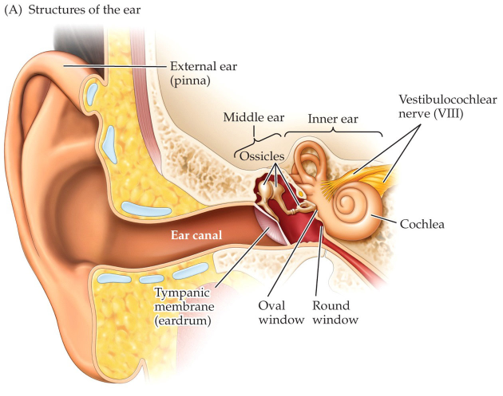
Ear
* Different to the retina - the ear cannot spatially map the locations of sound
* Vibrations travel from the tympanum to the middle ear where they’re amplified
* Hair cells are arranged tonotopically - they’re tuned to narrow range of sound frequencies by their location along the basilar membrane
* Vibrations travel from the tympanum to the middle ear where they’re amplified
* Hair cells are arranged tonotopically - they’re tuned to narrow range of sound frequencies by their location along the basilar membrane
8
New cards
Stereocillia (stiff hair)
Stereocillia helps to stretch open the ion channels:
1. Bending of stereocilia at input zone
2. Opening of nonselective ion channels that allow influx of K+ and Ca2+ ions
3. Depolarisation of hair cell open voltage-gated Ca2+ channels in the base of the hair cell at output zone
4. Neurotransmitter is released to excite afferent auditory interneurons (which form the cochlear nerve)
9
New cards
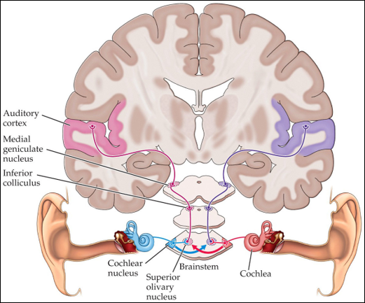
Auditory pathway
1. Cochlear projects to the contralateral cortex
2. Each superior olivary nuclei of brain stem receives inputs from both cochlear nuclei for first stage of binaural analysis of sound-source location
10
New cards
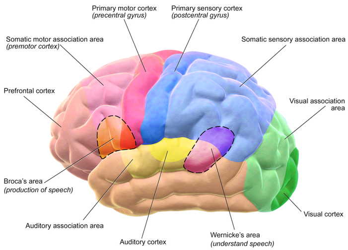
Multimodal integration
* Cortical areas receiving multimodal sensory inputs - see diagram
11
New cards
Direction of attention
* Selective attention is different from vigilance (global state of alertness)
* Overt direction of attention - gaze centred on area of interest coincides with the visual information selected for attention (e.g. when reading a text)
* Covert attention - gaze fixates on location in visual field whilst information from another part is selected for attention
* Magicians are masters in misdirecting and splitting attention, introducing uncertainties or multilevel tasks with multimodal sensory stimulation in addition to social cues
* Kuhn et al (2016) - using eye tracking, found gaze directed to face (when asked a question by magician) resulted in more change blindness
12
New cards
Cocktail party effect (Treisman, 1964)
The ability to consciously drive attention towards one stimulus filtered out from the noisy environment
* Better performance with binaural stimulation (both ears listen). Also demonstrated in other sensory modalities (e.g. fast visual sequence)
* Perceptual segregation of stimuli: brain filters and predicts through uni/multimodal processing and integration but exerts also top-down control to affect various stages and connections during sensory processing
* Some stimuli can be more salient (e.g. strongly contrasting colours). Some can be more semantic (e.g. one’s own name) and therefore elss susceptible to interference
13
New cards
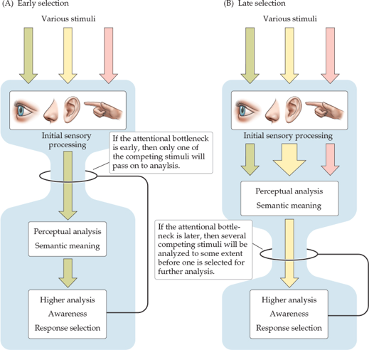
Models
* Bottleneck models - suggest that attention is largely dependent on association cortices
* Perceptual load models - propose that filtering could already influence activity in sensory pathways, e.g. if stimuli are complex
* Important regions for gaze control, attention and filtering: pulvinar, LIP/MIP, frontal eye fields
* Perceptual load models - propose that filtering could already influence activity in sensory pathways, e.g. if stimuli are complex
* Important regions for gaze control, attention and filtering: pulvinar, LIP/MIP, frontal eye fields