Homeostasis
the maintenance of relatively constant conditions for physiological variables in the internal environment
Involves detecting changes in masses within compartments and activating responses that alter inflow or outflow (or both) for that compartment.
Mass
The amount of a substance in a specific compartment
The same substance in different compartments represents different masses (because mass is specific to compartment)
Masses are regulated by the balance between inflow and outflow
A change in a mass means there is an imbalance between inflow and outflow
Compartment
An area/space/container where substances exist
Inflow vs Outflow
Inflow: the rate that the substance is being added to the compartment
Outflow: the rate that the substance is being removed
Masses may have multiple inflows and outflows
There is always an inflow and outflow for every mass in physiology
inflow and outflow are controlled by feedback, feed forward, and receptor mediated control
Inflow, outflow, and gradients
inflow and outflow have their own energy gradient and conductance
The regulated mass represents the low energy (ELO) for its inflow.
The regulated mass also represents the high energy (EHI) for its outflow.
Inflow and outflow each contain their own energy gradient (EHI – ELO).
the ELO for inflow and the EHI for outflow for the regulated mass IS the regulated mass
Increase in mass can occur from…
Inflow increases and outflow does not change.
Outflow decreases and inflow does not change.
Inflow increases and outflow decreases.
Outflow increases a little but inflow increases a lot.
Inflow decreases a little but outflow decreases a lot.
Decrease in mass can occur from…
Outflow increases and inflow does not change.
Inflow decreases and outflow does not change.
Outflow increases and inflow decreases.
Inflow increases a little but outflow increases a lot.
Outflow decreases a little but inflow decreases a lot.
No change in mass can occur from…
Inflow and outflow do not change.
Inflow decreases and outflow decreases at the same time (changes in inflow and outflow are equal).
Inflow increases and outflow increases at the same time (changes in inflow and outflow are equal).
Flow will increase if…
Conductance (K) increases
The energy gradient (ΔE) increases because the high energy (EHI) increased
The energy gradient (ΔE) increases because the low energy (ELO) decreased
The energy gradient (ΔE) increases because the high energy (EHI) increased and the low energy (ELO) decreased
Flow will decrease if…
Conductance (K) decreases
The energy gradient (ΔE) decreases because the high energy (EHI) decreased
The energy gradient (ΔE) decreases because the low energy (ELO) increased
The energy gradient (ΔE) decreases because the high energy (EHI) decreased and the low energy (ELO) increased
Flow will not change if…
Conductance (K) does not change
The energy gradient (ΔE) does not change because neither the high energy (EHI) or low energy (ELO) changed
Control system
Exist to maintain regulated variables within a normal range
Regulated variables
Certain variables which are vital to survival. They are relatively stable (within a range), but not static (unchanging)
Homeostatic set point
Ideal point for a substance to be at.
Conservation of mass
Preservation and restoration of an amount of substance within a compartment
How are mass and flow related?
What happens when inflow = outflow?
Mass remains stable
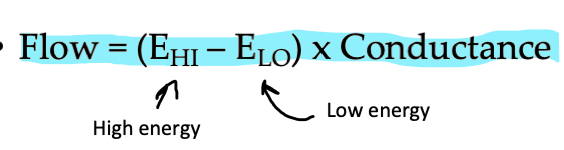
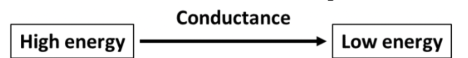
Flow
the rate at which a mass moves from one compartment to the next or the conversion of a substance from one form to another in a metabolic reaction.
determined by energy gradient and conductance
Flow = K x (EHI – ELO)
Flow models
Flow models are models that represent physiological flow
arrows represent flows
energy gradient is the difference in energy between the two sides of the arrow, the high energy at the flat end of the arrow and the low energy at the arrowhead end of the arrow
conductance is written on top of the arrow
Energy gradient (ΔE)
the “energy” difference between the masses of two different compartments that drives flow. Can be pressure, electrical charge, or concentration gradient.
Things tend to move down an energy gradient (high to low)
larger gradient causes more flow
flow continues until equilibrium is reached (ΔE=0)
ΔE = Ehi - Elo
Pressure Gradient (ΔP)
Pressure is the physical force exerted by a substance on the walls of its compartment. This gradient is created by the difference in pressure between two compartments. (PHI -PLO)
Concentration Gradient (ΔC)
This gradient is created by the difference in the concentration of a substance in one compartment compared to another compartment. (CHI -CLO) or ([HI] – [LO])
square brackets [ ] are often used to denote the concentration of a substance
Electrical Gradient (Δe)
This gradient is created by the difference in the electrical charge, which is determined by the number of charged particles (positively or negatively charged), of one compartment compared to another compartment. (eHI -eLO)
Point of equillibrium
point in which there is no difference between the high and low energy of a gradient
no net change, there is equal rates of change in the flow in both directions
Conductance
opposite of resistance; the ease with which a mass can flow, or move, between compartments
If resistance is HIGH, then conductance is LOW
If resistance is LOW, then conductance is HIGH
Explain the physiology of blood flow
Blood flows from the arteries (high pressure) to veins (low pressure), the pressure gradient drives flow. The arterioles (after arteries) constrict (close) or dilate (open) to control flow and determine conductance.
Explain the physiology of membrane transport flow
The membrane transports molecules in and out of the cell, acting as a flow. The concentration gradient is the difference between molecules outside and inside the cell. The conductance is the number of transport proteins on the cell wall that can let the molecules cross the membrane.
Feedback
Detecting a change in the regulated variable, error signal is sensed and a controlled response is triggered to correct the change and return to homeostasis. Can be positive or negative.
Feed forward
Detecting a disturbance that could lead to a change in the regulated variable. Happens before the error signal is detected; can be wild and uncontrolled. Acts to anticipate a change in the regulated variable.
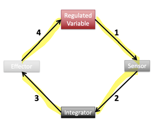
Feedback control loop
A change in the regulated variable results in altered activity of the sensor.
The sensor’s change in activity will be received and interpreted by the integrator.
The integrator alters the activity of one or more effector(s).
The effector(s) acts on the regulated variable to return it to it’s original state.
Integrator
what is computing the signal being sent from the sensor
Negative feedback
changes in the regulated variable are sensed and a response is triggered to bring the regulated variable back to normal (i.e. opposite direction)
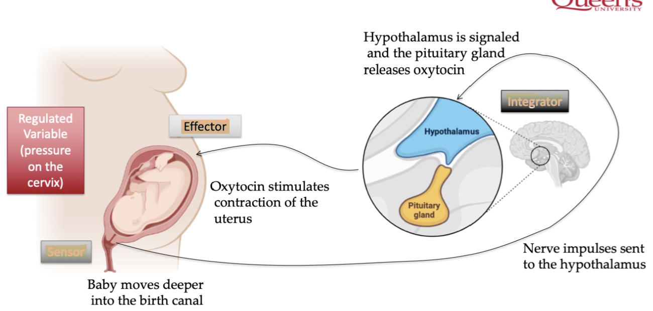
Positive feedback
changes in the regulated variable are sensed and a response is triggered to continue changing the regulated variable (i.e. same direction)
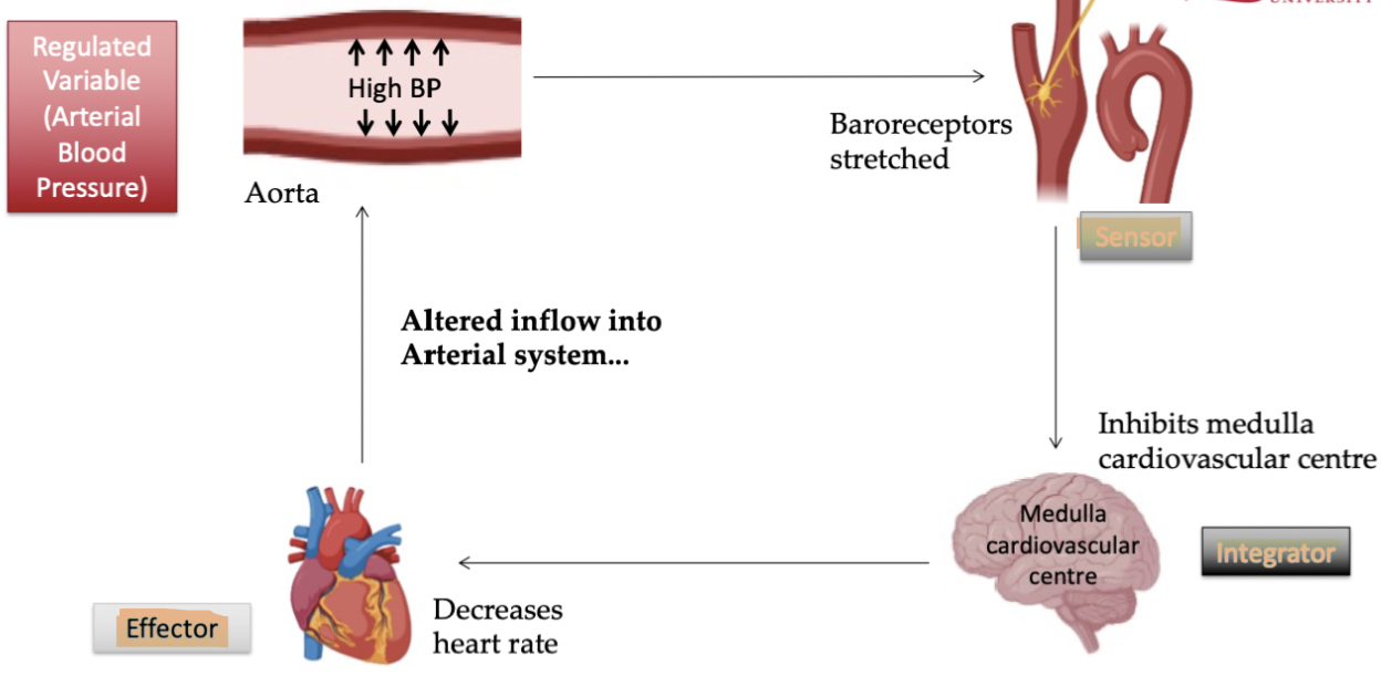
Physiological example of negative feedback
Aorta is the spot where bp is sensed as blood leaves heart, when high bp is sensed, system must bring it down. Baroreceptors sense the stretch of it's walls high bp, signal is sent to brain, brain acts as integrator, heart rate is the effector, heart rate decreases to lower the high blood pressure.
Physiological example of positive feedback
Sensors are in birth canal, it recognizes that cervix pressure increases, brain is integrator, effector is pituitary glans which releases oxytocin, increasingly until child is born
Feed forward control loop
Disturbance is measured prior to a change in the regulated variable and is detected by a sensor
The sensor signals the integrator
The integrator alters the activity of one or more effector(s)
The altered activity of the effector(s) act to create a response to try and prevent a change in the regulated variable
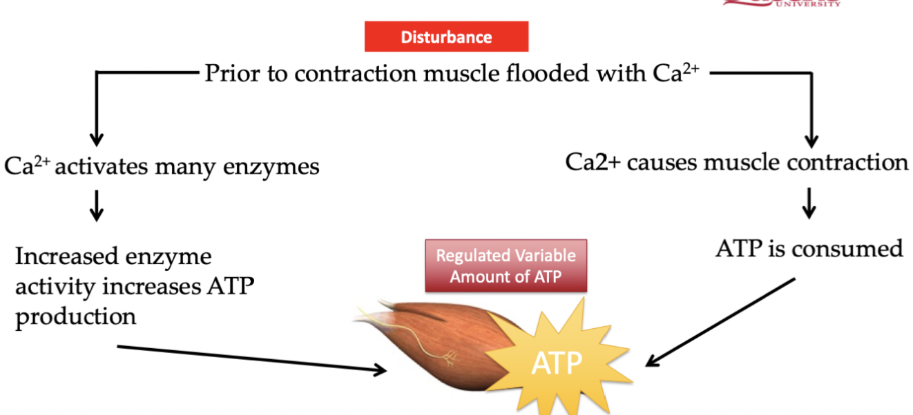
Physiological example of feed forward
Muscle becomes flooded with calcium before movement occurs, we can prevent an ATP shortage by detecting increase in calcium and increasing the enzymes that produce ATP
Receptor mediated cellular control
Controlling effectors via the process;
brain (or integrator) sends a signaling molecule
binds to receptor protein
intracellular signal molecule occurs
target proteins are made
cellular response occurs
Signaling molecule acts on target proteins in the cell and proteins then carry out a response in the cell.
What are the 3 types of signaling molecules?
hormones, neurotransmitters, paracrine
Hormones
Secreted by endocrine glands (integrator) or cells into the blood. Only target cells (effectors) with receptors for the hormone will respond to the signal.
Neurotransmitters
chemicals secreted by neurons that diffuse across a small gap (synapse) to the target cell.
Paracrines
Secreted by a cell to act on a neighbouring cell (cell to cell communication) and does not travel through blood. The receiving cell must have a receptor for the paracrine.
Specificity
a receptor can only bind one kind of messenger but messengers can bind to multiple receptors (as long as they are coded for it)
Affinity
Strength of (likelihood of) messenger binding to a receptor. Larger affinity = more likely to bind to that receptor than another receptor.
Types of membrane bound receptors
Channel-linked receptors, Enzyme-linked receptors, G-protein-linked receptors
Channel linked receptors
Signalling molecule binds to receptor causing an ion channel to open/close . Changes conductance for flow of a substance into or out of a cell
Once inside ions can:
Change the electrical properties of the cell (membrane potential)
Interact with proteins inside the cell and initiate a cellular response
A ‘closed channel’ opens when the receptor binds to allow substances to bind
Enzyme-linked Receptor
Signaling molecule binds to receptor activating an enzyme. Receptor then modifies another protein (an enzyme) activating an intracellular response.
G protein-linked receptor
Very common in our bodies and an important part of physiological regulation. When receptor is activated, GDP is exchanged for GTP, alpha subunit is released and can act on the effectors. G protein is activated to create signaling cascade.
G Proteins have 3 subunits: ⍺, β and γ
⍺-subunit bound to GDP
When the receptor is activated, GDP is exchanged for GTP
⍺-subunit released, and acts on effectors
What are the 3 types of G protein linked receptors?
Channel Affecters: The alpha subunit acts directly on slow ligand gated ion channels to open or close them
Stimulatory: Activate amplifier enzymes (enzymes that catalyze the production of a second messenger)
Inhibitory: Inhibit amplifier enzymes
Secondary messengers
Molecules acting in between the signaling molecule and the target response
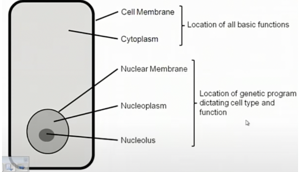
Cell
location of all basic function. in essence fat globules in water with the inside of the cell separated from the rest of the body.
Cytosol
the largest area of the cell that is considered ‘empty space’. It contains the organelles.
Endoplasmic reticulum
Organelle made up of a network of lipid membrane, forming an intracellular compartment that is separate (distinct storage) from the cytosol.
Calcium handling in muscle (release of calcium from sarcoplasmic reticulum); the calcium can be released from ER in the cell upon contraction
Important for intracellular signalling; movement of proteins that are synthesized in ER to other areas of cells
Adjacent to the nucleus; allows for transport of mRNA and nuclear info from nucleus to ER
Granular Endoplasmic Reticulum
AKA rough ER, covered with ribosomes to act as the site of protein synthesis (mRNA translation at ribosomes)
Smooth ER
responsible for intracellular transport (from ER to golgi) and lipid synthesis (forming membranes)
Golgi Apparatus
forms a network with the ER, responsible for transport of substances from the ER to other parts of the cell.
Involved in budding, fusion, and vesicles
Budding, fusion, and vesicles
‘Stuff’ moved from golgi apparatus to other areas of the cell via ‘budding’ and ‘fusion’
Budding: formation of a vesicle from the membrane on the golgi via blubbing (formation of a bubble on the membrane) eventually turning to an independent vesicle that integrates the membrane protein into the cellular membrane and expels waste product to extracellular space
Integral membrane proteins go on the membrane of the vesicle, waste product goes inside the vesicle
Fusion: opposite of budding, attachment of the vesicle to the cell membrane upon contact
Summary of cell transport
Substances formed in the ribosomes in ER, vesicle transport moves substances from ER to Golgi, vesicles more from Golgi to target area where substance is needed
Mitochondria
contains an inner and outer membrane, creating in intra-membrane space called the matrix, with the foldings of the inner membrane being called cristae.
produce ATP (powerhouse of the cell)
inner mitochondrial membrane has ETC where H+ is pumped
recent evidence that mitochondria form a network that allows for transport
Tends to be localized around areas of the cell that use energy (i.e; muscle cells)
Mitochondrial DNA is an index of mitochondrial content
Mitochondria and exercise
Mitochondria increase with exercise training
More mitochondria = more ATP = greater exercise performance
Mitochondria and health
Fatty acid can be oxidized by mitochondria or stored, evidence that storing fat in muscle impairs insulin signaling, causing type 2 diabetes
More mitochondria metabolism, there is lower fatty acid storage, and higher insulin sensitivity
Nucleus
component of cell containing genetic material, consists of nuclear membrane, nucleoplasm, and nucleolus. Dictates cell type and function.
Location of DNA (controls protein synthesis)
Nuclear membrane is continuous with ER (allows RNA direct access to ribosomes)
Nucleolus
location of transcription in the nucleus, initiation of protein synthesis
Organization of the cell
Chemical reactions
Formation of new substance(s) (product) from existing one(s) (substrate). There are 2 types; require energy or produce energy.
Free energy
amount of energy a molecule contains
Free energy change (∆E)
change in free energy following a reaction
∆E = Free energy in products – Free energy in substrates
Endergonic
Require energy to proceed (positive ∆E)
Must be coupled to exergonic reaction (cannot occur spontaneously)
ENDER energy ENTERS
ex; photosynthesis
most reactions are endergonic
Exergonic
Give off energy (negative ∆E)
Can occur spontaneously
EXER energy EXITS
ex; lighting a match
ATP vs ADP
When high energy bonds are broken; ATP goes to ADP + Pi. This releases energy that is the underlying energy source for cellular work.
The energy required for reforming ATP comes from the breakdown (metabolism) of fat, carbohydrate, and protein
Regulated variable; amount of ATP
Masses: ATP and ADP + Pi
Inflow; amount of energy needed for cellular work
Outflow; amount of ATP broken down
Activation energy
Most exergonic reactions do not occur spontaneously, even for exergonic reactions energy is required before a reaction can start
Enzymes
Large high specific proteins that act on substrate to convert them to product and catalyze chemical reactions.
decrease activation energy and increase the rate of reaction without changing net change in free energy
determines conductance
Specific binding sites for specific substrates
These reactions would occur spontaneously at a slower rate
Enzyme-substrate binding
Enzyme brings pieces closer together so we can achieve the product. Enzymes facilitate the combining of substrate and thus decrease energy required to initiate reaction. it occurs at the active site.
How efficient are enzymes?
In general, enzymatic reactions take between 0.02 and 0.002 seconds.
~10^15 times faster than the reaction would proceed spontaneously
1000000000000000 times faster or 1 quadrillion times faster
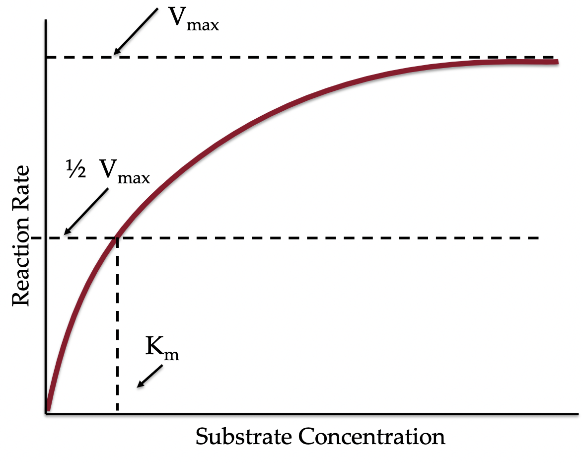
what is Vmax and why does it occurs?
Maximum velocity a reaction can proceed
At a certain point no matter how much substrate you add the reaction will not speed up because all enzymes are occuppied.
Michaelis Constant (Km )
Substrate Concentration required to meet ½ V max
How do enzymes increase rate of reaction?
Higher probability that the substrate will end up at the enzyme or the appropriate substrates will end up together
Increasing the Ehi for the flow
increasing amount of substrate will increase the rate of reaction (more ability to bind to enzyme)
increasing amount of enzyme will increase the rate of reaction (more binding sites available), increases conductance
Saturating substrate
At a certain point no matter how much substrate you add the reaction will not speed up (Vmax). If all enzymes/conductance's are active increasing substances will not increase the rate of reaction.
What is the flow model of enzymes?
Substrate affects the energy gradient, enzyme affects conductance
Transcription
Production of RNA from DNA in the nucleus. Endergonic process.
DNA
Deoxyribonucleic Acid; Contains information to make every protein in the body. Has a double helix shape with each strand containing 1000’s of gene.
There's a full copy of your DNA in every cell
Every cell has the same DNA, but how the DNA is used and which proteins are made determines cell function
Each protein is coded by a specific area of DNA
Gene
Contains the encoded information to form a single protein
Promoter
where the activation of gene is controlled
Coding region
contains the information to make protein
3 types of protein action
Repressor protein: inhibits coding of gene
Activator: activates coding of gene
Negative feedback: if gene is copied and used a lot, it binds to it's own promoter to slow it's own production to prevent overproduction of the product
Transcription steps
Polymerase (The Zipper) binds to promoter, when bound gene becomes active, when active, RNA is made.
As polymerase moves down coding region the two strands of DNA are separated
separated strand is transcribed and a strand of RNA is formed (Exact copy of DNA). Formation of RNA requires ATP.
The polymerase moves to the end of gene and unbinds. DNA is put back together. Result is RNA strand.
Types of RNA
Messenger RNA (mRNA): copy of DNA used to form new protein
Transfer RNA (tRNA): brings building blocks (amino acids) to ribosome 60s subunit during translation to create polypeptide chain.
Ribosomal RNA (rRNA): forms most of a ribosome
Translation
Endergonic process of producing protein from mRNA, happens in the ribosomes of the ER. Binding of amino acids together to form a new protein.
mRNA codes for a specific sequence of amino acids that determines protein function
Amino acids are brought to the ribosome by tRNA
Ribosome structure
Ribosome is made up of 2 parts:
40 s subunit: binds mRNA
60 s subunit: builds and releases protein
Both subunits are made up of rRNA
Translation steps
Initiation: mRNA is bound to the 40s subunit, with the use of energy from ATP, mRNA starts to move through the ribosome
Elongation: As mRNA moves through ribosome, protein is formed
Termination: When the end of the mRNA is met, the newly formed protein is released. This also uses ATP.
New protein moved to its proper cellular location and contributes to cellular function
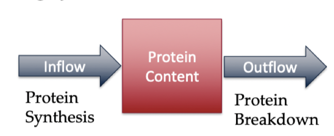
Protein synthesis flow
Protein content is mass
Protein synthesis is inflow
Protein breakdown is outflow
Ricin
Ricin is a substance that inhibits ribosomes and protein synthesis, so decreases the inflow to protein content.
Leads to malfunction because no new proteins replace the broken down proteins
Cell eventually dies, and organism does too
The training effect
An acute bout of exercise increases the rate of mitochondrial transcription; causes increased mRNA for protein involved in ATP production
An acute bout of exercise increases mitochondrial translation capacity; causes increased proteins available to bind to mRNA and stimulate translation
overall; more protein synthesis = more protein/enzyme = greater function of mitochondria = more ATP = better exercise performance
Cell membrane
cells are surrounded by a phospholipid bilayer that provides barriers between inside and outside of cells. Phospholipid Bilayer forms a hydrophobic barrier between inside and outside of cell
Phospholipids
composes membranes; phosphate head is hydrophilic, fatty acid tail is hydrophobic.
Hydrophobic vs hydrophyllic
Hydrophobic: Repels water (does not dissolve). Lipid portion.
Hydrophyllic: Dissolves in water. Phosphate portion.
What can and can’t pass the phospholipid bilayer?
Water soluble substances don’t pass (Carbohydrate, Protein, Ions); allows content to be controlled, but can be transported by channels that are always open.
Fat soluble substances do pass (Oxygen, Carbon Dioxide)
What are the two types of simple diffusion?
membrane is permeable for substances to pass (fat soluble)
diffusion through channel proteins that are always open (water soluble), so no work needs to be done
Fat transporters
Until recently it was generally assumed that fat uptake into tissue was achieved by simple diffusion, when actually occurs via facilitated diffusion.
Facilitated diffusion
Accomplished by transport proteins via conformational change (work must be done, but energy is not used). Maximal rate of transport limited by number of transporters.
Diffusion across membranes is flow. The flow will plateau eventually if all the channel proteins are being used.