Cognitive Neuroscience Ritchey Exam 1
1/94
There's no tags or description
Looks like no tags are added yet.
Name | Mastery | Learn | Test | Matching | Spaced | Call with Kai |
|---|
No analytics yet
Send a link to your students to track their progress
95 Terms
Willis (1650)
Noticed that brain damage caused changes in behavior - "Neurology"
Phrenology
Gal (1810) Aspects of oneself was represented based on shape of the skull --> Psuedoscience
Distributed views of brain function
- Cognitive functions are supported by distributed sets of neurons across the brain
- Impairments are proportional to total damage
Localizationist view
- Cog functions are supported by specific localized areas of the brain
- Impairments are proportional to damage to a specific region
Distributed views theories/papers
Aggregate field theory and Flourens (1824)
Localizationist view theories/papers
Functional mapping of the aphasias, Broca (1860) and Wernicke (1876)
Two Philosophical Approaches
Rationalism (Descartes, mind-body 1644)
Empiricism (Locke, tabula rasa 1697)
Experimental Approaches (late 1800s)
RT differences (Donders, 1869)
Forgetting Curve (Ebbinghaus, 1885) (Associationism)
Three Behaviorist and their Beliefs (early 1900s)
Thorndike: law of effect
Watson: mental activity cannot be observed
Skinner: operant conditioning, reward and punishment
Central Nervous System
Brain + Spinal Cord
Peripheral Nervous System
Nerves & nerve cell ganglia —> autonomic & somatic nervous systems
Nucleus
Relatively compact arrangement of nerve cell bodies and their connections
Grey matter
Neurons arranged in layers form a sheet of tissue
Most the outer region of the brain
White matter
Axons and glial cells form tracts inter-connecting the brain
Much of the inner part of the brain
Cerebral Cortex
the outer layer of your brain's surface, located on top of the cerebrum
Sylvian fissure
Divides Frontal and Temporal Lobes
Central Sulcus
Divides Frontal and Parietal Lobe
Corpus Chalosm
Divides Right and Left hemisphere
Sensory Cortices
brain regions specialized for processing sensory information, such as vision, hearing, touch, taste, and smell
Association Cortices
regions of the brain that integrate information from different sensory modalities or sensory and motor regions. Unlike sensory cortices, they are not dedicated to processing just one type of information but are involved in more complex tasks such as perception, memory, attention, and decision-making
Visual Cortex
Located in the back of the occipital lobe (Area 17 or V1)
Frontal Cortex
Divided into Medial prefrontal cortex(also includes ACC) and motor cortex
4 Major divisions of the Brainstem
Pons, Midbrain, Medulla, and Spinal Cord
Cingulate Gyrus
Wraps around corpus callosum like a belt
Component of the limbic system, it is involved in processing emotions and behavior regulation. It also helps to regulate autonomic motor function
What are the three major subcortical areas and their function
Hippocampus - memory
Basal Ganglia - controls body’s voluntary movements
Amygdala - Emotional memory
Subtraction method
Compare tasks or task conditions that differ by only one cognitive process
i.e., Experimental —> Make a decision about a word on the screen
Baseline —> press button when word appears on the screen
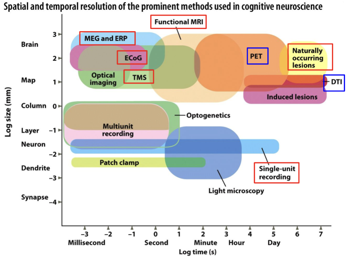
Spatial and Temporal Resolution of Methods (Show 8)
Three major degenerative causes neurological damage
Alzheimer’s, Parkinson’s, Huntington’s
The lesion approach
Location of lesions + neuropsychological testing
Some limitations of the lesion approach
Other changes might be correlated with brain damage
Damage to one region might lead to dysfunction in other connected regions
Need really good control conditions to isolate process
Examples of Pharmacological manipulations
L-DOPA, Haloperidol —> altering amount of dopamine and testing reward based learning
Transcranial Magnetic Stimulation
Magnetic field generator to induce electrical currents through the scalp & skull
Currents stimulate action potentials —> firing disrupts responses in that area
Can target small areas of cortex, but only cortical regions and only one region at a time
Online vs. Offline TMS
Online —> TMS performed during the task
Offline —> TMS given before the task
Transcranial Direct Current Stimulation
Low-level currents, electricity, depolarization of anode site
Affects bigger area, does not necessarily drive action action potentials
Electroencephalography(EEG)
Oldest cognitive neuroscience method
Detect electrical current from pyramidal cells in cortex measured with electrodes on the scalp
Electrical signals could be from anywhere in the head not just underneath (volume conduction)
Magnetoencephalography(MEG)
Similar to EEG but measures currents from a magnetic field; magnetic fields are less distorted by the skull
Event-Related potentials
Averaging of scalp potentials time-locked to the stimulus; Get the cognitive signal out of the noise
Electrocortogram (ECoG)
Excellent temporal resolution and spatial resolution
limited to testing surgical patients who volunteer their time
little control over electrode location
Electrodes sit directly on the brain
Magnetic Resonance Imaging(MRI)
Uses magnetic fields to detect hydrogen atoms in the brain, can scan the brain slice-by-slice
Diffusion Tensor Imaging (DTI)
Use directional information to identify probable axons tracts - “tractography”
Measures the diffusion of water molecules in the brain
What is a voxel?
3-D Pixel in brain image as they all have four dimensions
Blocked-Design Trial
Similar stimuli are grouped together
lower temporal resolution
focused on sustained period
Overall activation patterns during task blocks
e.g. Block 1: Happy Faces (10 faces)
Rest (10 seconds)
Block 2: Sad Faces (10 faces)
Rest (10 seconds)
Block 3: Neutral Faces (10 faces)
Rest (10 seconds)
Event-related Trial
Random ordering of stimuli or determined by the subject; better temporal resolution
Face 1: Happy
Face 2: Sad
Face 3: Neutral
Face 4: Happy
Face 5: Sad
Face 6: Neutral
(This continues for a total of 30 faces, with a mix of emotional expressions)
Sensation
Early processing of stimulus in environment
Perception
Formation and experience of the mental representation of the stimulus
Pathways Sensation and Perception
Receptor cells —> nerve pathways —> brainstem —> thalamus —> primary sensory cortex and secondary sensory cortex
Coding Principles
Receptor cells - limited range and tuned to info in the receptive field
Acuity - how well we can distinguish two stimuli
Adaptation -adjusting to the sensitivity of the sensory system to the current environment
Rods (photoreceptors)
rhodopsin
sensitive to low light
deplete quickly
distributed throughout
no color sennsitivity
Cones (photoreceptor)
Photopsin
require more intense light
replenish quickly
most in fovea
color sensitivity(RGB)
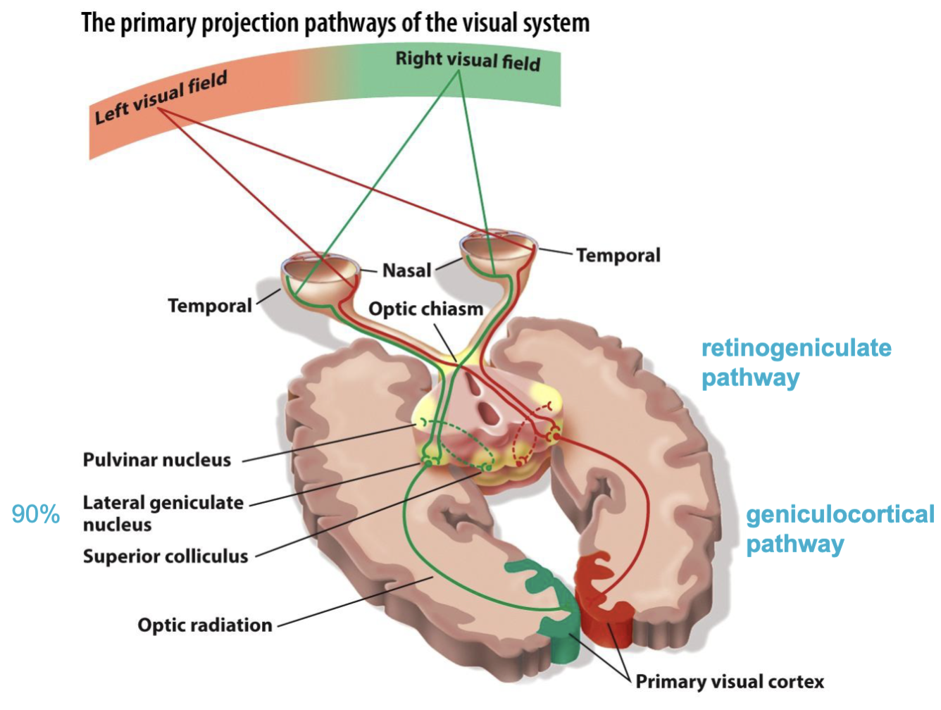
Pathway of Visual System
Receptive Field
Region of space in which a stimulus will
trigger a response in a particular neuron
○ Ordered continuously
○ Arranged in an ordered way
Retinotopy
Ordered mapping of the visual field onto visual areas (retinal projection)
An example of topographical organization within the brain
Topographical Organization
The ordered mapping of the external world to its representation in the brain
Nearby neurons represent similar aspects of the world
Retinotopy, tonotopy, somatotopy
Ocular dominance and orientation selectivity
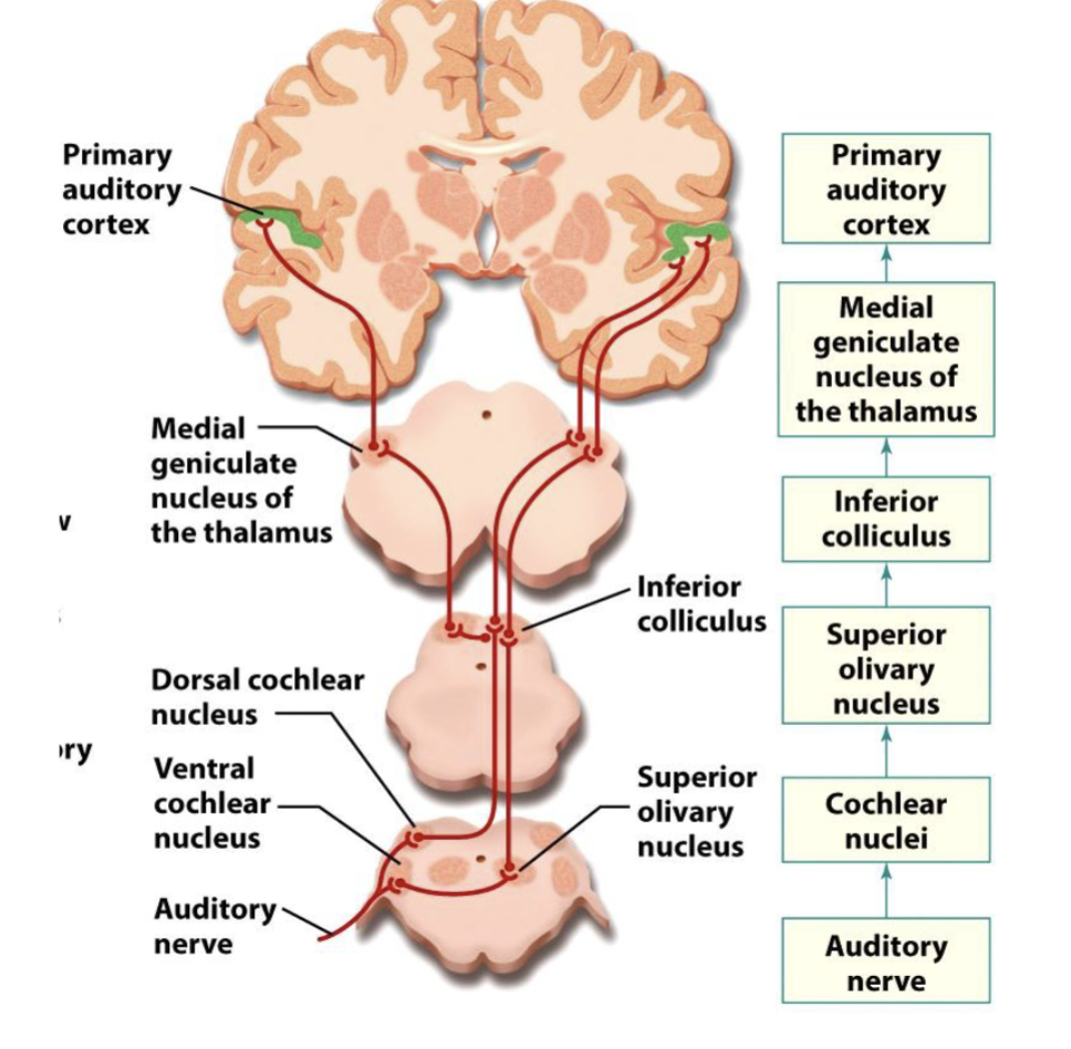
Pathway of the Auditory system
Two aspects of Sound Localization
Interaural time - difference in timing between ears
Differences in intensity between ears
Olfaction and Stimuli
Sensed Stimuli: Odorant
Perception: Smell
Gustation and Stimuli
Sensed Stimuli: Tastant
Perception: Taste
Gustatory Transduction Pathway
Taste receptor cells → Vagus cranial nerve → Thalamus → Cortex
Somatosensory in S1
McGurk Effect
Hearing different audio based off what your eyes see them pronouncing
Where does multisensory integration occur?
Premotor cortex, inferior prefrontal cortex, posteriors superior temporal sulcus
What is machine learning?
An algorithm that learns to make predictions; given a new set of data, similar to other data is has seen before, what will be the outcome?
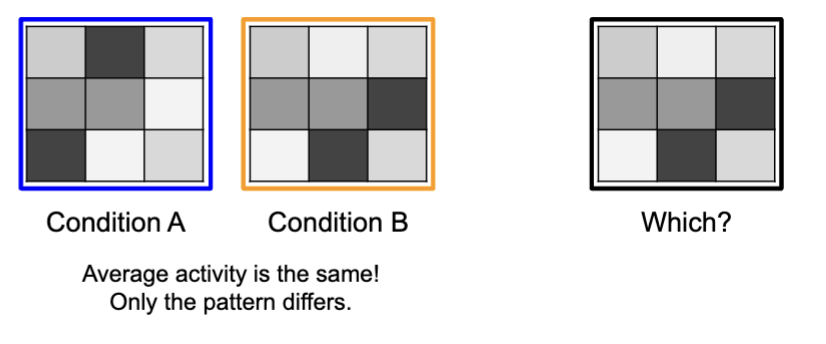
Multi-Voxel Pattern Analysis
Apperceptive Agnosia
Difficulty with object constancy (e.g. unsual views)
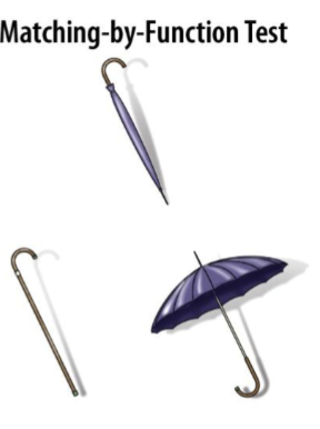
Associative Agnosia
Difficulty linking perception to the object’s identity/function
Integrative Agnosia
Difficulty integrating parts into a whole
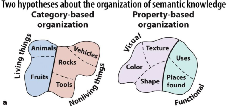
Evidence for category specificity
Agnosia’s for specific categories
Brain areas for specific categories (e.g FFA, Lateral occipital cortex(objects))
Evidence against Category specificity
Category membership is correlated with certain characteristics(properties)
Representations may be organized around object properties
Two Hypothesis about the organization of semantic knowledge
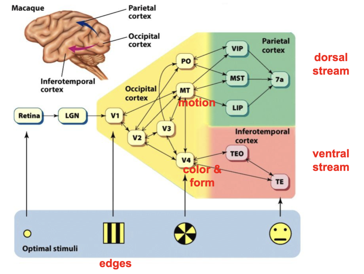
Ungerleider and Mishkin (1982)
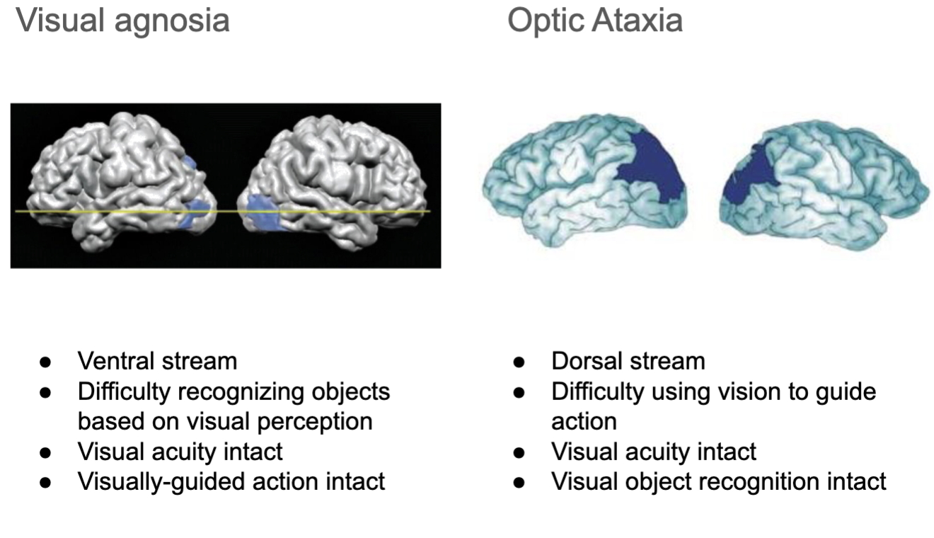
Double dissociations discovered the What(ventral) pathway and the Where(dorsal) pathway. Both stem from V1
Monkeys were tasked with what(non match to sample) or where(close to sample) related tasks
Lesions to ventral and dorsal areas of the brain showed double dissociation
Difference between the Two What and Where pathways
Temporal Lobe Neurons(What)
Foveal focus, selectivity for complex features, especially in anterior regions(
Center of vision field and more details
Parietal Lobe Neurons(Where)
Foveal and peripheral, less selective
Entire visual field
Optic Ataxia( Balint’s Syndrome)
a condition that causes difficulty with visually guided arm movements, such as reaching for objects
Can you tell you what an object is but cannot reach for it
Dissociating ventral & dorsal pathways through visual agnosia and optic ataxia
Sound perception pathways(Analogous to visual)
Anterior auditory regions —> sounds identification
Posterior auditory regions —> sound localization
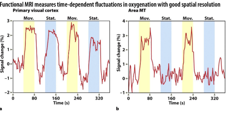
The Buckner study pushed the fMRI field forward by introducing a new way to look at brain activity. What was their innovation?
Single-trial(event-related) designs can be used for fMRI research, no longer limited to blocked designs
What are the advantages to being able to look at fMRI data in this way? (Buckner 1996)
Now able to mix trials of different conditions, sorted by behavioral performance and can see changes in brain activity with much better temporal resolution
You should be able to interpret a plot of fMRI data (activity on y-axis, time on x-axis) similar to those presented in the Buckner article and in the Methods lecture slides. (Buckner 1996)
How does this study relate to the idea that sensation and perception reflect two different aspects of sensory processing? (Haynes 2005)
The Haynes (2005) study illustrates that sensation and perception are distinct processes, as the brain's primary visual cortex (V1) can detect and encode sensory stimuli even when the stimuli are not consciously perceived(masked or subliminal)
This demonstrates that sensation (the detection of stimuli) can occur without perception (conscious awareness), highlighting the brain's ability to process sensory information unconsciously
Is activity in the primary visual cortex sufficient to drive conscious awareness of visual information? (Haynes 2005)
Based on the results of the Haynes (2005) study, activity in the primary visual cortex (V1) is not sufficient to drive conscious awareness of visual information.
While V1 encodes detailed sensory information (such as the orientation of invisible stimuli), this activity alone does not lead to conscious perception, suggesting that additional processing in higher-order brain regions beyond V1 is required for conscious awareness.
What is the advantage of studying multivariate patterns of brain activity across voxels, compared to looking at the magnitude of responses in individual voxels or areas? (Haynes and Haxby)
Easier to detect distributed, fine-grained patterns of activation that can reveal how information is encoded across the brain, which may be missed by examining the magnitude of activity in individual voxels
Provides greater sensitivity to subtle differences and enables the decoding of complex information from broader brain regions, offering deeper insights into representational content and neural processing.
Did Haxby et al. find evidence for the distributed view or for a modular view (i.e., domain specificity)? Briefly describe the main finding that supported their conclusions.
They showed that overlapping, distributed patterns of activity in the ventral temporal cortex represent different object categories, such as faces, houses, and tools.
Even though certain regions like the fusiform face area (FFA) and parahippocampal place area (PPA) showed category-specific peaks, the broader patterns of activity across the entire region contained information about various categories.
This finding indicated that object categories are represented by distributed neural patterns, not confined to specialized, isolated regions.
Did Pitcher et al. find evidence for the distributed view or for a modular view (i.e., domain specificity)? Briefly describe the main finding that supported their conclusions
provided evidence for both a modular role of the FFA (between the rLO, rEBA, and rOFA) in face recognition and a distributed processing approach, indicating that while the FFA is specialized for faces, it can also encode information about other object categories
This suggests a more nuanced understanding of visual processing, where certain regions are specialized but still participate in broader, overlapping networks.
What were the three areas examined in the Pitcher study, and how did they relate to the three stimulus categories?
Right Occipital Face Area (rOFA) → Faces
Right Lateral Occipital Face Area (rLO) → Objects
Right Extrastriate body area (rEBA) →Bodies
What is a double dissociation? (Also covered in lecture) What is an example of a double dissociation in this paper? (Pitcher 2009)
Examining two functions and two lesions or impairments and the differences to them
Lesion to Location X
Impairment of function A but preserve B
Lesion to Location Y
Impairment of function B but Preserve A
They had three double dissociations in the Pitcher example. For experiment 1 they lesioned the the rOFA and found impairments for face but not lesions of rLO. And they found the oppsite resutls for object discrimination over the rLO
Combining Haxby & Pitcher: Why did Pitcher et al. use TMS to test their hypothesis, rather than fMRI or EEG? Why did Haxby et al. use fMRI to test their hypothesis?
Pitcher was the first of its kind to use TMS to examine the difference between modular and distributed view and allowed for a more casual approach than TMS
Haxby used fMRI
Dissociation between perception linked to awareness and perception linked to action
Explicit Matching task vs Action Task
Patients with visual agnosia had trouble visually orienting the piece of paper to fit the slot but had no trouble when told to just insert the paper
Sympathetic Nervous System
Norepinephrine based; fight or flight response system
Parasympathetic Nervous System
Acetylcholine based; rest and digest system
Cognitive Psychology
Understanding how the brain represents and manipulates objects or ideas; identifying mental operations that are required to perform tasks
Single-cell recordings
record from individual neurons and correlate increases and decreases in activity to behavior or stimulation
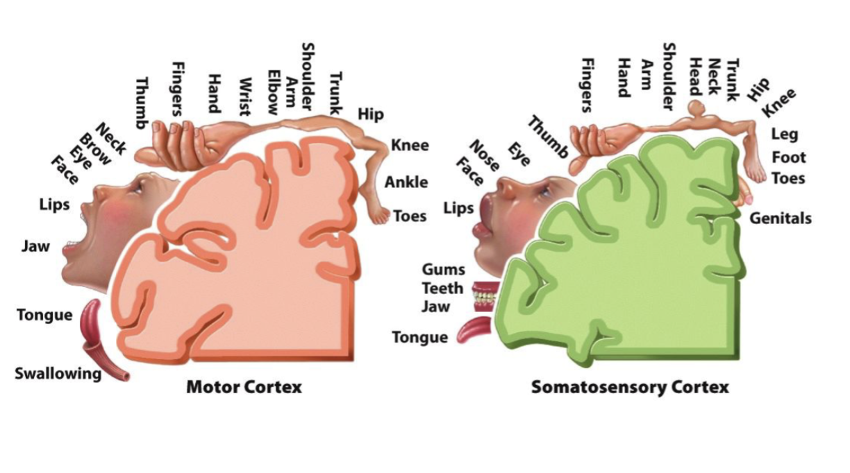
Primary somatosensory cortex (S1)
Contains homunculus of the body; more sensitive = larger areas of cortex
Optic Nerve
formed from the axons of the ganglion cells; they cross of from visual fields
Retinotopic Maps
Visual cortex is made up many distinct regions defined by their distinct maps; funcitonal differences e.g. V4 = color and V5 = motion processing
What region is critical for recognition of an object’s shape?
Lateral Occipital Cortex