Microbiology: Microbial Growth 1.2 A
1/105
There's no tags or description
Looks like no tags are added yet.
Name | Mastery | Learn | Test | Matching | Spaced |
|---|
No study sessions yet.
106 Terms
what nutrients are required for cells to build macromolecules
all cells need access to large amounts of carbon, nitrogen, phosphorous, sulfur, and oxygen (macronutrients) to build macromolecules
Organelles are NOT a general feature of prokaryotic cells but are common in eukaryotic cells
True
True or False. There are no examples of bacterial organelles because organelles are strictly found in eukaryotic cells.
False (ex. Magnetosome)
what micronutrients are required by microbes
- several metal ions (like Na+, Mg2+, Mn2+, etc.)
- often required for protein structure/activity
why can't we cultivate most microorganims
because you have to know their nutrient requirements which is hard/we don't know each microorganism's nutrient requirements
what are essential nutrients
nutrients that must be supplied through the environment to the microorganism
different microbial species require different essential nutrients (ex. essential amino acids are not the same for all microorganisms)
what are prototrophs
they are microbes that can synthesize macromolecular precursors such as amino acids from a single carbon source (ie. glucose) and inorganic molecules (like salts)
- ex. E. coli can make everything it needs if given the correcy sugar
do prototroophs always synthesize everything themsevles
prototrophs are capable of synthesizeing everything themselves, but it still takes energy to synthesize these precursors so prototrophs will preferentially use those found in the environment even though it is not essential for them to do this
what are auxotrophs
they are microbes that cannot synthesize all the needed precursors from a single carbon source and must aquire nutrients from the enviroment
what are the common growth mediums
- liquid broth culture
- plates: broth (which provides the necessary nutrition requirmnets) and agar (which makes it solid/solidifying agent) which produces circular compact colonies = colonies arise from a single bacteria on a plate
microbial growth chart based on nutrient concentrations
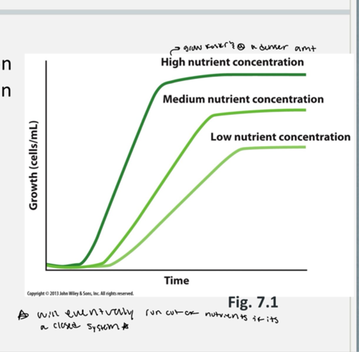
how does nutrient concentration affect microbial growth rate
- growth rate depends on the amounts of nutrients in the environment
- one key nutrient will be available in the lowest amount and will be the limiting factor for growth
- eventually all of the nutrients will run out if it is a closed system of growth
what methods do microbes grow by
with binary fission
how is the growth rate with binary fission
- there is 2 offspring per generation produced each generation time (ex. 1 cell --> 2 cells --> 4 cells --> 8 cells etc.)
- this is exponential growth
what is generation time
the time it takes for a population to double
generation time of e. coli
20 minutes
binary fission equation
Nt = N0 x 2^n
- Nt = final cell number
- N0 = original cell number
- n is the number of generations
how is the generation of E. coli if grown with no limiting conditions for 48 hrs
E. coli generation time is 20 minutes (in optimal conditions)
- in 48 hrs = 1 cell = 10^7 tons of bacteria
growth curve for microbes
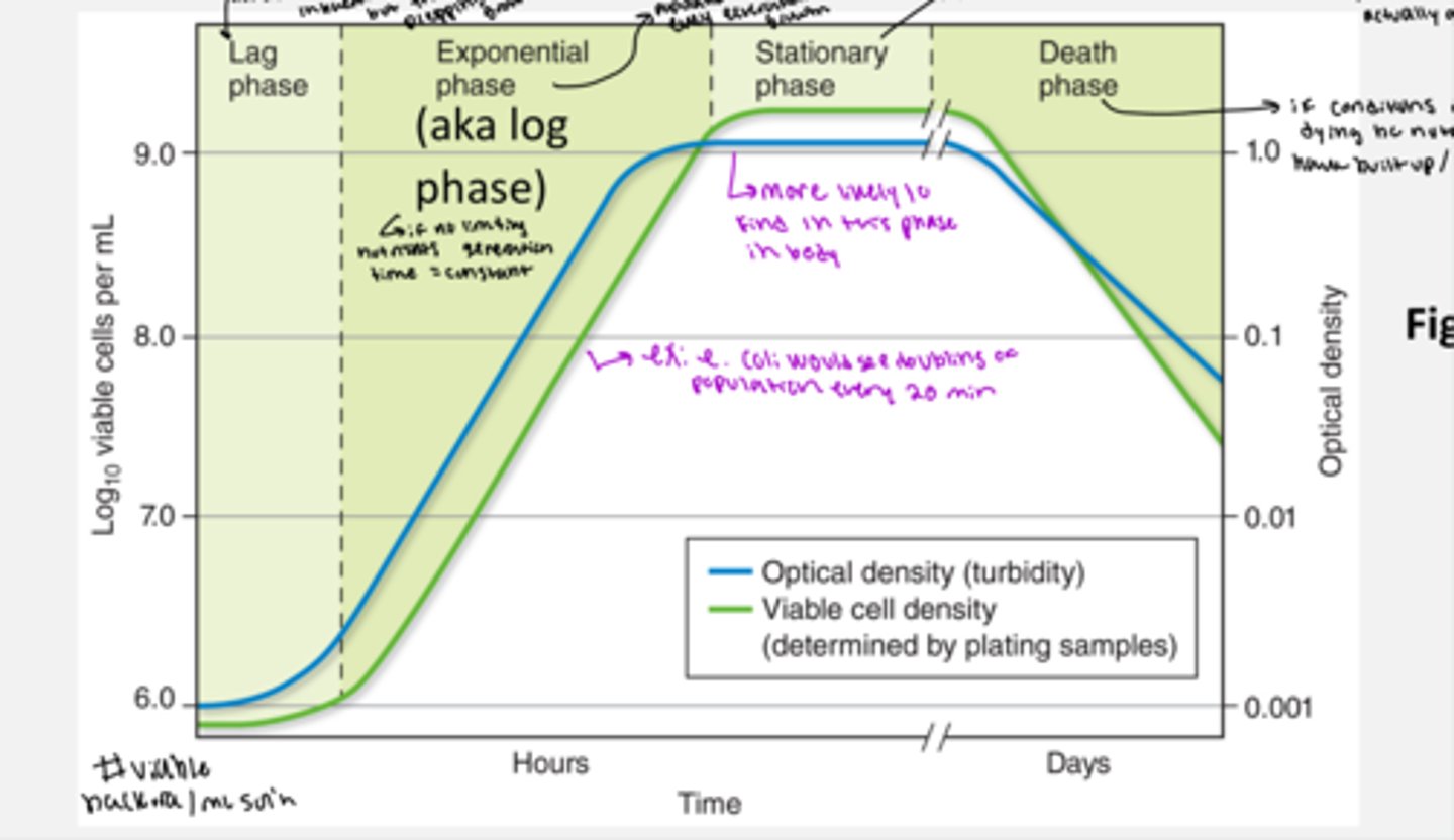
explain the phases of microbe growth curve
1. lag phase (like is low at beginning) = no real increase in bacterial numbers but the bacteria are prepping for growth for example the bacteria might need to synthesize more ribosomes or other protein necessary for rapid DNA repair and cell division
2. exponential (aka log) phase = population doubles with every generation time and there is a rapid growth. ~ really steep increase in line
3. stationary phase = more likely to find this phase in the body; this is when nutrients run out and toxins are starting to build up. the bacteria are alive but are just trying to survive, but are not actually growing (~ flat line on graph)
4. death phase = if conditions do not change bacteria start dying bc nutrients are scarce and toxins have built up/everything starts to die (~ line goes down)
what are standard growth conditions of bacteria
- sea-level
- temp = 20 - 40 degrees C
- neutral pH
- 0.9% salt; and ample nutrients
what are extremophiles
organisms inhabiting ecological niches outside of the window of standard growth conditions.
- have certain adaptations allowing for growth in extreme conditions
which extremophile conditions are best understood
extremophile conditions allowing for growth in extreme temperatures
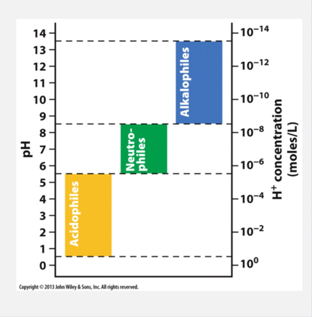
what are the extremophiles based on pH
pH 0.5 - 5.5 = acidophiles
pH 5.5 - 8.5 = meutrophiles
pH 8.5 - 13.5 = alkalophiles
what are the O2 requirements of microbial growth
some microorganisms are OBLIGATE AEROBES (oxygen required) and some are OBLIGATE ANAEROBES (oxygen absence required)
however, many microbes are FACULTATIVE or AEROTOLERANT meaning they tolerate oxygen, but do not need it to survive so the presence or absence of oxygen does not effect them/ can grow in either condition
so the types of facultative microbes = anaerobes (no oxygen required), micro-aerophilic (low oxygen required), and aerobes (high oxygen required)
why is O2 beneficial in microbe growth
beneficial for metabolism
why is oxygen not alway desirable for microbe growth
O2 is not alway desirable due to formaion of reactive oxygen species (ROS) ex. H2O2, superoxide (O2^-), and hydroxyl radical (OH with a single electron so its radical)
- so anaerobes do not have the defenses to neutralize reactive oxygen which is why O2 is toxic to them
what structures in a bacterial cell are part of the cell envelope
cell membrane, cell wall(peptidoglycan) and outer membrane (if gram negative)
what does the cell enevelope do for bacteria
protects bacertia from an unpredictable and often hostile envrionment
label a bacterial cell diagram
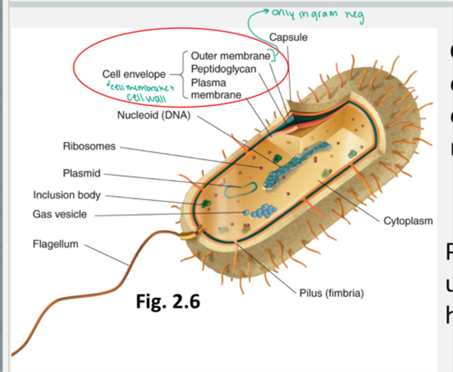
what are the functions of a cell membrane in bacteria
- maintains homeostasis of the interior of the cell
- phosopholipid bilayer with embedded proteins
- proteins provide support, transport, and communication (ie. receptors)
- is able to provide energy with the proton motive force
which cells have a cell membrane
all cells have a cell membrane
do cell membranes give structure to the cell
the cell membrane does not give structure to the cell, only the cell wall gives structure to the cell
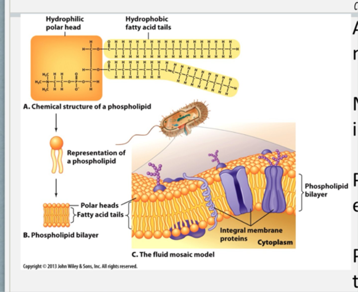
how is the structure of the cell membrane
made of phospholipids that have hydrophobic fatty acid tails that interact with one another and hydrophillic heads which interact with the outside enviroment
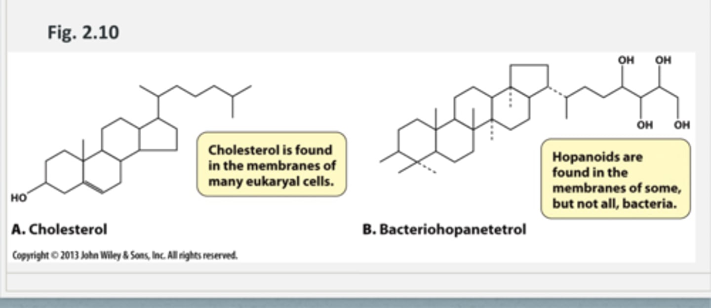
what are the sterol molecules of the cell membrane
cell membrane may have sterol molecules called HOPANOIDS in it which help with stabilty across temperature ranges
ex. bacteriohopantetrol (hopanoids are found in the membranes of some, but not all, bacteria)
- hopanoids are related to cholesterol found in membranes of many eukaryal cells/have same function
how is passive diffusion across the cell membrane
- passive diffusion can occur with small, uncharged molecules like O2, CO2, and water.
- weak acids and bases in their uncharged form are also passively transported (example HA not H+ and A- form) so like weak acids and weak bases
what do cell membranes to help with water diffusions
have aquaporins which are protein channels that facilitate diffusion of water across the membrane (passively)
which molecules require transporters to cross the cell membrane
- polar molecules or charged molecules require transport through specific protein transporters (ex. ions, amino acids, vitamins, sugars)
- this can happen via facilitated diffusion (passive) from high to low conc across membrane or can happen aactively with cotransport mechanisms
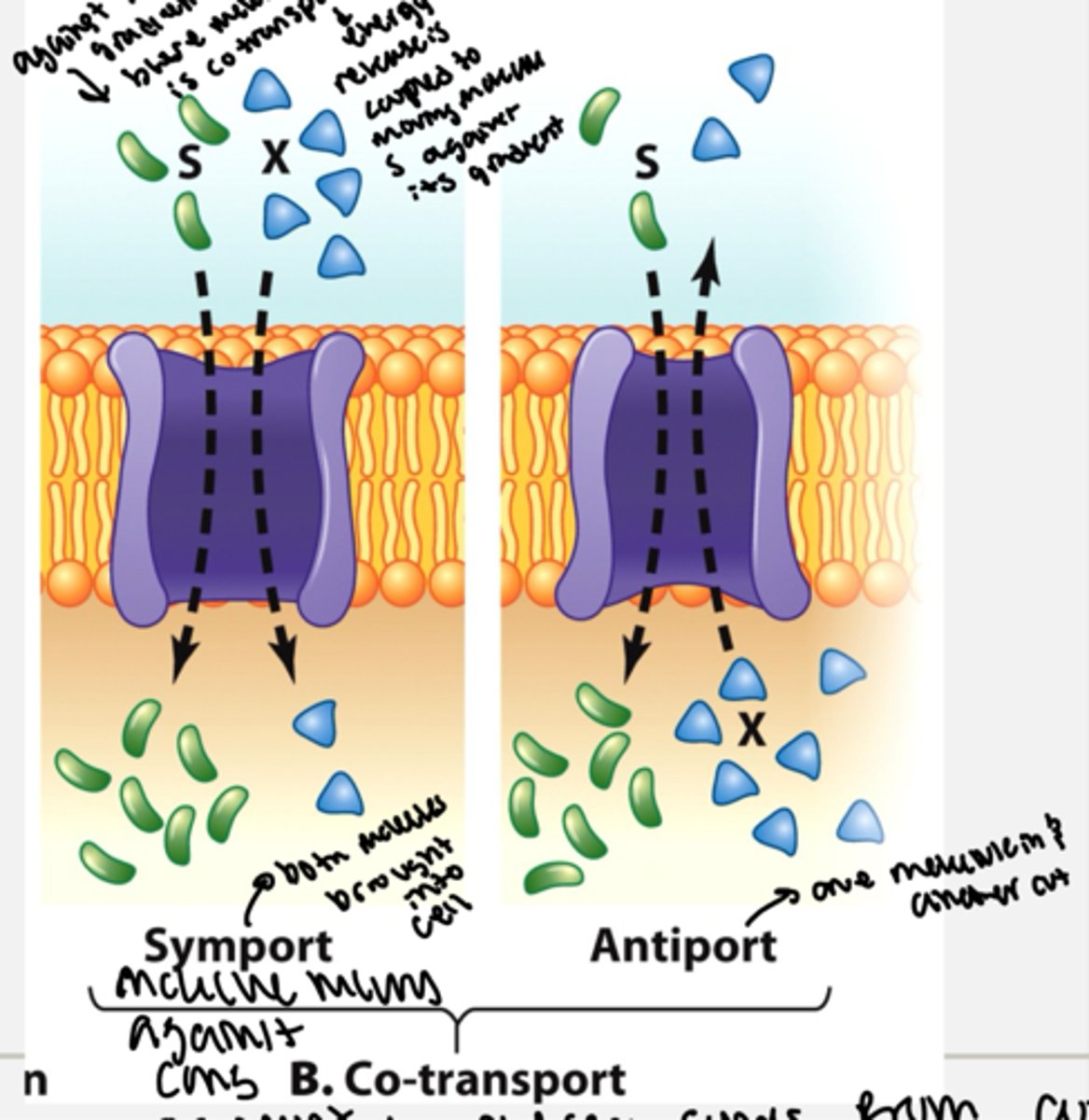
what is cotransport mechanims across cell membrane
this is a form of active transport and moves molecules against their concnetration gradients
- symport = both molecules are brought into the cell with one moving with its conc gradient to provide energy to move another molecules against its conc gradient
- antiport = one molecule moves into the cell against it's conc gradient while one molecule moves out of the cell with it's conc gradient which provides energy to transport the molecules
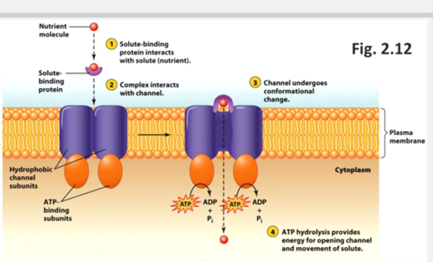
what are ABC transporters
ATP Binding Cassette transport system
- type of active transport
- can do influx or efflux
1. there is a nutrient molecule outside the cell that a solute binding protein interacts with
2. the solute binding protein then binds to the ABC trasporter (interacts with the hydrophobic channel subunits embedded in the membrane that have ATP binding subunits attached to them on the inside of the cell membrane)
3. the channel undergoes a conformational change which allows for the hydrolysis of ATP which provides energy to open the channel and moves the solute (nutrient) into the cell cytoplasm through the plasma membrane through the channel.
ATP hydrolysis = ATP --> ADP + Pi
how many different ABC transporters are in E. coli
70
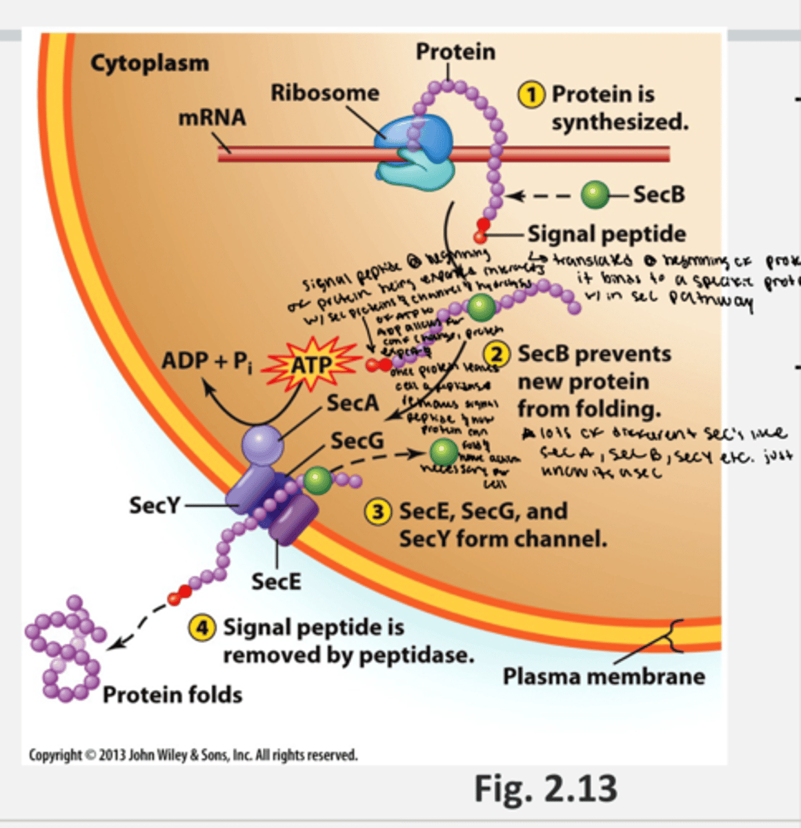
what is the sec pathway
- form of active transport
- sec general excretory pathway exports proteins across cell membrane (ex. toxins, enzymes, etc)
- only happens for protein export across a cell
- proteins are identified by the signal peptide
1. a protein is synthesized by the ribosome bound to an mRNA strand in the cytoplasm that contains a signal peptide at the beginning
2. a secB protein (specific protein in the sec pathway) binds to the protein and prevents the new protein from folding in the cytoplasm
3. the secB protein bound to the peptide (new protein being exported) also interacts with a sec channel (which is made of a series of sec proteins like SecA, sec G, and sec Y) on the cell membrane which triggers atp hydryolysis (ATP --> ADP + Pi), opening the sec channel and releasing the signal peptide end of the protein into the outside of the cell and releases the SecB protein back into the cytoplsm.
4. outside of the cell, the peptidase enzyme removes the signal peptide and the protein is allowed to fold outside of the cytoplasm
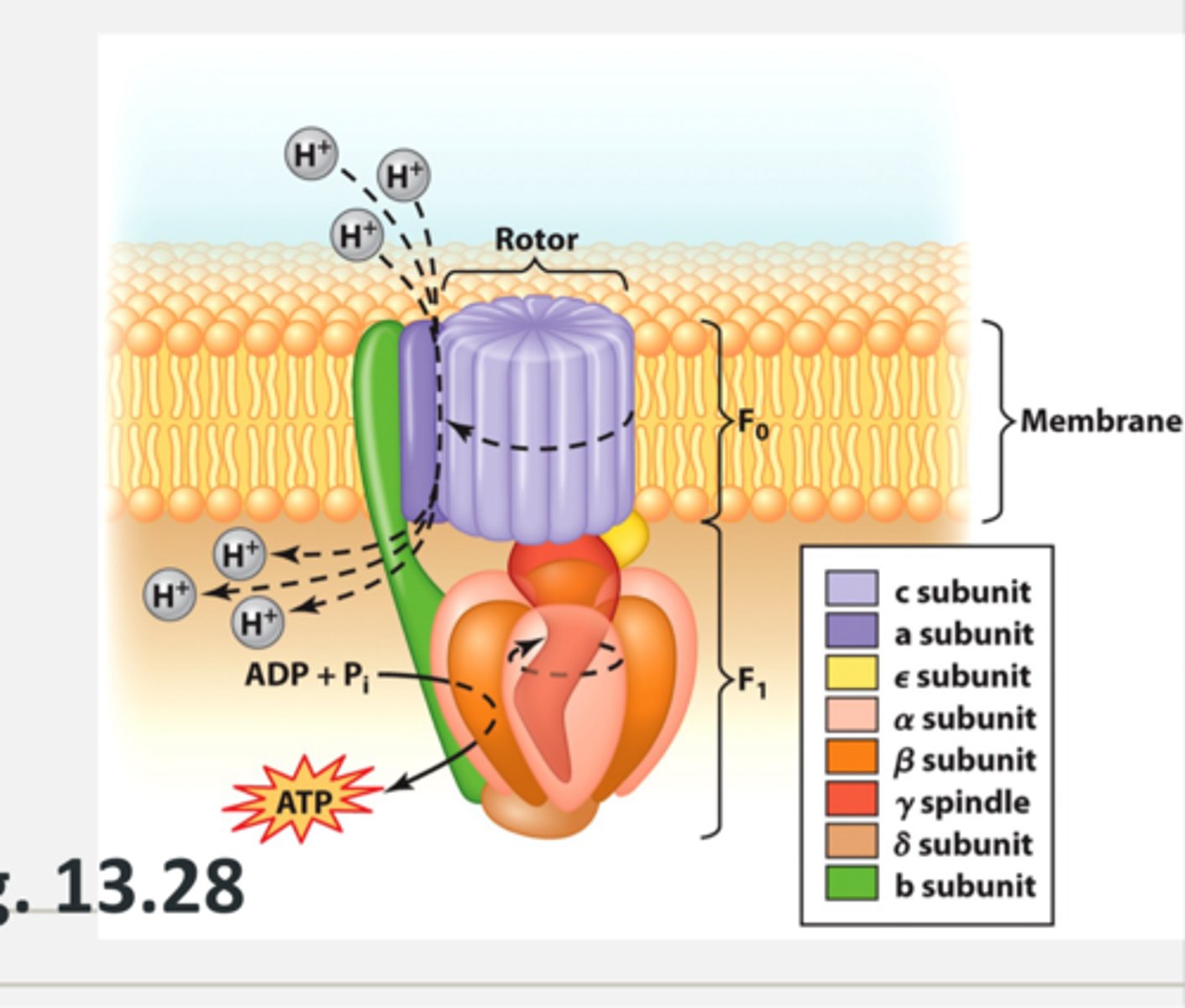
do prokaryotes have mitochondria
no, prokaryotes do not have mitochondira, so their cell membrane must work to generate ATP
what membrane functions occur to generate ATP
the bacterial cell membrane is used for capturing energy
- embedded electron transport chains can help create more proton motive force (PMF) which will be used for respiration/photosynthesis and used to derive energy for motion (flagella)
flagella motor with ATP
what is the bacterial cell wall composed of
- crosslinked strands of peptidoglycan (aka murein)
- gives the cell its shape
- major function is to protect bacterium from osmotic lysis
- the cell wall is essential in nearly all bacterial species
which molecules make up the cell wall of bacteria
N-Acetylmuramic acid (NAM) and N-Acetylglucosamine (NAG) which are linked with peptide cross links
which bacteria has an exception to the cell wall
mycoplasma, an obligate intracellular pathogen; only found inside other cells where the cytoplasmic conditions stay the same/don't have to fight osmotic lysis
what is peptidoglycan made of
protein and sugar
each disacharide subunit contains:
- N-acetylglucosamine (NAG)
- N-acetylmuramic acid (NAM) with a small peptide chain
the peptide chain is linked to NAM
- some amino acids found in D form (D forms are steroisomers (mirror images) of the L forms normally found in biological proteins
- composition of amino acids in peptide chain varies by species
structure of disacharide backbone
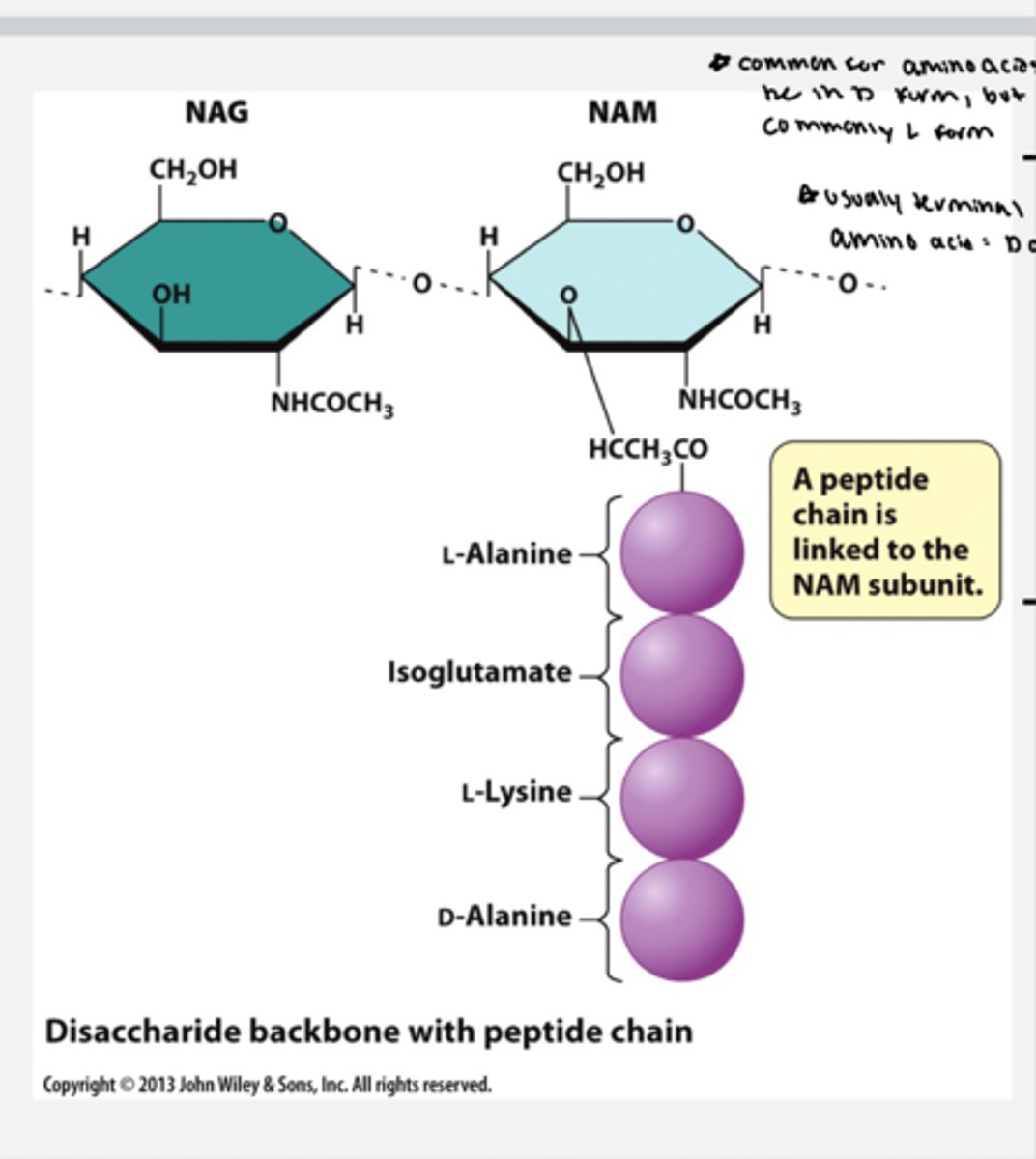
what form are amino acids most commonly in
D form but most commonly L form
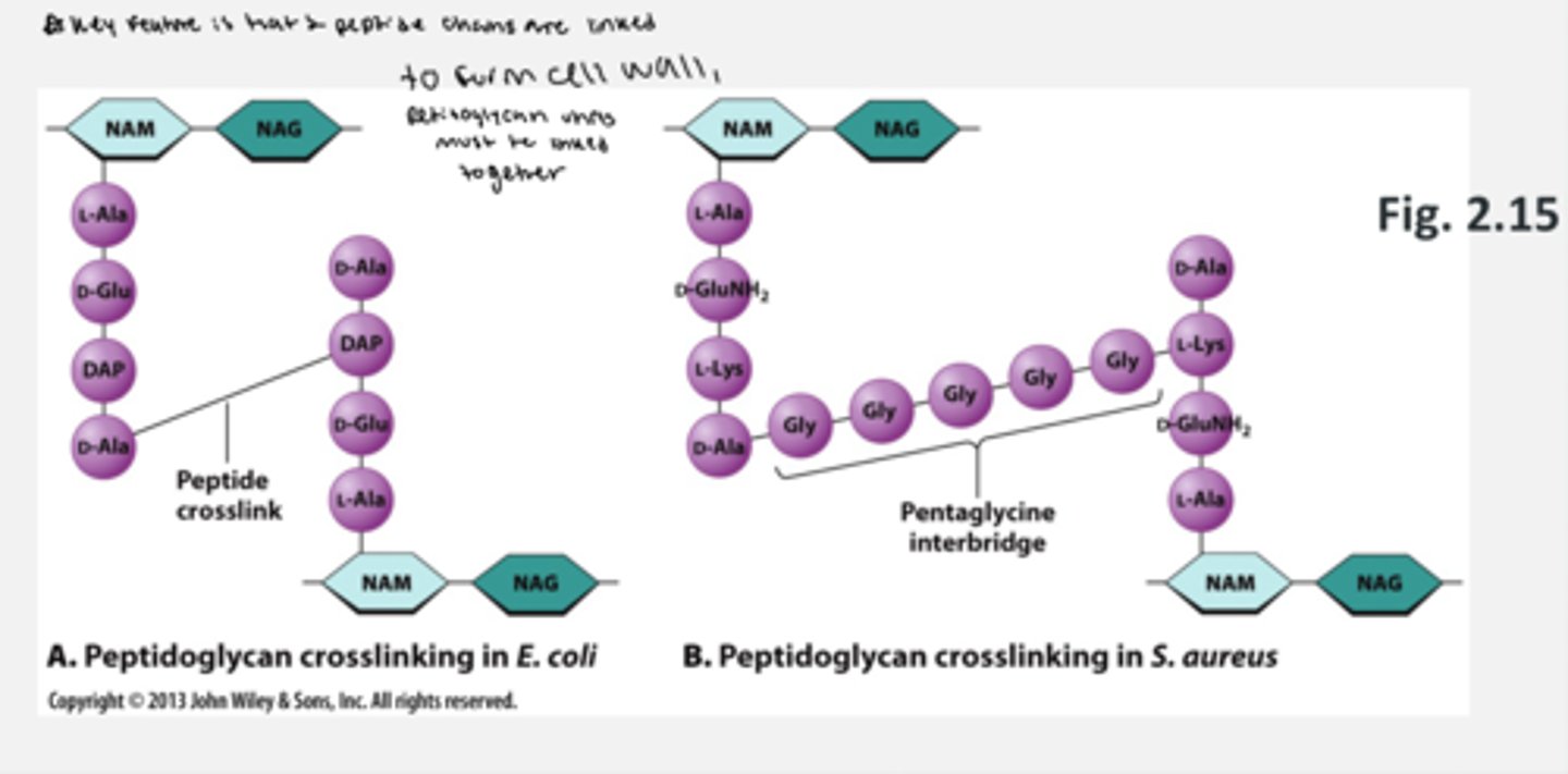
how is peptidoglycan cross-linking
- amino acids on the peptide on NAM can vary from species to species
- the way the peptides are cross-linked in the cell wall can also vary
the key feature is that the 2 peptides are linked
how does the cell wall form
1. NAM (with a peptide chain made) is synthesized in the cytoplasm and linked to UDP
2. NAM is linked to bactoprenol which is embedded across the cell membrane
3. NAG is added to NAM
4. Bactoprenol flips NAM-NAG to periplasm so now the peptidoglycan unit is on the outside
5. now that the disacharide is on the oustide, it is added to the existing chain and crosslinking occurs. peptides are linked and new sugars are linked
6. once the unit of peptidoglycan is linked to a growing chain, bactoprenyl can flip back into the cytoplasm and accept the next unit of peptidoglycan
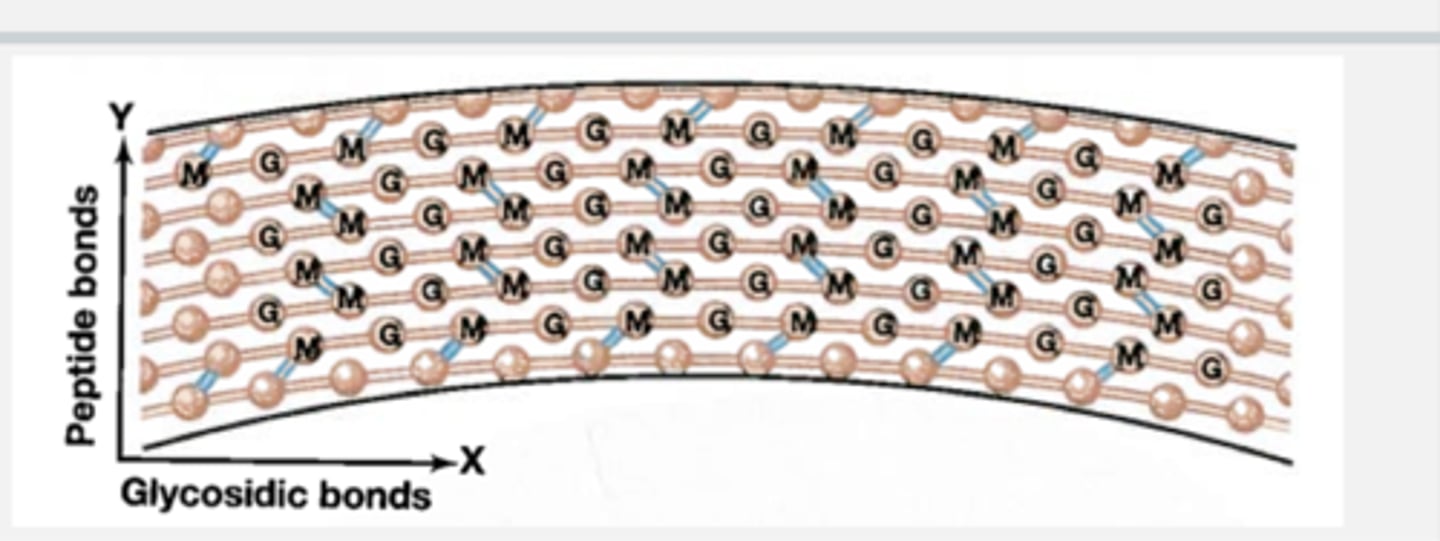
where does the strength of the cell wall come from
- the strength of the cell wall comes from cross linking (between peptides and sugars)
- bonds also form in 2 directions
if one of these phenomenons didn't happen then the cell wall would fall apart
in what direction do peptide bonds and glycosidic bonds move
peptide bonds move out (protrude the cell wall vetically) while glycosidic bonds extend the length of the cell wall horizontally
how does cross linking occur in the cell wall
- cross linking (aka transpeptidation) adds strength to cell wall
- transpeptidase recognizes D-Ala-D-Ala and helps form bond to glycine "bridge" (these are the last 2 terminal alanines)
how does penicillin antibiotic affect a cell
- penicillin interferes with the rection of transpeptidase by binding transpeptidase and blocks its ablility to interact with D-alanine-D-alanine preventing cross links from forming thus destabilizing the cell wall of the bacteria because cross links are not formed
- transpeptidases binds penicillin but cannot release it thus inactivating the transpeptidase
what is the normal function of transpeptidase
- transpeptidases bind D-ala-D-ala and releases product when finishes
- normally catalyzes the reaction where glycine is linked to D-alanine-D-alainine and the terminal D-alanine-D-alinine leaves and transpeptidase goes onto catalyze the next reaction
why does trasnpeptidase bind penicillin
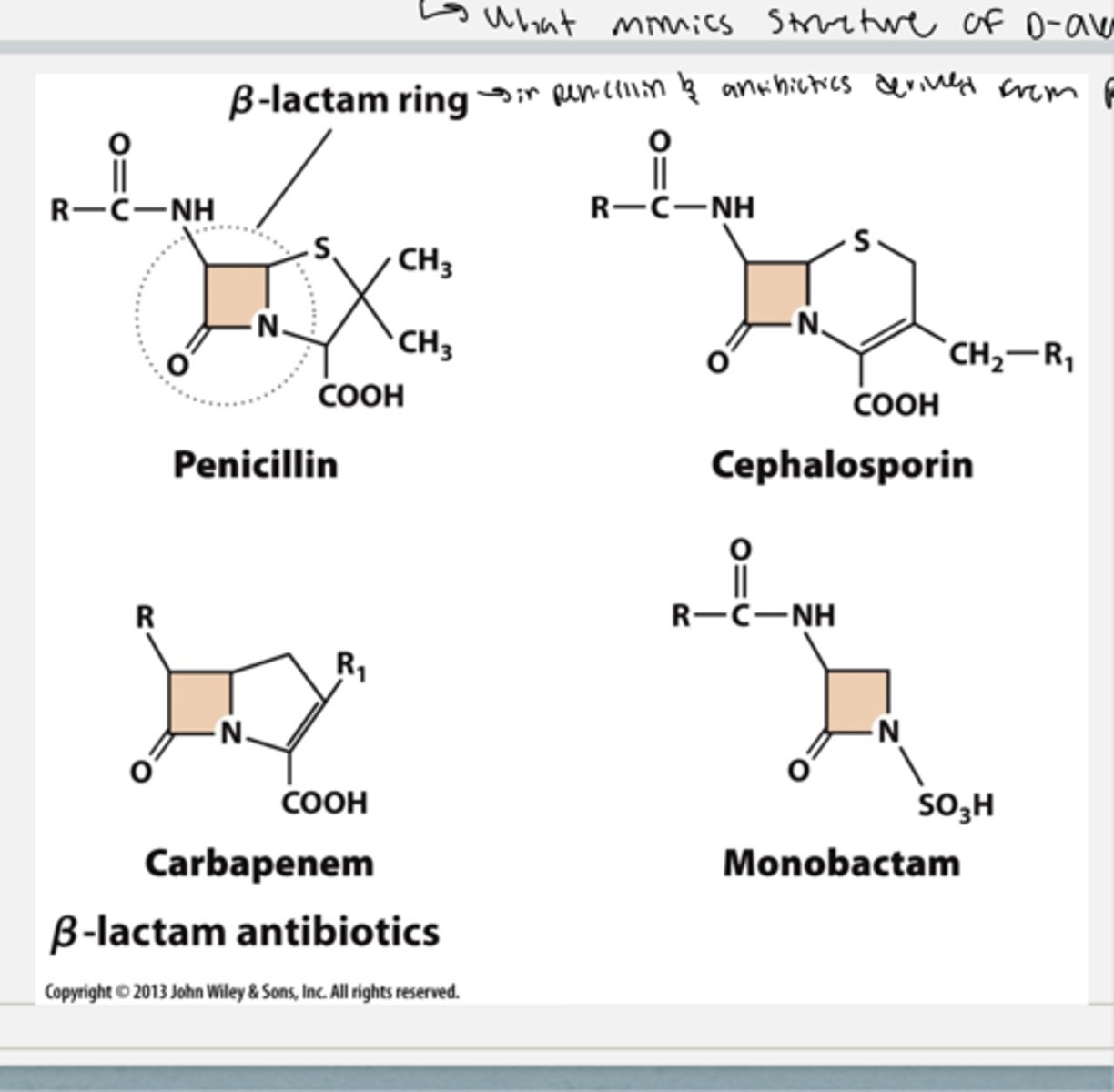
penicillin has a Beta lactam ring that is very similar to that of D-Ala-D-ala. Thus, transpeptidase can bind penicillin, but transpeptidase will not release it so it is inactivated and prevents further catalyzation of other reactions
identify the beta lactam ring in penicillin
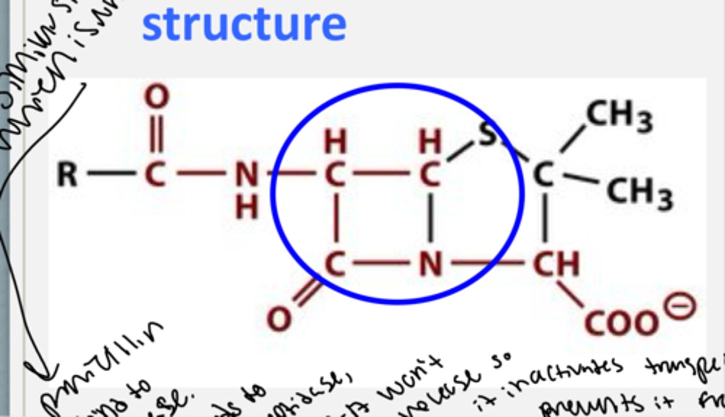
what antibiotic family is penicillin a part of
beta lactam family which also includes ceopahlosporin, carbapenem, and monobactam because all of them contain a beta lactam ring
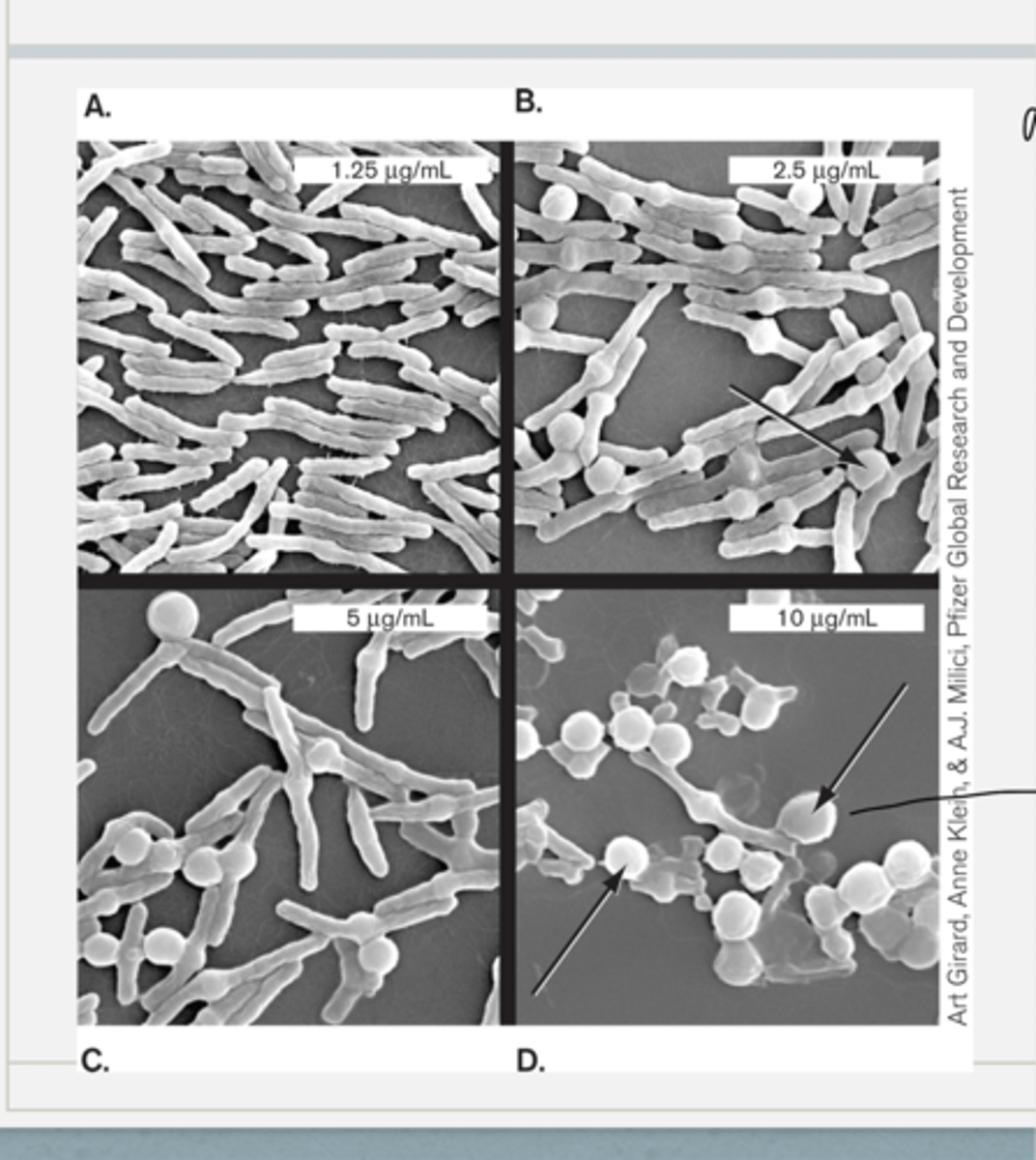
what is the effect of penicillin on bacterial cells
- cells were incubated for one our with the displayed concs of penicillin
- what happened to the cells is that buldges formed bc the cells are growing, but they cant form cell wall to make those cross links so now osmotic pressure buldged the cells because there is no cell wall to protect them/keep the rigidity and the shape
- the buldging increases as conc of penicillin increases leading to osmotic lysis
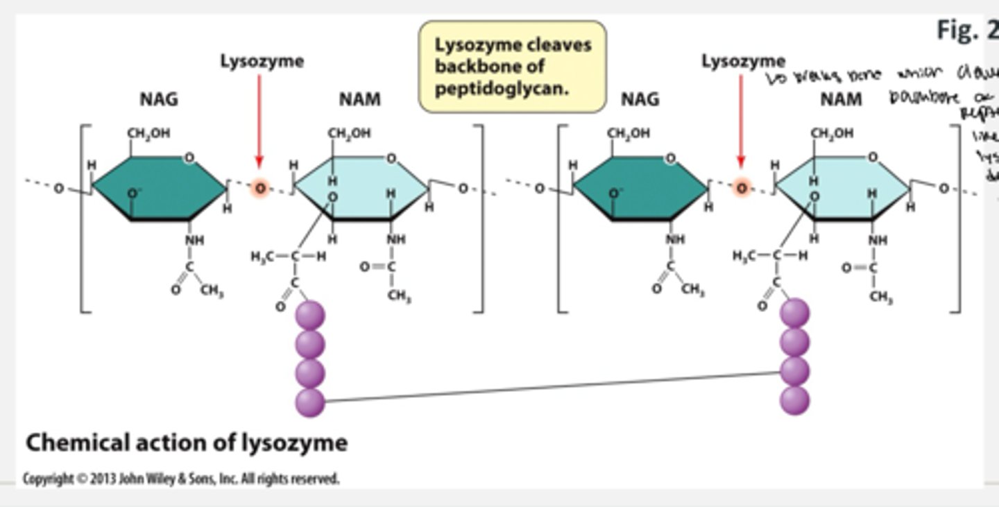
how does the lysozyme affect the cell
- lysozyme targets the cell wall of a bacteria
- lysozyme is a host antimicrobial protein found in saliva and tears
- it recognizes and cleaves the beta-1,4-glycosidic bond
- it breaks the bond which cleaves the backbone of peptidoglycan and like penicillin, lysozyme will degrade the cell wall
which antimicrobial agents target the cell wall
antibiotics like penicllin in the beta lactam family and lysosymes
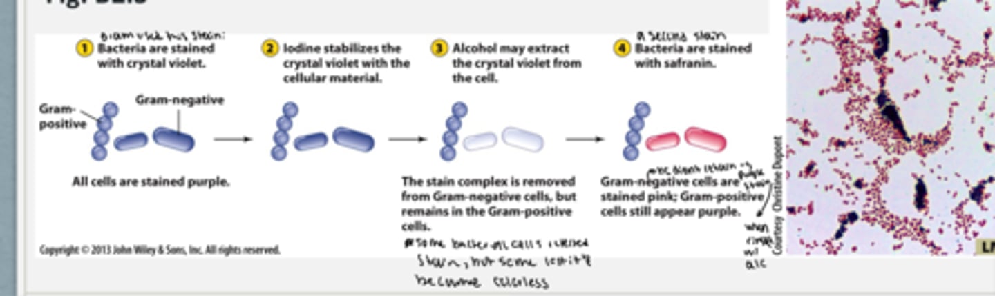
how are microbes separated based on the cell wall
- a stain method developed in 1884 by Hans christian gram can separate the microbes into two classes
1. bacteria are stained with crystal violet stain which turns all bacteria (+ and -) purple
2. iodine stabilizes the crystal violet with the cellular material
3. alcohol is added which will may extract the crystal violet from the cell = the stain complex is removed from gram negative cells, but remains in the gram positive cells.
4. bacteria are stained with safranin. gram negative cells are stained pink and gram positive cells still appear purple
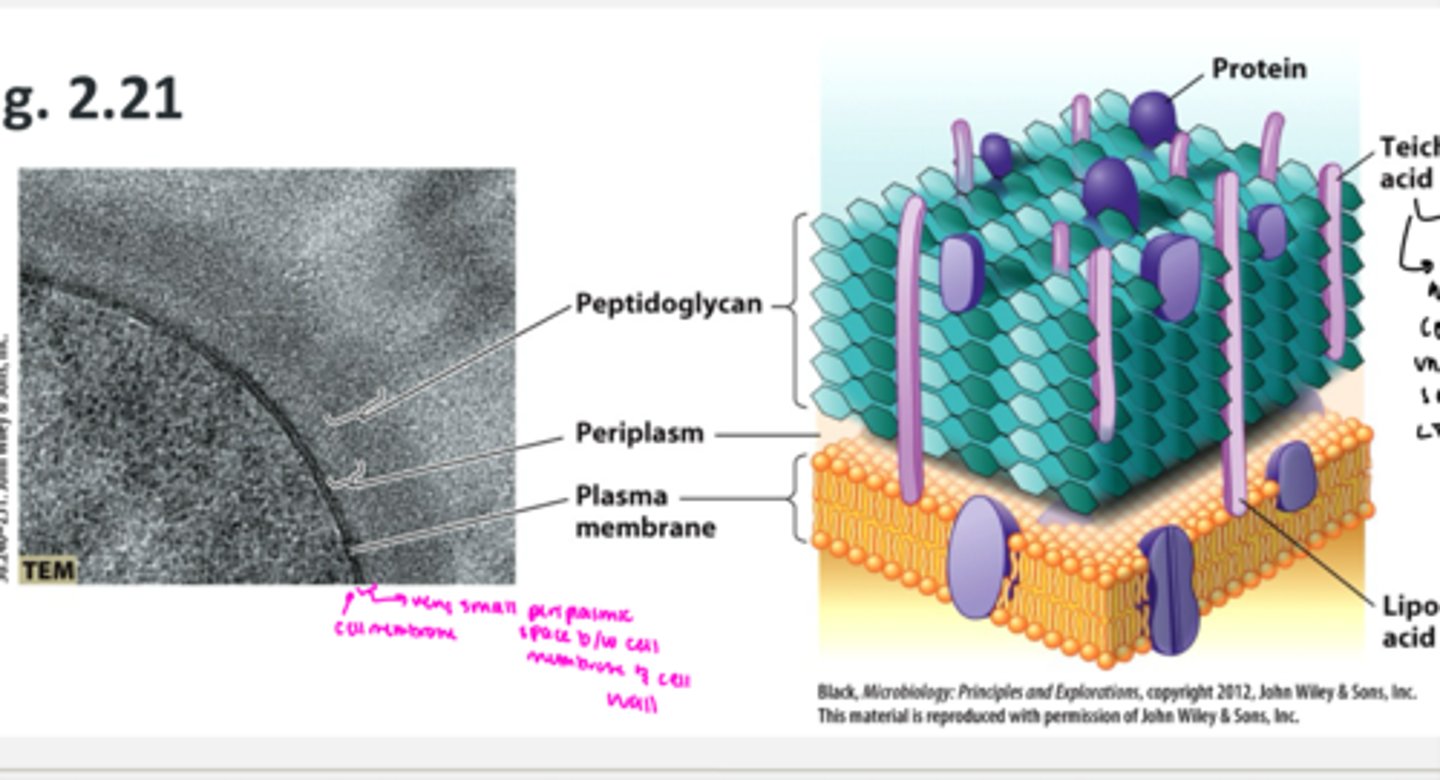
what are the characteristic of gram positive cells
1. have a thick outer layer of peptidoglycan
2. a narrow periplasmic space
3. techoic acids in the peptidoglycan make them negatively charged
also have Lipotechoic acid (LTA) which can induce a strong inflammatory response in the host cell
what are techoic acids and lipotechoic acids
- major function of both is structural support of the cells (form something similar to concrete rods)
- Techoic acid = cannot see these in a micrograph but these are proteins usually embedded in the cell wall only.
- however LTAs (lipotechoic acids) are sometime satatched to the cell membrane through the cell wall. these can induce a strong inflammatory response in the host. they are recognized as foreign by the host immune system and induce an immune response, damaging the host (ex. human host)
gram positive bacteria cell wall
what are the characteristics of gram negative cells
gram negative cells have:
- a varying width periplasmic space containing a very thin layer of peptidoglycan (super thin cell wall)
- an outer membrane composed of lipopolysachrides (LPS)
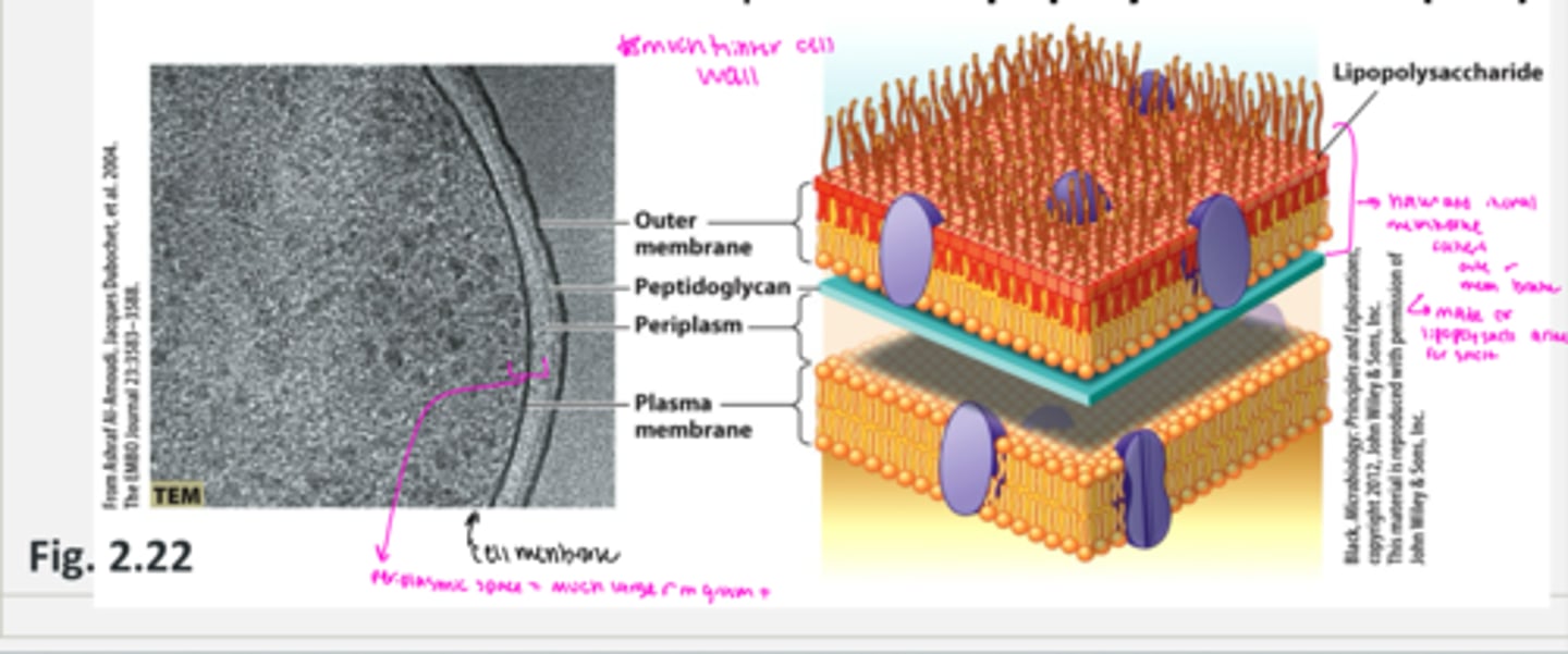
what are lipopolysachrides (LPS)
LPS are from gram negative cells which can be harmful
- structural compoennt of the outer membrane
- unique to bacteria
- lipid A portion induces a strong inflammatory response
- O (outer) side chain of polysachrides can vary by species
LPS is very foreign to hist so human immune system has a very strong response that can cause harm to the host
- they are composed of an O side chain (which is different for each species) and this is very hydrophillic and negatively charged because of all the sugars (makes it difficult for hydrophobic compounds to get near the membrane)
- this is linked to a core polysaccharide (same in all species) which contains ethanolamine
- this is linked to lipid A which is a fatty acid allowing it to interact with the membrane
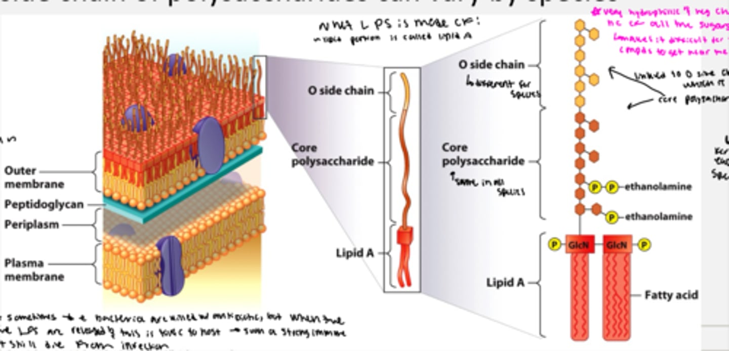
why are lipopolysacharides so dangerous to hosts
Problem with LPS is that when bacteria are killed with antibiotics, the cell starts to degrade, and the LPS are released and this is toxic to the host (releases such a strong immune response that host might still die from infection)
how does having an outer membrane benefit a gram negative bacteria
outer membrane of gram negative protects agaianst many toxic compounds including some antibiotics and lysoszymes
why is it harder to develop antibiotics against gram negative bacteria
- harder to develop antibiotiocs to kill gram negative bacteria bc of the outer membrane (ex, vanconyocin is a major antibiotic to treat gram + baacteria like spah infectiosn but it targets the cell wall and inhibits cell wall synthesis, but it cannot be used against gram negative bacteria bc vanomyocin cannot penetrate the cell wall)
are lysozymes effective against gram negative bacteria
no because they cannot penetrate the cell wall to get to the membrane
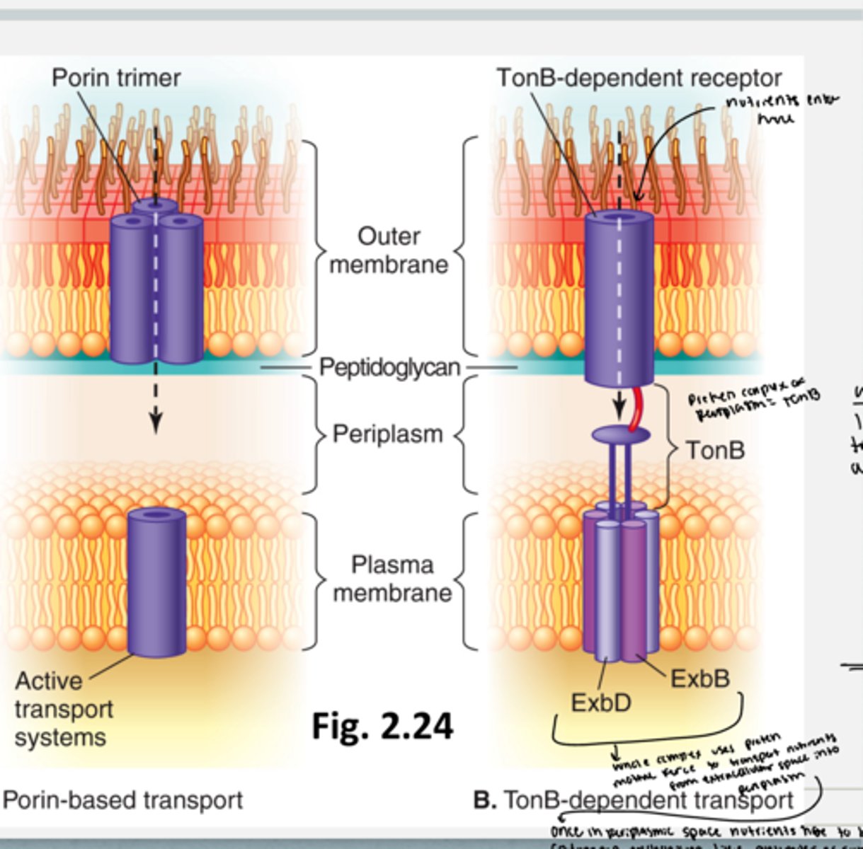
how does import across the outer membrane happen
have porin trimers in the outer membrane that connect to active transport systems in the plasma membrane
- porin channels: facilitate diffusion of small polar nutrients including sugars into the periplasm... the sugars are more abundant nutrients outside of the cell so they enter the periplasm through facilitated diffusion and are put into the cell memrane through active transport systems)
TonB dependent receptor: transports scarce nutrients using active transport into the periplasm
- there is a tonB dependent receptor on the outside of the outer membrane where nutrients enter and travel into the periplasmic space with the help of the proton motive force. in the periplamis space, the ton B dependent receptor form a protein complex with Ton B embedded in the plasma membrane. the ton B contains exbD and ExB which is a complex that transports the nutrients in the periplasmic space into the cytoplasm with active trasnport (like an ABC trasporter or co transport system liek anti or symport which is this protein in the cell membrane)
can the sec protein export system be used in gram negative bacteria
no because the proteins have to be able to cross the outer membrane too
what protein export systems are used in gram negative bacteria
type II and type III secretion systems
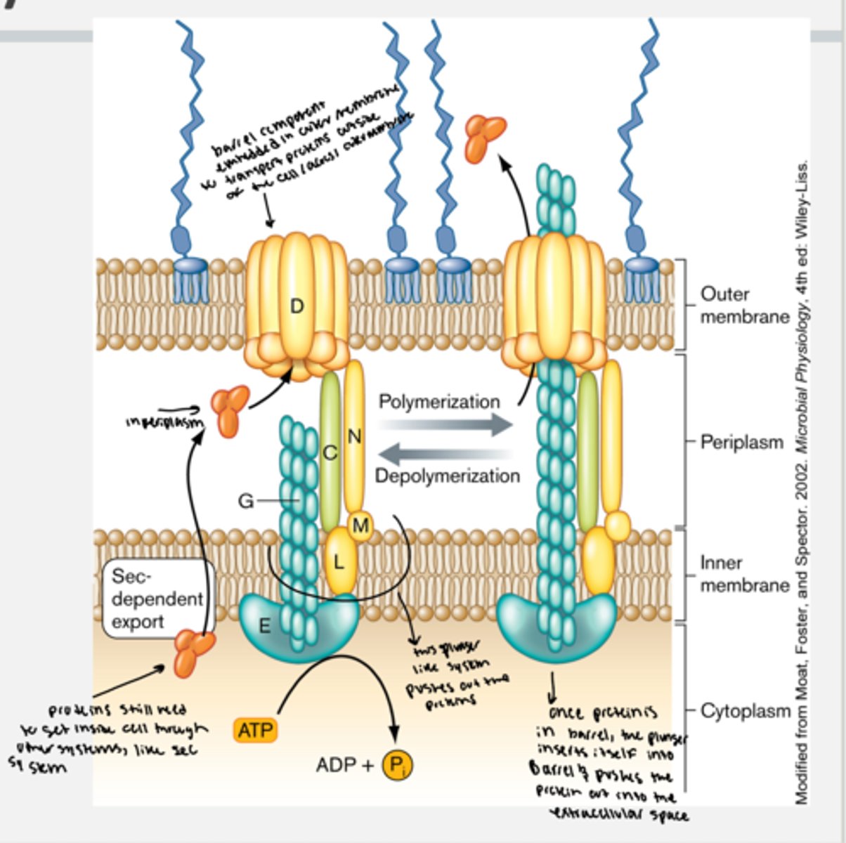
how is type II gram negative secretion system
- in gram negatives, some proteins use the sec system to get in the periplasm and then various outer membrane export systems like type II secretion
- proteins still leave the cell membrane and enter the periplasmic space through the sec system
1. to transport proteions out, a protein embedded on the innerface of the membrane that is also in the cytoplasm hydrolyzes ATP and is attackjed to a barrel protein in the outer membrane. when the protein from periplasm enters the barrel protein from periplasm, the cytoplamic protein hydrolyzes ATP and the prteined polymerize.
2. once the protein is in the barrle, the plunger insets intside into the barrel and pushes the protein out ijnto the extracellular space and then depolymerizes again
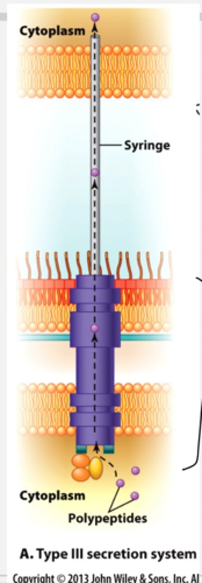
what are the type III sectretion systems for protein export in gram negative bacteria
- this is a sec pathway independent system
- in type III, proteins (toxins) are exported directly from the cytoplasm of the bacterial cell and injected directly into the host cytoplasm
- polypeptides (toxins) enter and the syringe like structure injects them into the cytoplasm of the host
- it has a transport barrel that spans both the cell membrane and the outer membrane. in addition to transporter that crosses the bacterial membrane, there is the syringe itself that will extend through the extracellular space and will insert through the cell membrane into the cytoplasm of another cell (usually its host like eukaryotes)
- so any proteins synthesized in the cytoplasm can travel through protein channel all the way into cytoplasm of eukaryotic cells = never come in contact with the periplasmic or extracellular space.
what kind of microbe typically uses the type III secretion system
- this is used a lot by bacterial pathogens that inject toxins --> interfere with the eukaryotic cell and burts functioning in a way that benefits the bacteria and allows it to easily infect the host
what are the optional bacterial structures
- pili
- flagella
- nonflagellar motiloities
- capsule
- s-layer
- endospores and exospores
- biofilms
what do pili allow bacteria to do
- adhere to surfaces - ____ are most commonly used to aid the bacterium in attaching to a surface
what are pili made of
- ___ are made of repeating subunits of the protein pilin
what kind of bacteria are pili found in
- ___ are found in both pathogenic and non-pathogenic bacteria
what are pili also called
- ___ are also known as fimbria
what do pili let bacteria do
- ___ look like hair and extend from the bacterial cell from all parts or just from one of the poles. at the end of the pillus are adhesive proteins that interact with a target molecule on the surface tightly so this lets bacteria stay attached to the place they want to be in.
- often bacterial environments have fluid washing over them/environment changes a bunch so this is helpful
- in pathogenic bacteria, pili are often virulence factors bc they establish an infection by adhering to certain places
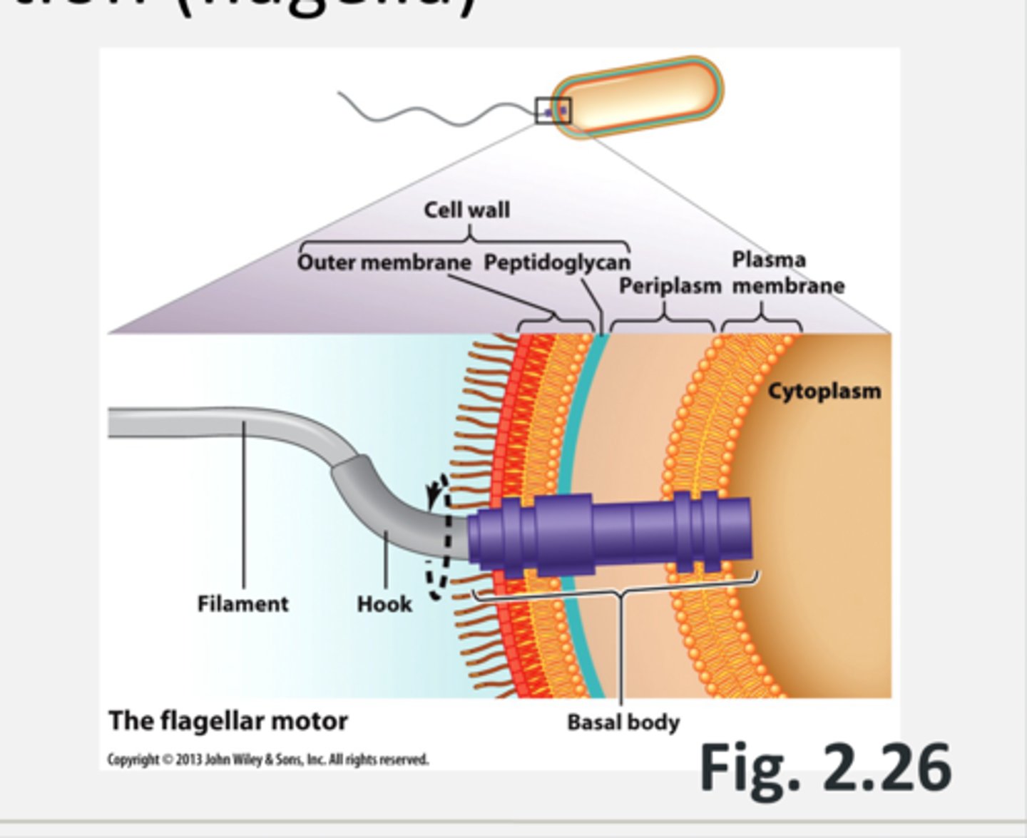
what structure allows for bacterial movemnet
flagella
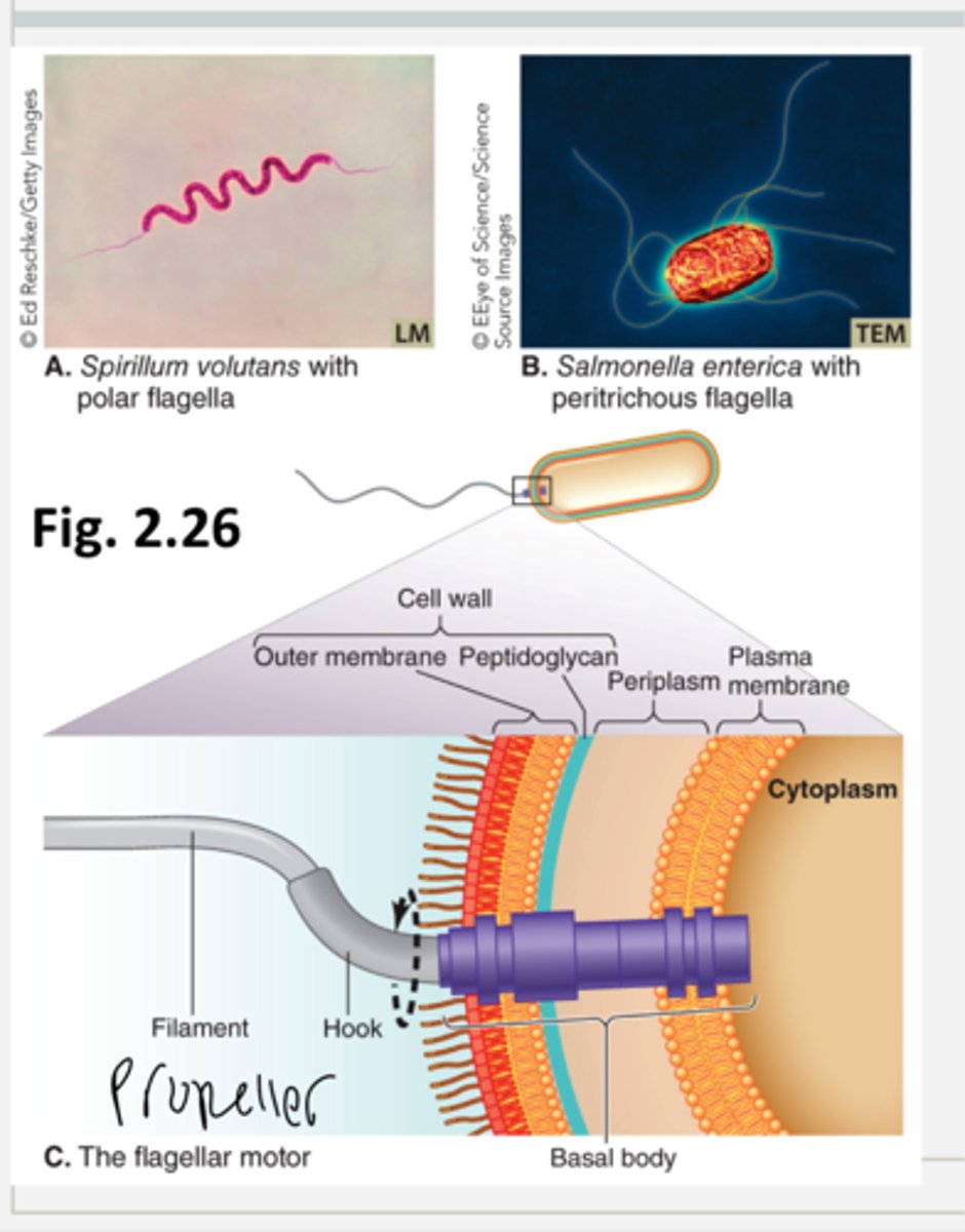
what do flagella do for bacteria
- allow bacteria to move for example towards nutrients or away from a toxin
- ___ are only at the poles or only in one spot or extend from several different places on bacteria
- they are propellers whose rotation is driven by movement from the proton motive force = as hydrogen ions flow in through the channel from high to low conc. energy is released which drives the flagella
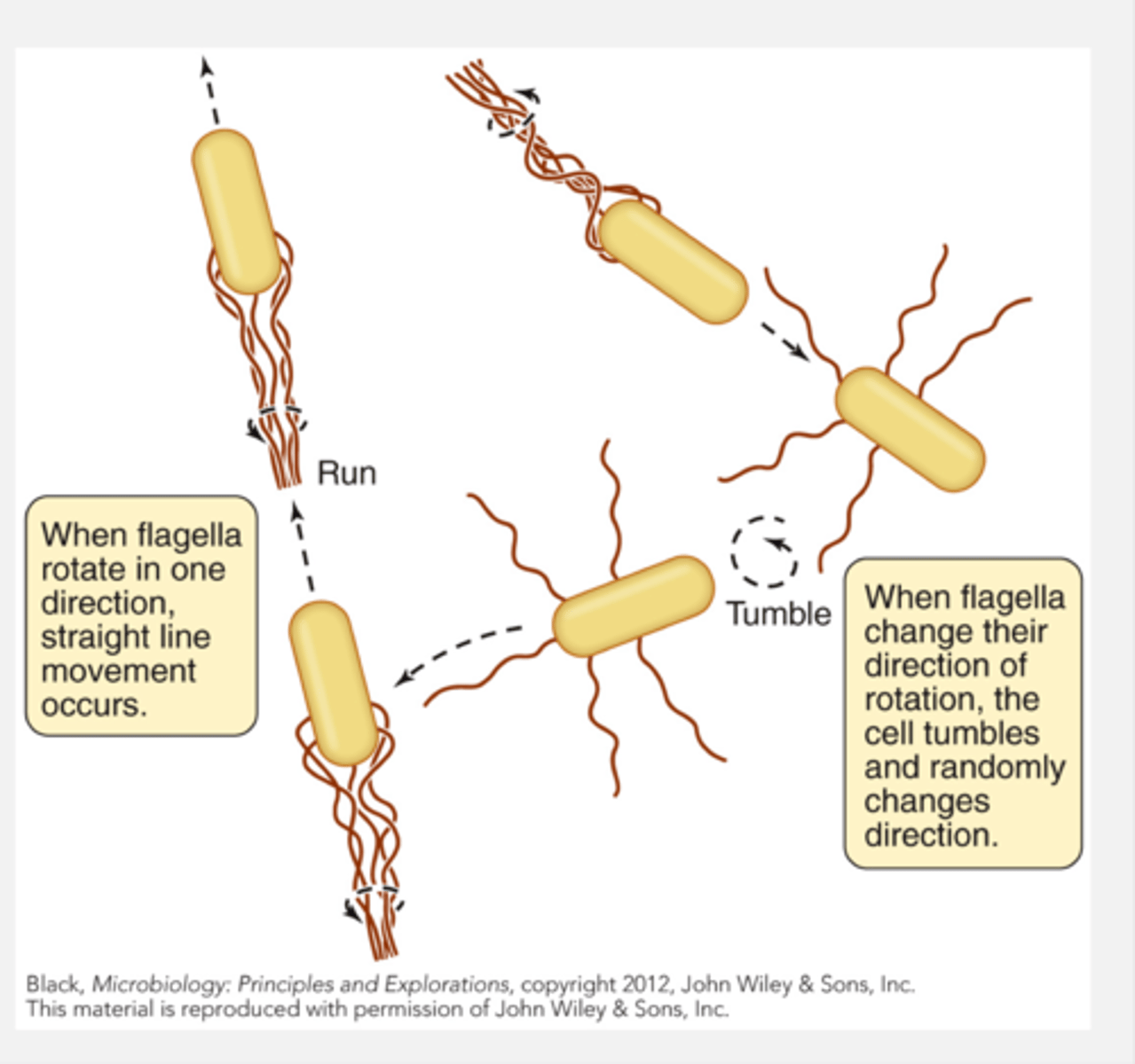
what is run/tumble of flagella
- when flagella. rotate in one direction they have straight line movement that occurs called a run
- when flagella change their direction of rotation, the cell tumbles and randomly changes direction
falgellar motor diagram
what are the types of bacterial movemnet without a flagella (nonflagellar movemnet)
- gliding motility: smooth sliding over a surface, not well understood but is done by myoxbacteria and cyanobacteria
- twitching motility: slow, jerky process using pili that can be extended, attach to a surface and then pull bacterium forward (ex. N. meningitidis, P. aeruginosa) = not common bc motsly pili are used for adhesion
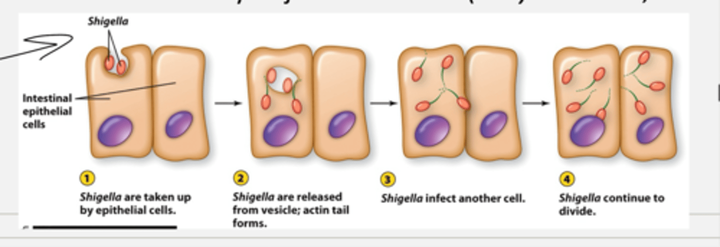
- actin based motility: hijack host actin (S. dysenteriae, L. monocytogenes)
= example: shigella protein being taken up into intestinal epithelial cells.
- shigella are taken up by epithelial cells bc they express proteins that are similar to actin causing actin fulamnets to form behind it and additional acyin units form behind it pusing the shigella forward
- shigella are released from vesicles and actin tail forms
- shigella then infect another cell connected to that cell
- shigella continue to divide
what part of bacteria do capsules affect q
cell surface
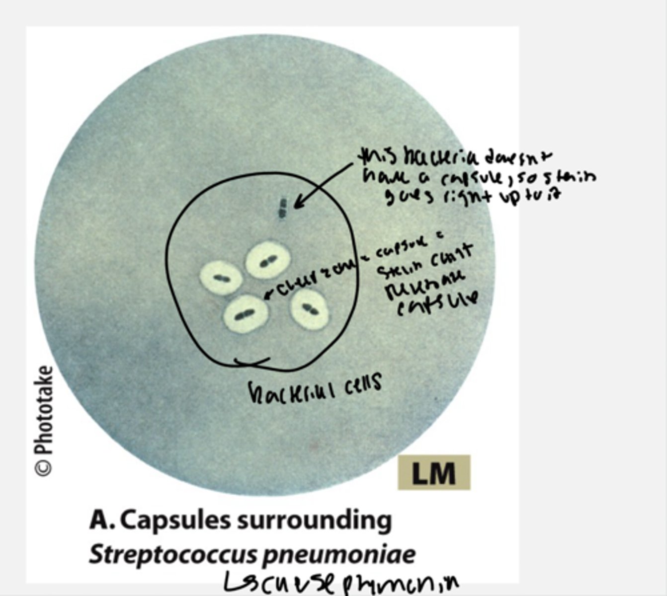
what is a capsule
- thick layer of polysacharides surrounding some cells
- helps bacteria adhere to a surface and protects against dessication
- major virulkence factor for many pathogens bc it inhibits phagocytosis by immune cells.
- if a bacteria loses the capsule, it loese the genes that encode the capsule. often times the capsule genes are encoded in a plasmid which can sometimes be lost. if they lose the ability to make/have a capsule then bacteria become avirulent and cannot cause an infection (type of bacillicus anthracis worked with in TCU labs bc it isnt harmful and is also the strain that Pasteur used to make his first vaccinations)
how is staining of bacteria that have capsules
- in bacteria that have capsules, the stain cannot penetrate the capsule and they have a big white glob around them. if a bacteria does not have a casule, the stain goes right up to the bacteria
what part of bacteria does the s-layer effect
cell surface
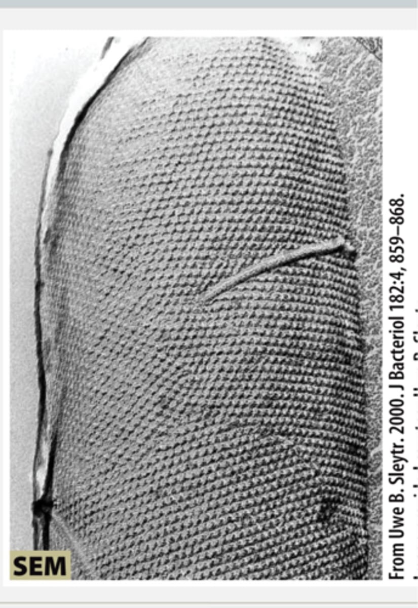
surface arrays (S-alyers) =
- crystalline array or interlocking proteins
-found in gram positive and gram negative cells
- "bacterial armor" protecting a cell against predation, infection with bacertiophage or host defenses
- very resource costsly = syntheisis stops when not needed ex. when a bacteria is in perfect conditions, the S- layer diappears because it isnt needed
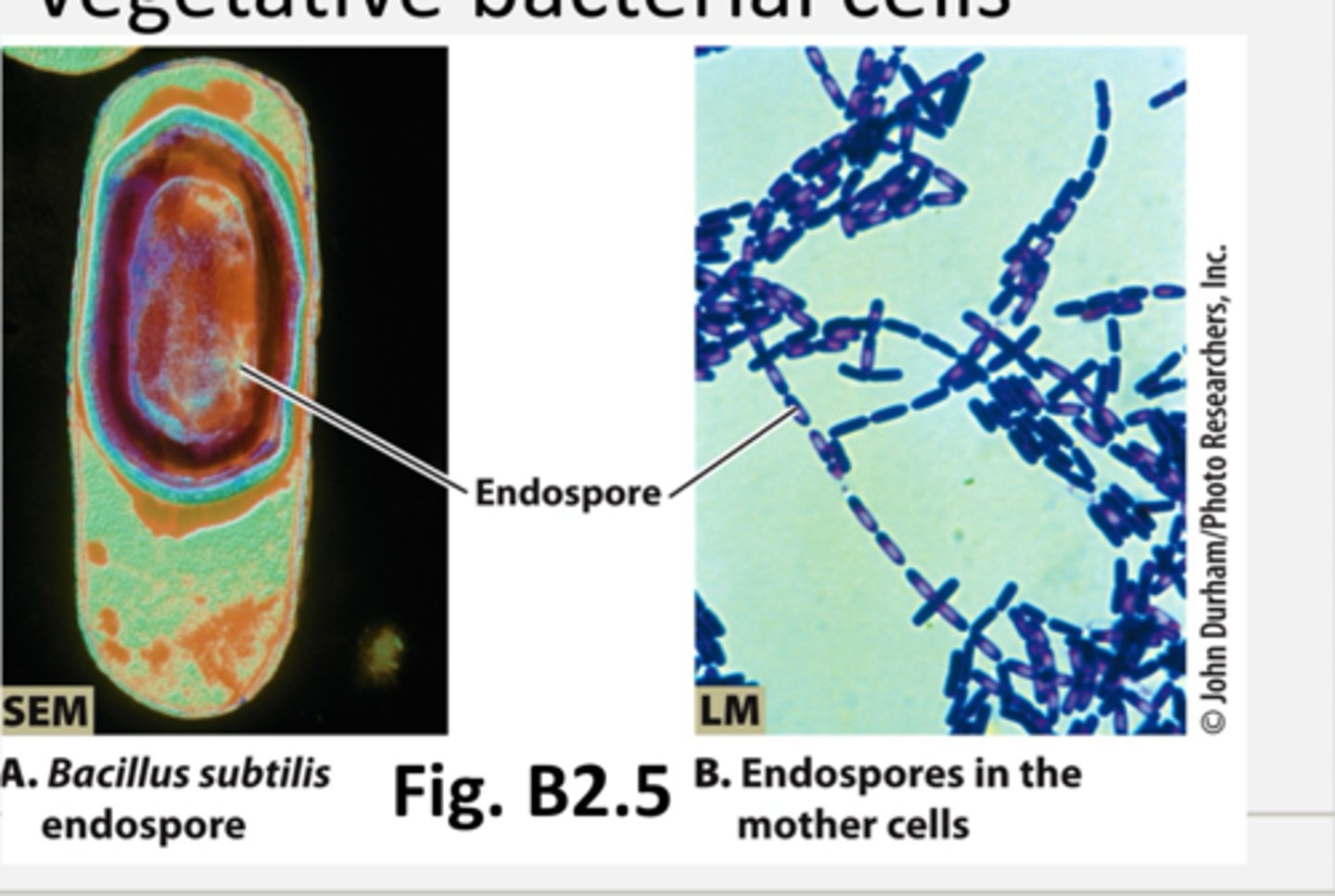
what are endospores and exospores of bacteria
some bacteria undergo sporulation when conditions deterioate
- metabolically inert (do not need nutrients/no energy production/no chemical biosynthesis/dont need much to survive and have a hard exoporium coat that protects them from chemical stress)
- very hardly happens but spores can exist for decades (like anthrax tower)
- once conditions improve, spores will germinate back to vegetative bacterial cells
- endospores are inside of the mother cell
- exospore = happen once the spore is released from the mother spore (like mom cell dies) and the spore is now found in the environment
ex. is bacillus anthracis developed as a biological weapon. v ery hard to deconmaniate spores bc they can exist for so long)
what are examples of spore forming bacteria
- bacillus anthracis = anthrax
- clostriduium botulinum = food poisoning
- clostridium tetani = tetanus
what are microbial communities
biofilms
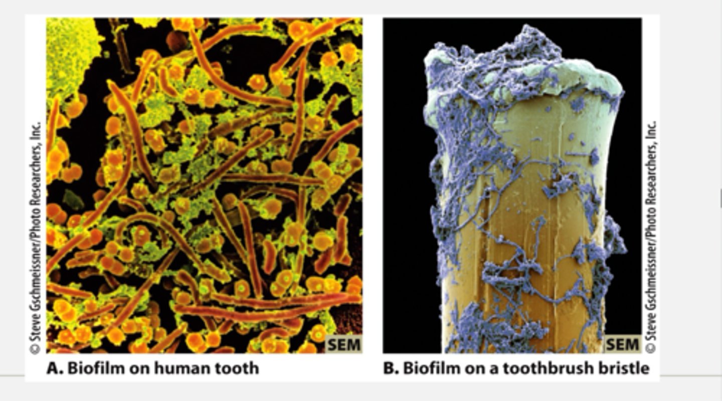
what are biofilms
- specialized, surface attached communities (ex. dental plaque and mold on bathroom surfaces)
- provide protection/enhanced survival
- often contain multiple microorganisms (like bacteria archea and eukaryotes all in one community)
- once the targeted critical mass of the community is reached, they will start to secrete the polysacharide that will form coating over the community to protect it and attachmnet will provide defenses against immune systems (like an animal host for example)
ex. of biofilms = decoloration on shower curtains
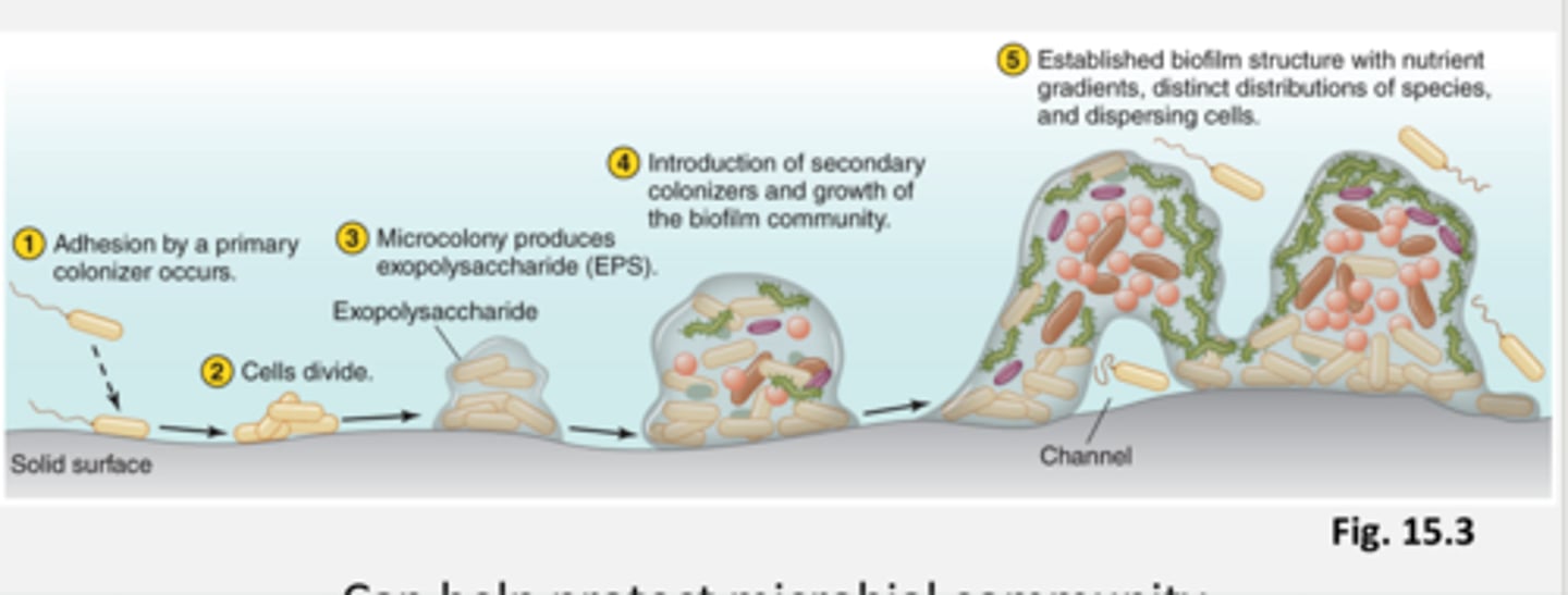
how does biofilm form
- formed in response to environmental signals
- can help protect a microbial community (for example antibiotic resistance)
1. adhesion by a primary coloizer occurs and attaches to the solid surface
2. cells of the coloizer divide a lot
3. microcolony produces expolusacharide (EPS)
4. introduction of secondary colonizers and growth of the biofilm community
5. established biofilm structure with nutrient gradients, distinct distributions of species, and dispersing cells \
these microbes will now behave differently as a biofilm than they would have as individual cells or a small clumping of cells.