6.6: Skeletal muscles are stimulated to contract by nerves and act as effectors
1/15
There's no tags or description
Looks like no tags are added yet.
Name | Mastery | Learn | Test | Matching | Spaced | Call with Kai |
|---|
No analytics yet
Send a link to your students to track their progress
16 Terms
How do muscles work in antagonistic pairs?
The agonist muscle contracts, pulling on the bone and producing force
The antagonist muscle relaxes
This happens against the incompressible skeleton
So the muscle transmits force to the bone
What is the advantages of muscles working in antagonistic pairs?
A second muscle is required to reverse the movement caused by the first muscle
As muscles can only pull
So both muscles contract to maintain posture and stabilise joints
What is a skeletal muscle?
The muscle that contracts to move bones

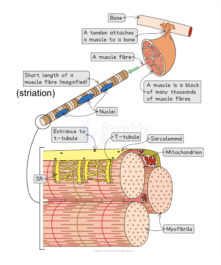
Describe and label the gross and microscopic structure of a skeletal muscle
Made of many bundles of muscle fibres packaged together
Attached to bones by tendons
Muscle fibres contain:
Inward folding sarcolemma to form transverse tubules
Sarcoplasm
Multiple nuclei
Many myofibrils
Sarcoplasmic reticulum
Many mitochondria
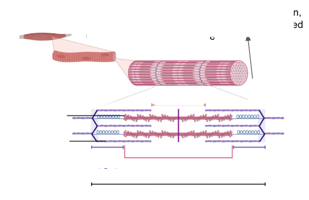
Describe and label the ultrastructure of a myofibril
Many protein filaments are arranged in a parallel pattern
Containing myosin (thick filament) and actin (thin filament)
Proteins are arranged in functional units called sarcomeres
Z disc: ends of sarcomeres where actin filaments attatch
M line: central point of sarcomeres where myosin filaments attach
H zone: region only containing myosin filaments
A band: region where myosin filaments extend
I band: region containing only actin filaments
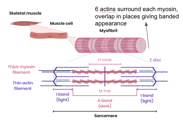
Why do sarcomeres form banding patterns?
I bands contain only actin filaments so they are light
H bands contain only myosin filaments so they are darker than I bands
A bands contain overlapping myosin and actin filaments so they are the darkest
What is the overall process of muscle contraction?
Myosin heads slide actin filaments along myosin, causing the sarcomere to contract
Simultaneous contracton of many sarcomeres cayses myofibrils and muscle fibres to contract
When sarcomeres contract, H zones get shorter, I band gets shorter, A band stays the same and Z lines get closer
What is the role of actin in myofibril contraction?
Calcium ions bind to tropomyosin, causing it to move
Binding sites on actin are exposed
This allows the myosin head with ADP attached to bind to binding sites on actin
Actomyosin crossbridge is formed
Myosin heads change angle, pulling actin along myosin using energy from ATP hydrolysis
New ATP binds to myosin head, causing it to detach from binding site
Myosin attaches to a different binding site further along actin
Process is repeated as long as calcium ion concentration is high
What is the role of myosin in myofibril contraction?
The myosin head with ADP attached binds to binding sites on actin
Actomyosin crossbridge is formed
Myosin heads change angle, pulling actin along myosin using energy from ATP hydrolysis
New ATP binds to myosin head, causing it to detach from binding site
Hydrolysis of ATP by ATP hydrolase, activated by calcium ions, releases energy for myosin heads to return to original position
Myosin attaches to a different binding site further along actin
Process is repeated as long as calcium ion conc
What is the role of calcium ions in myofibril contraction?
Depolarisation spreads down sarcolemma via t tubules
So calcium ions release from sarcoplasmic reticulum
Calcium ions diffuse to myofibrils
Calcium ions bind to tropomyosin, causing it to move
Binding sites on actin are exposed
This allows the myosin head with ADP attached to bind to binding sites on actin
Actomyosin crossbridge is formed
Hydrolysis of ATP by ATP hydrolase, activated by calcium ions, releases energy for myosin heads to return to original position
What is the role of tropomyosin in myofibril contraction?
Calcium ions bind to tropomyosin, causing it to move
Binding sites on actin are exposed
This allows the myosin head with ADP attached to bind to binding sites on actin
Actinomyosin crossbridge is formed
What is the role of ATP in myofibril contraction?
Myosin heads change angle, pulling actin along myosin using energy from ATP hydrolysis
New ATP binds to myosin head, causing it to detach from binding site
Hydrolysis of ATP by ATP hydrolase, activated by calcium ions, releases energy for myosin heads to return to original position
What is the process of muscle relaxation?
Calcium ions are actively transported back into the endoplasmic reticulum using energy from ATP
Tropomyosin moves back to block the myosin binding site on actin again, so no actinomyosin cross bridges can be formed
What is the role of phosphocreatine in muscle contraction and when is it used?
Acts as a source of inorganic phosphate
So rapidly phosphorylates ADP to regenerate ATP
Short lasting so only used in short bursts of vigorous exercise
Aerobic and alactic process
What is the role of ATP in muscle contraction?
Provides energy to reset the position of myosin heads for contraction to repeat
Provides energy for active transport for calcium ions to return to the sarcoplasmic reticulum after contraction
Breaks the actin-myosin cross bridge by attaching to the myosin head and causing it to detach from the actin filament
Compare the structure, location and general properties of slow and fast skeletal muscle fibres
Structure
Slow twitch has high conc of myoglobin which stores oxygen for aerobic respiration, fast twitch has low levels of myoglobin
Slow twitch has many mitochondria so has a high rate of aerobic respiration, fast twitch has lots of glycogen that is hydrolysed to provide glucose for glycolysis/aerobic respiration
Slow twitch has many capillaries so has a high concentration of oxygen and glucose for aerobic respiration
Fast twitch has a high concentration of enzymes for anaerobic respiration
Fast twitch stores phosphocreatine
Location
Slow twitch has a high proportion in muscles used for posture, fast twitch has a high proportion in muscles used for fast movement such as biceps and eyelids
General Properties
Slow twitch are specialised for slow, sustained contractions, fast twitch are specialised for brief, intensive contractions
Slow twitch obtain ATP from aerobic respiration, fast twitch obtain ATP from anaerobic respiration
Slow twitch release energy slowly and so fatigue slowly, fast twitch obtain release energy quickly and so fatigue quickly due to high lactate concentration