Lab 3: Introduction to Skeletal System and Axial Skeleton
1/84
There's no tags or description
Looks like no tags are added yet.
Name | Mastery | Learn | Test | Matching | Spaced | Call with Kai |
|---|
No analytics yet
Send a link to your students to track their progress
85 Terms
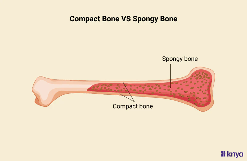
Compact Bone
This is the HARD, EXTERNAL LAYER OF BONE
Gives STRENGTH AND STRUCTURE

Spongy Bone
It’s DEEP INSIDE THE COMPACT BONE
This allows for SUPPORT AND PROTECTION TO DIFFERENT CELLS that MAKES THE BONE MARROW
Looks like a sponge
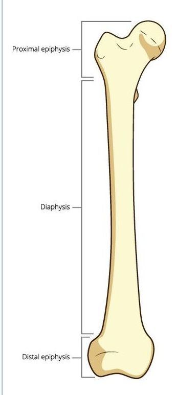
Diaphysis
The SHAFT of a long bone that’s MADE OF COMPACT BONE with SMALL AMOUNT OF SPONGY BONE
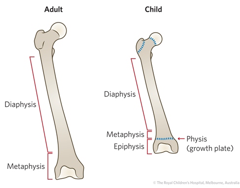
Epiphysis (plural: epiphyses)
ENDS OF LONG BONE
CONTAINS SPONGY BONES with THIN LAYER of COMPACT BONE
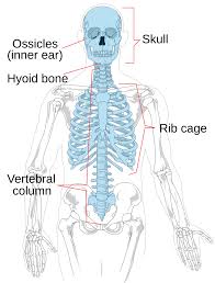
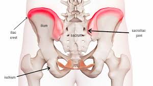
Axial Skeleton
INCLUDES BONES ALONG THE MIDLINE OF THE BODY
Contains the Cranial, Thoracic, and Pelvic Regions
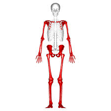
Appendicular Skeleton
UPPER AND LOWER LIMB BONES
The Arm, Hips, and Leg bones
Meatus
LARGE OPENING IN THE BONE that LEADS INSIDE THE BODY
Any opening that allows something to flow in/out of the body
e.g, external auditory _____ allows sound to go inside the ears and lets the brain process it
Foramen
OPENING (HOLES) IN THE BONE that GIVES PATH FOR BLOOD VESSELS/NERVES
Fossa
HOLLOW (Ditch-like) AREA IN A BONE that can FORM JOINTS or GIVE AREA OF ATTACHMENTS
It’s like a small dip in the bone
A spot where muscles can attach
Fossa = TRENCH/DITCH in Latin
Condyle
It’s a ROUND, SMOOTH BUMP that’s TYPICALLY AT THE END OF A BONE (really at the end)
Think CONnecting joint surface
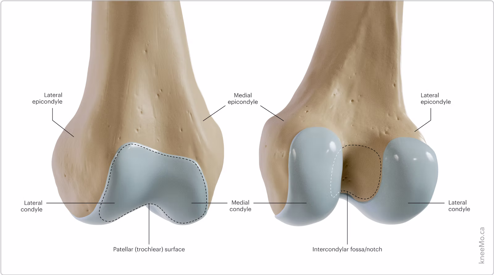
Epicondyle
RAISED BUMP ABOVE the condyle
Epi = above

Crest
RIDGE/BUMP THAT STICKS OUT of a bone
This is SLENDER RIDGE OF A BONE
Process
BROAD TERM FOR PROJECTION or OUTGROWTH on a bone
This could include things like the condyle, trochanter, coracoid, ETC
Facet
SMOOTH, NEARLY FLAT (or generally flat) SURFACE ON A BONE
this typically FORMS A JOINT with another bone
Spine
FLEXIBLE COLUMN of BONES IN THE BACK that SUPPORTS AND PROTECTS THE SPINAL CORD
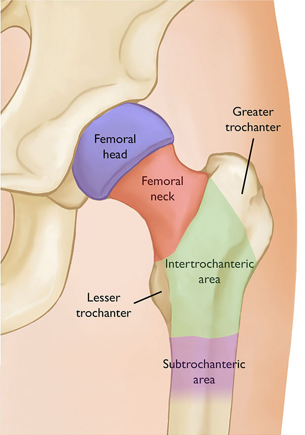
Trochanter
LARGE, BLUNT BUMP ONLY on the FEMUR (there’s 2)
BONY PROJECTION
Typically in the appendicular skeleton
Think TROJAN for the TRO part and Trojan is kinda BIG/LARGE
Tubercle
SMALL, ROUNDED BUMP
Tiny bumps for attachment
Typically in the appendicular skeleton
Tuberosity
MEDIUM-SIZED ROUGH BUMP
It’s in the appendicular skeleton
refers to a prominent, often rough, bony projection, and these are common features on bones of the limbs and the girdles that support them
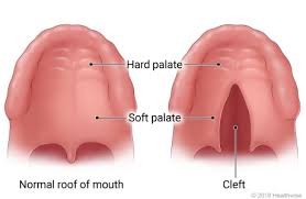
Cleft Palate
a BIRTH DEFECT where the ROOF OF THE MOUTH DOESN’T FULLY CLOSE
CAUSED GENETICALLY AND ENVIRONMENTALLY
Osteocytes
MATURE BONE CELLS in the BONE MATRIX
Allows for EXCHANGES in gas, nutrients, ETC
It’s WITHIN THE LACUNAE

Lacunae (singular: lacuna)
These are SMALL, EMPTY SPACES within the bone matrix
These are where OSTEOCYTES LIVE IN
Think like an UNFINISHED NEW YORK APARTMENT

Osteon
FUNCTIONAL UNIT of the COMPACT BONE
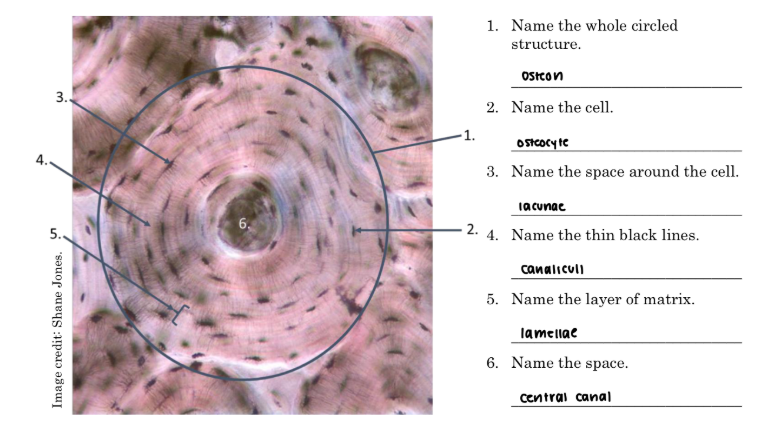
Central Canal
This is WITHIN THE OSTEON
It HOUSES BLOOD VESSELS AND NERVES

Lamellae (singular = lamella)
THIN, FLAT STRUCTURE (like a sheet or plate or layer)
This is the LAYER OF THE BONE MATRIX
GIVES STRENGTH AND RIGIDITY

Canaliculi (singular: canaliculus)
SMALL CANALS WITHIN LAYERS OF BONE MATRIX
it connects osteocytes together, which ENCOURAGES EXCHANGES between the osteocytes

Frontal Bone
FORMS FRONTAL and ABOVE ORBITAL
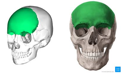
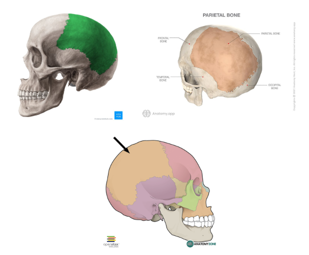
Parietal Bone
SIDES AND TOP OF THE HEAD
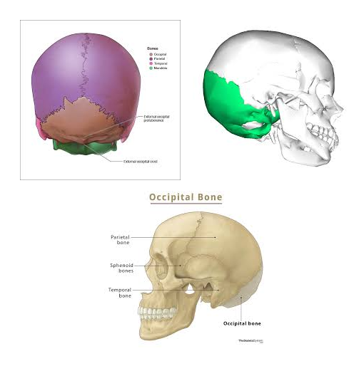
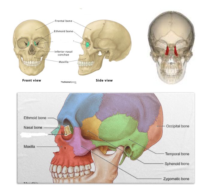
Occipital Bone
Forms the BACK AND BASE OF THE SKILL
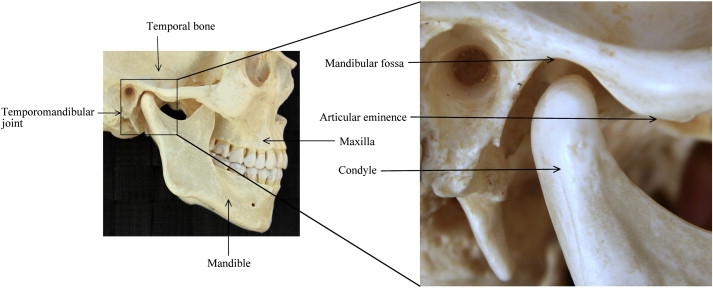
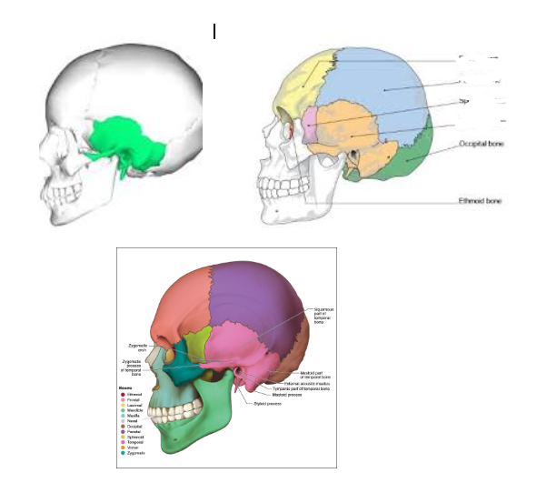
Temporal Bone
Towards the SIDES AND BASE (bottom) OF THE SKULL (kinda near the ear area)
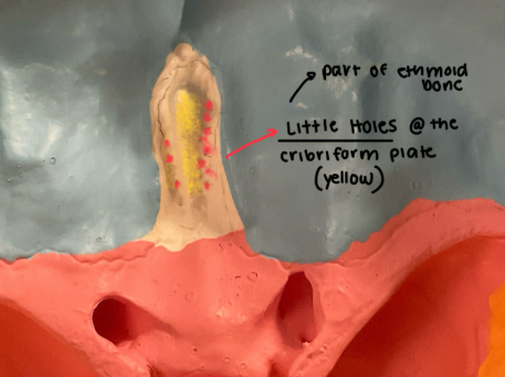
Ethmoid Bone
FLAT CRANIAL BONE that PROTECTS DELICATE BRAIN
BETWEEN THE EYES
It FORMS THE ROOF of the NASAL BONE
SUPPORTS OLFACTORY NERVES
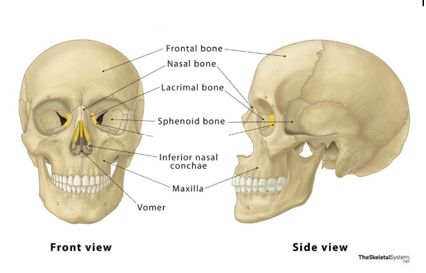
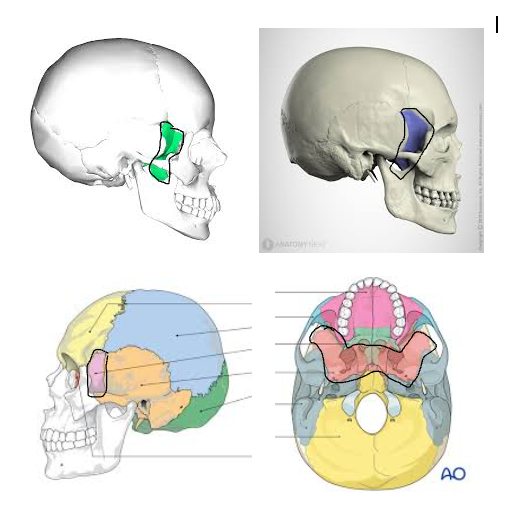
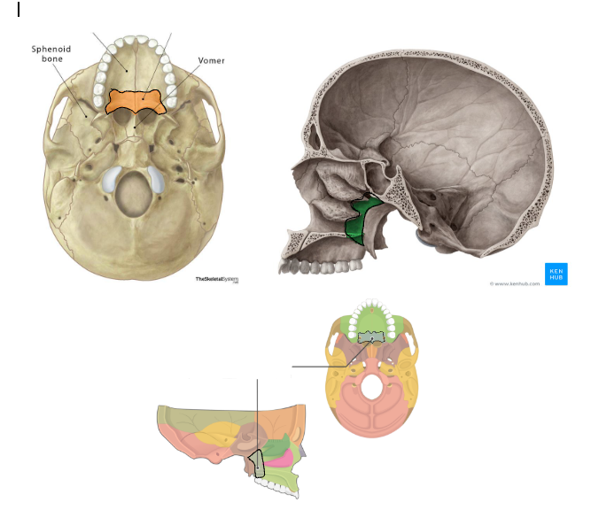
Sphenoid Bone
BUTTERFLY/BAT SHAPED BONE in the MIDDLE of the SKULL BASE
NEXT TO TEMPORAL BONE
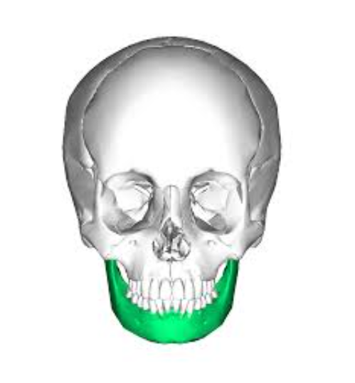
Mandible
JAW
Vomer Bone
Small, thin, PLOW SHAPED bone that’s in the MIDLINE OF NASAL CAVITY
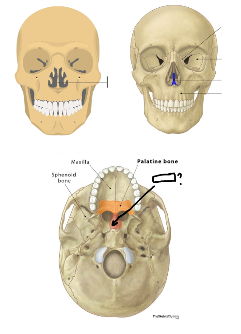
Maxillae (sing. maxilla)
UPPER JAW that FORMS PART OF NOSE AND EYE SOCKET
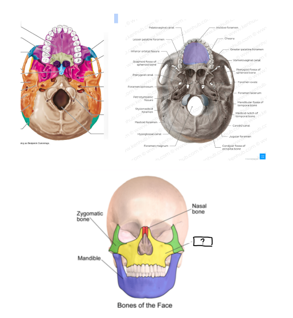
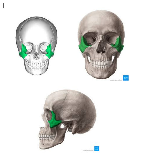
Zygomatic Bones
CHEEKBONES
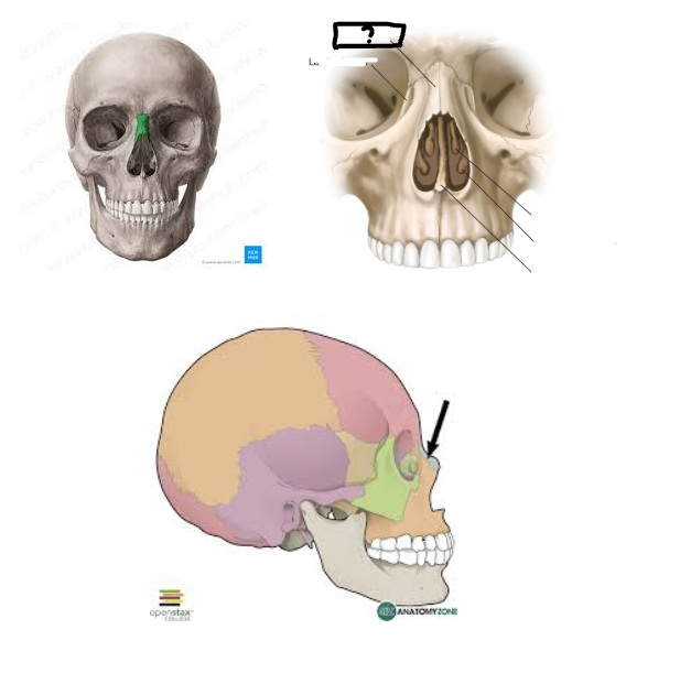
Nasal Bones
NOSE BONE
Inferior Nasal Conchae (concha)
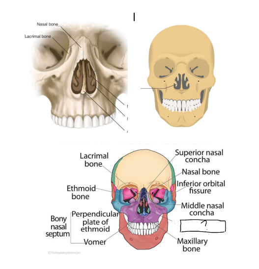
Lacrimal Bones
INNER WALL OF EYE SOCKET
Palatine Bones
Olfactory Foramina
The little holes within the cribriform plate
Those little holes and the cribriform plate are PART OF THE ETHMOID BONES (which is deep compared to the lacrimal bone within the eyes)
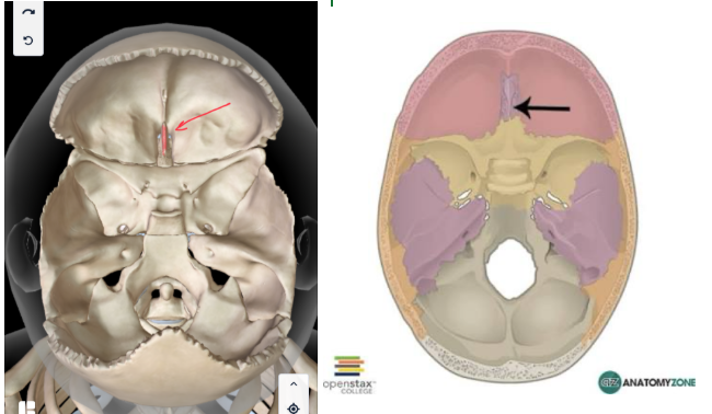
Coronal Suture
This is the suture that DIVIDES THE FRONTAL AND PARIETAL bones
It’s another word for the frontal plane that cuts ANTERIOR AND POSTERIOR
This picture is FACING TOP
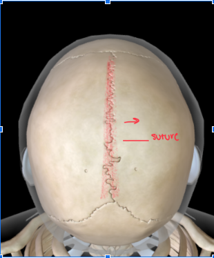
Sagittal Suture
This DIVIDES THE left and right PARIETAL BONES
This DIVIDES LEFT AND RIGHT…what other plane divides the body left and right?
This is the TOP of the skull
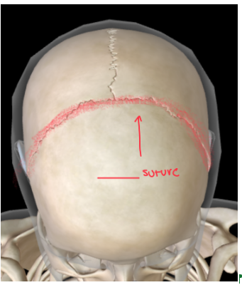
Lambdoid Suture
DIVIDES PARIETAL and OCCIPITAL BONE
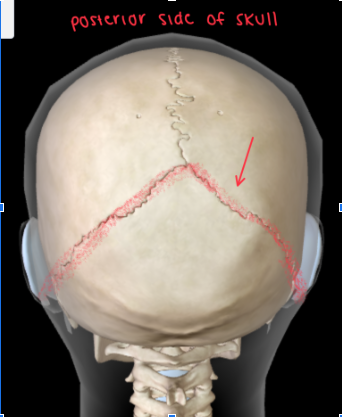
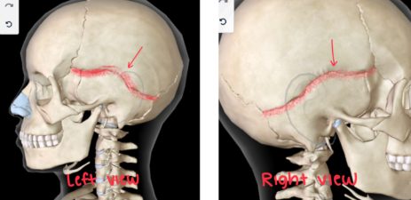
Squamous Suture
DIVIDES the PARIETAL and TEMPORAL BONES
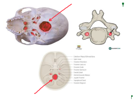
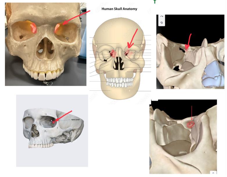
Optic Foramen
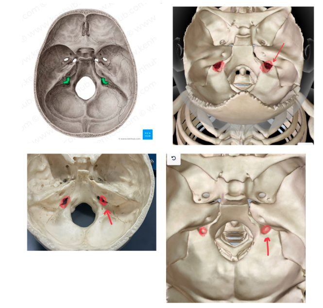
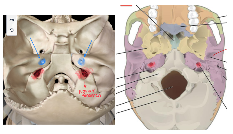
Jugular Foramen
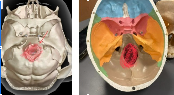
Foramen Magnum
Carotid Canal (Carotid Foramen)
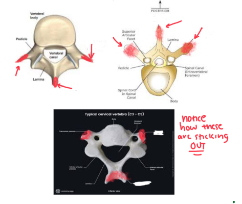
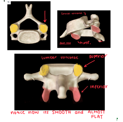
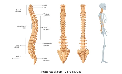
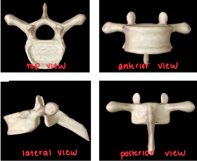
Cervical Vertebrae
Thoracic Vertebrae
Looks like a STINGRAY
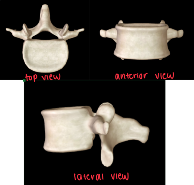
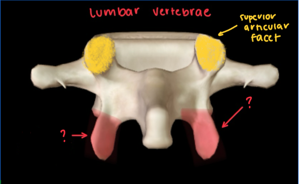
Lumbar Vertebrae
It’s SMALLER
Looks like a PLANE
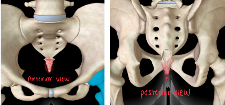
Sacrum
TRIANGULAR SHAPE that’s INFERIOR to the different vertebras
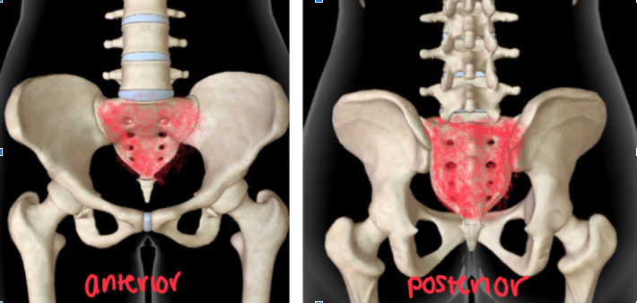
Coccyx
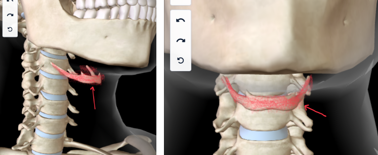
Hyoid Bone
BELOW THE MANDIBLE
True Ribs (1-7)
These are the BONES DIRECTLY CONNECTED TO THE STERNUM
The individual ribs are connected to the sternum
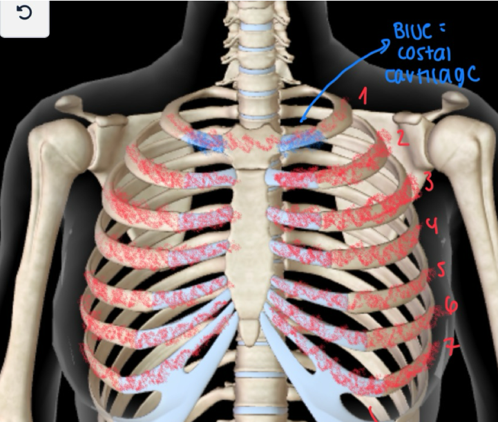
False Ribs (8-12)
These ribs are NOT DIRECTLY ATTACHED TO THE STERNUM
They’re not individually attached unlike the true rubs
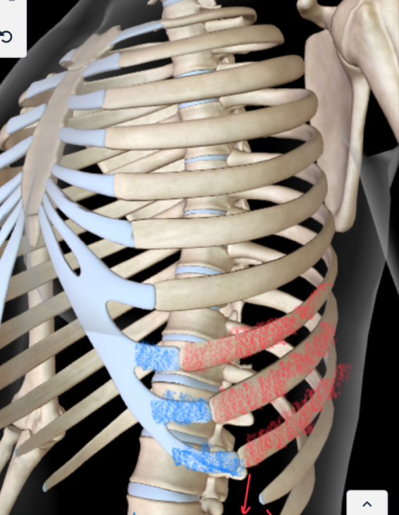
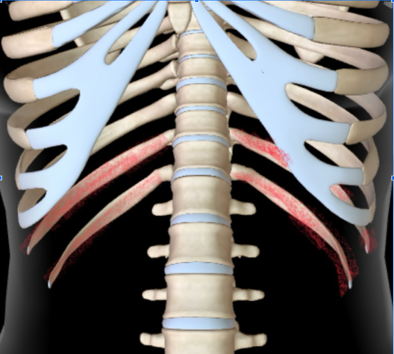
Floating Ribs (11-12)
These are ribs that DOESN’T HAVE COSTAL CARTILAGE
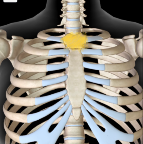
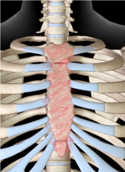
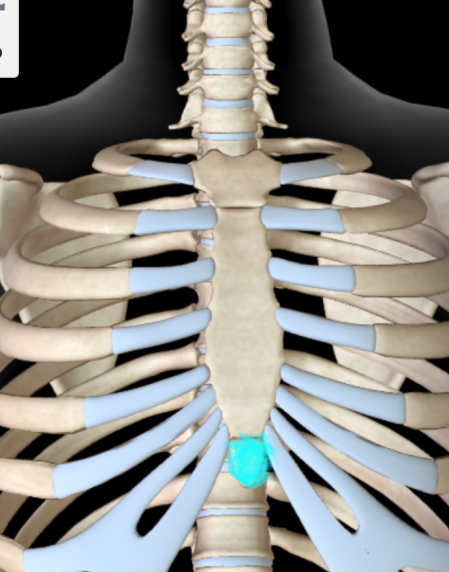
Sternum
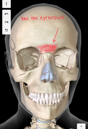
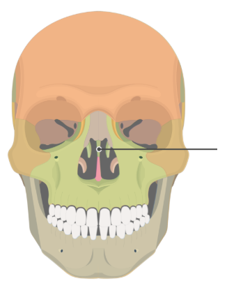
Glabella
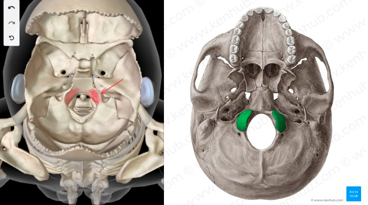
Occipital Condyle
Mastoid Process
BELOW THE EAR
There’s TWO SIDES (left and right)
External Auditory Meatus (L/R)
This is an OPENING IN THE EAR that LEADS TO INSIDE THE BODY
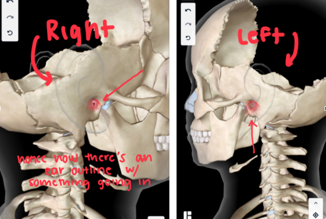
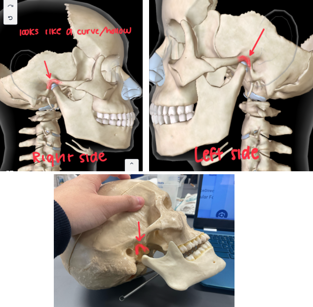
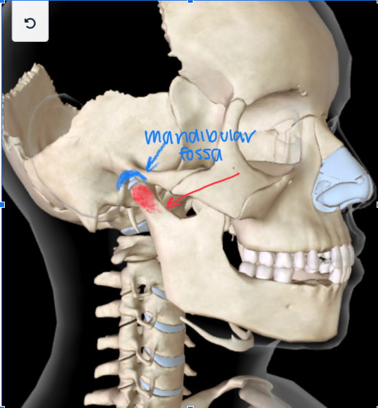
Mandibular Fossa
Cribriform plate
Part of the ETHMOID BONE
OLFACTORY FORAMINA SURROUNDS IT
Perpendicular Plate
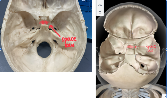
Sella Turcica
Part of the SPHENOID BONE
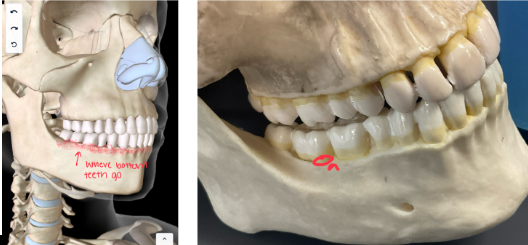
Alveolar Socket of Mandible
This HOLDS THE TOOTH ROOT
Angle of Mandible (mandibular angle)
JAW ANGLE (in both sides of the head)
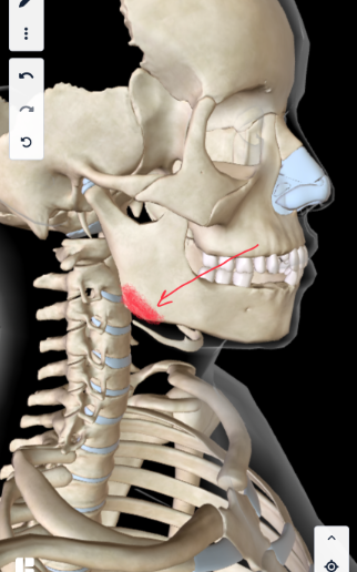
Ramus of Mandible
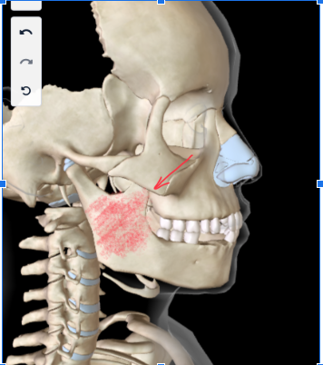
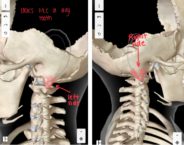
Mandibular Condyle
Alveolar Socket of the Maxilla
This HOLD THE TOOTH ROOT of the UPPER TEETH
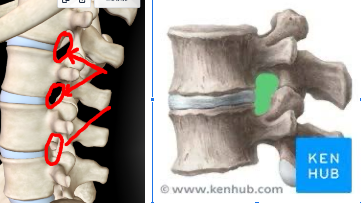
Intervertebral Foramen
HOLES BETWEEN EVERY VERTEBRAE
Vertebral Body
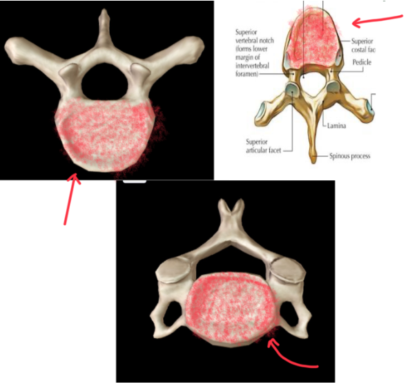
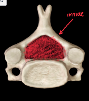
Vertebral Foramen
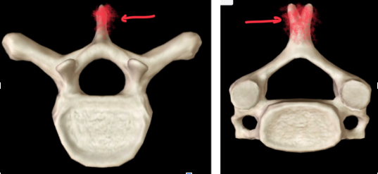
Spinous Process
Looks like MERMAID TAILS
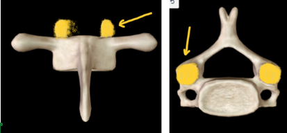
Superior Articular Facet
Inferior Articular Facet
This is BELOW the superior ________ _____
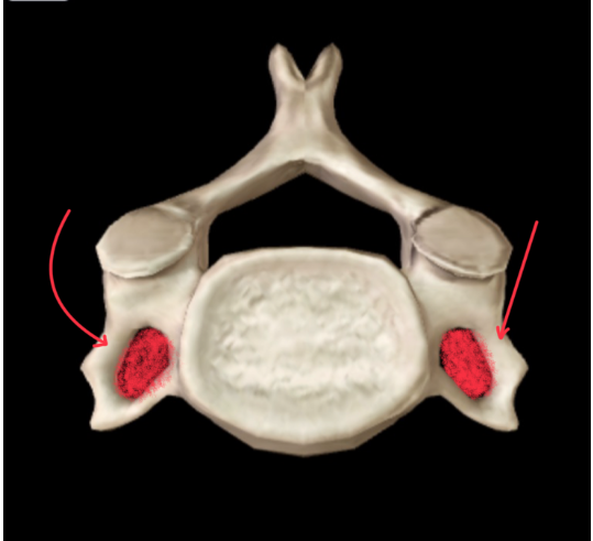
Transverse Process
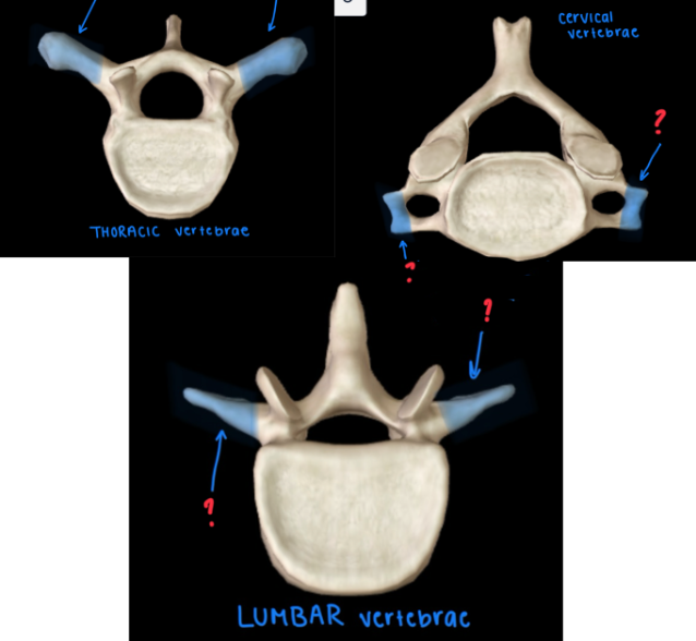
Transverse Foramen
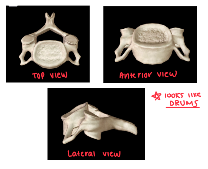
ONLY CERVICAL HAS THIS
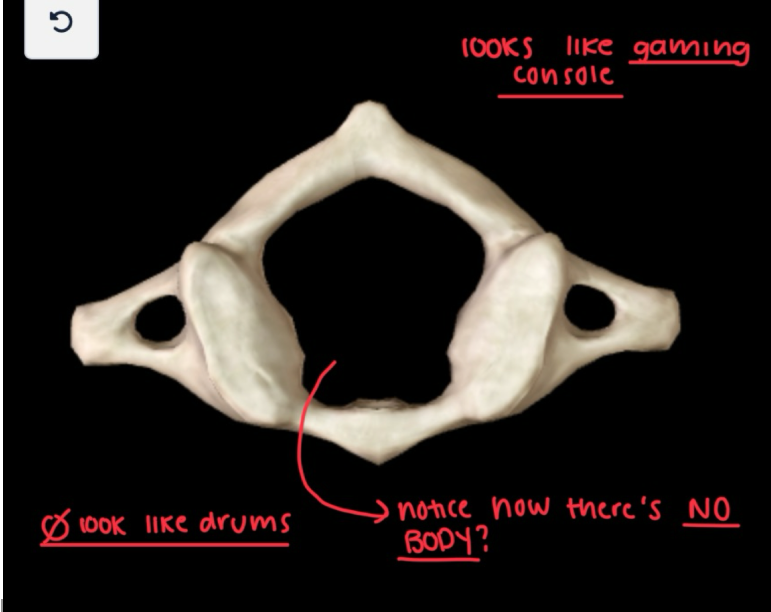
Atlas (C1)
Looks like a STEERING WHEEL/GAME CONSOLE AT TOP VIEW
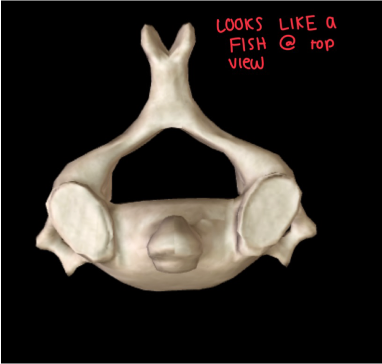
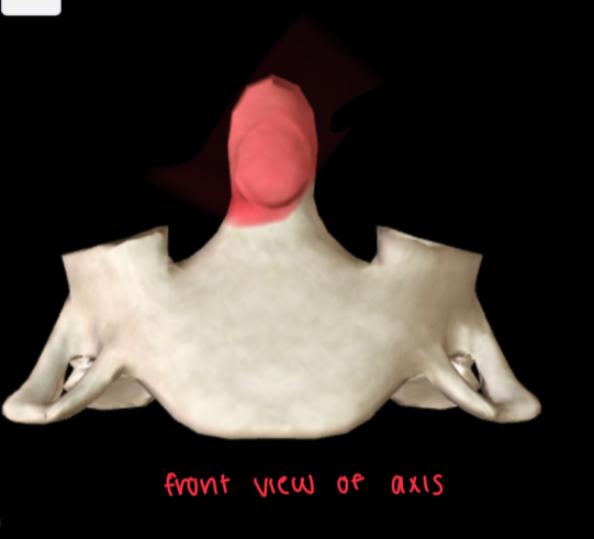
Axis (C2)
Looks like a PUFFERFISH from TOP VIEW
Cervical Dens
ONLY IN CERVICAL VERTEBRAE
Looks like a HEAD on anterior view
Body of Sternum
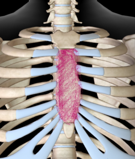
Xiphoid Process
Manubrium