nervous coordination flashcards
1/49
There's no tags or description
Looks like no tags are added yet.
Name | Mastery | Learn | Test | Matching | Spaced | Call with Kai |
|---|
No analytics yet
Send a link to your students to track their progress
50 Terms
What initial event occurs at the synaptic knob of the presynaptic neurone?
An action potential arrives at the synaptic knob of the presynaptic neurone.
What does the action potential stimulate in the presynaptic neurone?
The action potential stimulates voltage-gated calcium ion channels in the presynaptic neurone to open.
What happens after the calcium ion channels open?
Calcium ions diffuse into the synaptic knob. They are pumped out afterwards by active transport.
What is the effect of the influx of calcium ions into the synaptic knob?
The influx of calcium ions causes the synaptic vesicles to move to and fuse with the presynaptic membrane.
What neurotransmitter is released into the synaptic cleft, and what is this process called?
The vesicles release the neurotransmitter acetylcholine (ACh) into the synaptic cleft. This process is called exocytosis.
What happens to ACh after it diffuses across the synaptic cleft?
ACh diffuses across the synaptic cleft and binds to specific cholinergic receptors on the postsynaptic membrane.
What occurs on the postsynaptic membrane when ACh binds to its receptors?
The influx of sodium ions into the postsynaptic membrane causes depolarisation. An action potential on the postsynaptic membrane is generated if the threshold is reached.
How is ACh removed from the synaptic cleft, and why is this important?
ACh is removed from the synaptic cleft by the enzyme acetylcholinesterase (AChE) to prevent continuous response. The products are re-absorbed by the presynaptic neurone and used to make more ACh.
Describe the additional features of a myelinated motor neuron.
Schwann cells: wrap around axon many times.
Myelin sheath: made from myelin-rich membranes of
Schwann cells.Nodes of Ranvier: very short gaps between neighbouring Schwann cells where there is no myelin sheath.
Name 3 processes Schwann cells are involved in.
electrical insulation
phagocytosis
nerve regeneration
Explain why myelinated axons conduct impulses faster than unmyelinated axons
Saltatory conduction: Impulse 'jumps' from one node of Ranvier to another. Depolarisation cannot occur where myelin sheath acts as electrical insulator.
So impulse does not travel along whole axon length.
What is resting potential?
Potential difference (voltage) across neuron membrane when not stimulated (-50 to -90 mV, usually about -70 mV in humans).
How is resting potential established?
• The resting potential is maintained by a sodium-potassium pump, involving active transport and therefore ATP.
• The pump moves 2 Ktions in and 3 Nat ions out.
• This creates a electrochemical gradient and results in K+ diffusing out and Nat diffusing in, however because the membrane is more permeable to K+ more are moved out resulting in the -70mV.
Name the stages in generating an action potential.
Depolarisation
Repolarisation
Hyperpolarisation
Return to resting potential
What happens during depolarisation?
Stimulus→facilitated diffusion of Nations into cell down electrochemical gradient.
p.d. across membrane becomes more positive.
If membrane reaches threshold potential (-50mV), voltage-gated Nat channels open.
Significant influx of Nations reverses p.d. to
+40mV.
What happens during repolarisation?
Voltage-gated Na* channels close and voltage-gated K* channels open.
Facilitated diffusion of K* ions out of cell down their electrochemical gradient.
p.d. across membrane becomes more negative.
In the nerve pathway explain how, synapses ensure that the nerve impulses travel towards the muscle fibre
neurotransmitter is only made / stored in pre - synaptic neurone
Neuroreceptors only on the post synaptic membrane
Axon P was found to conduct impulses much faster than other axons in the nerve pathwaY. Describe and explain one feature of axon P that might cause this difference.
axon P is myelinated
So shows saltatory conduction / impulses jump nodes of Ranvier
Or
Axon p has a larger diamter
So less resistance to flow of ions
How does the inhibitory synapse work to inhibit an action potential
causes chloride ions to move inot the postsynaptic neurone and potassium ions to move out
The combined effect of negative ions moving in and positive ions moving out makes the membrane potential increase to -80mV, hyperpolarisation and therfore an action potential is highly inlikely
Why are inhibitory synapses needed
so that the synapse doesn’t respond to everything
So that your senses aren’t overwhelmed
So that your body can react to the most vital stimuli
What is the purpose of summation at a synapse?
Summation allows for the integration of multiple stimuli, enabling a finely tuned nervous response even from individually weak signals that might not independently reach the postsynaptic membrane's threshold for an action potential.
Explain the general concept of summation at a synapse.
Summation is the process by which a postsynaptic neuron integrates the effects of multiple excitatory and/or inhibitory neurotransmitter signals from one or more presynaptic neurons, determining whether an action potential is triggered.
Describe spatial summation.
Spatial summation occurs when multiple presynaptic neurons simultaneously release neurotransmitters onto a single postsynaptic neuron. The combined effect of these simultaneous inputs can collectively reach the threshold, triggering an action potential.
What happens in spatial summation if some presynaptic neurons release inhibitory neurotransmitters?
In spatial summation, if some presynaptic neurons release an inhibitory neurotransmitter, their hyperpolarizing effect can counteract the excitatory neurotransmitters, preventing the postsynaptic neuron from reaching the threshold and thus inhibiting an action potential.
Describe temporal summation.
Temporal summation occurs when a single presynaptic neuron fires multiple times in quick succession, releasing neurotransmitter repeatedly. This rapid accumulation of neurotransmitter in the synaptic cleft makes it more likely for the postsynaptic neuron to reach its threshold and generate an action potential.
How do agonist drugs affect synaptic transmission? Provide an example.
Agonist drugs mimic the action of natural neurotransmitters by having a similar molecular shape, allowing them to bind to and activate specific receptors. This binding leads to an increased activation of postsynaptic receptors. For example, nicotine acts as an agonist for acetylcholine at nicotinic cholinergic receptors in the brain.
How do antagonist drugs affect synaptic transmission? Provide an example.
Antagonist drugs block specific neurotransmitter receptors, preventing the natural neurotransmitter from binding and activating them. This reduces or eliminates the activation of these receptors. For instance, curare blocks acetylcholine receptors at neuromuscular junctions, leading to muscle paralysis.
How do drugs that inhibit enzyme breakdown of neurotransmitters affect synaptic transmission? Provide an example.
These drugs inhibit the activity of enzymes responsible for breaking down neurotransmitters within the synaptic cleft. By preventing enzymatic degradation, they prolong the presence and action of neurotransmitters on postsynaptic receptors. Nerve gases, for example, inhibit acetylcholinesterase, causing prolonged acetylcholine activity and loss of muscle control.
How do some drugs stimulate the release of neurotransmitters from the presynaptic neuron? Provide an example.
Some drugs increase the exocytosis of neurotransmitters from the presynaptic neuron into the synaptic cleft. This leads to a higher concentration of neurotransmitters available to bind with postsynaptic receptors, increasing receptor activation. Amphetamines are an example of drugs that stimulate neurotransmitter release.
How do some drugs inhibit the release of neurotransmitters from the presynaptic neuron?
Some drugs reduce or prevent the release of neurotransmitters from the presynaptic neuron into the synaptic cleft. This results in fewer neurotransmitters binding to postsynaptic receptors, thereby decreasing receptor activation and the likelihood of an action potential.
Describe and explain how the membrane potential changes during depolarisation (4 marks)
• A stimuli causes more sodium ion channels in the membrane to open so the membrane becomes more permeable to sodium ions
• As a result the sodium ions move into the axon through open channels via facillitated diffusion
• This results in an imbalance of positive ions, with more ions inside the axon than outside
• The resulting membrane potential is positive, reaching a peak of about +40mV
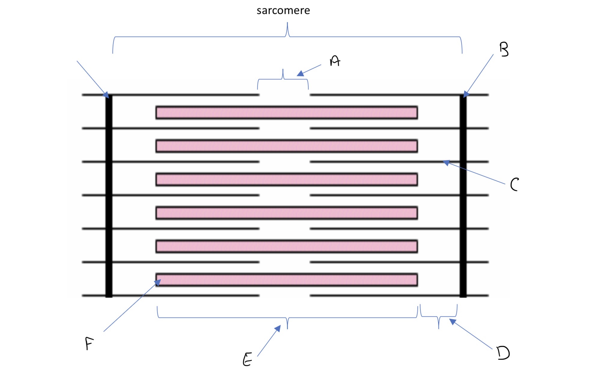
label the diagram
A H zone - the width of just the myosin
B - Z lines
C - actin
D - I band ( just the actin)
E - A band - total width of the myosin
F- myosin
State the steps in the sliding filament theory
when an action potential reaches a muscles it stimulates a response
Calcium ions enter and cause the protein tropomyosin that block binding sites for the myosin head of the actin to move and uncover the binding sites
Whilst ADP is attached to the myosin head, it can bind to the binding sites Whilst ADP on the actin to form a cross - bridge
The Angle created in this cross bridge creates tension and as a result the actin filament is pulled and slides along the myosin. In doing so the ADP molecules is released
An ATP molecule can then bind to the myosin head and causes it to change shape slightly and as a result it detaches form the actin
Within the sarcoplasm there is the enzyme ATPase which is activated by the calcium ions, to hydrolyse the ATP on the myosin head into ADP and releases enough energy for the myosin head to return to its original position
Which bands stay the same length and which bands change shape when a muscle contracts
the A band stays the same shape because the myosin isn’t moving, it’s pulling the actin together
The H zone decreases
The I band decreases in length
A lines get closer together
What are the two types of skeletal muscles
slow twitch
Fast twitch
What is the structure if slow twitch muscles
Contains a large store of myoglobin, a rich blood supply ( many capillaries) and many mitochondria
What is the structure of the fast twitch muscle fibres
Thicker and more myosin filaments, a large store of glycogen, a store of phosphocreatine to help make ATP from ADP and a high concentration of enzymes involved in anaerobic respiration.
Where are slow twitch muscle fibres stored
Calf muscles
Where are fast twitch muscle fibbers stored
Biceps
What are the general properties of slow twitch muscle fibres
Contract slower, can respire aerobically for longer periods due to the rich blood supply and myoglobin oxygen store. These muscles are adapted for endurance work, like marathons
What are the general properties of fast twitch muscle fibres
Caontract faster to provide short bursts of powerful contraction. These are adapted for intense exercise, such as sprinting or weight - lifting
What is the function of myoglobin
Stores lots of oxygen ( found In slow twitch fibres)
Why is the ATP binding site needed in muscle contraction
The site binds to ATP, the myosin head also acts as an enzyme, ATPase, which hydrolyses this ATP to ADP and an inorganic phosphate to release energy for the ‘recovery stroke’
What is the Actin - myosin binding site blocked by
Tropomyosin
Neuro muscular junction steps
When an impulse travels along the axon of a motor neurone arrives at the presynaptic membrane, the action potential causes calcium ions to diffuse into the neurone
• This stimulates vesicles containing the neurotransmitter acetylcholine (ACh) to fuse with the presynaptic membrane
• The ACh that is released diffuses across the neuromuscular junction and binds to receptor proteins on the sarcolemma (surface membrane of the muscle fibre cell)
• This stimulates ion channels in the sarcolemma to open, allowing sodium ions to diffuse in
• Calcium ions diffuse out of the sarcoplasmic reticulum (SR) and into the sarcoplasm surrounding the myofibrils
• This depolarises the sarcolemma, generating an action potential that passes down the T-tubules towards the centre of the muscle fibre
• These action potentials cause voltage-gated calcium ion channel proteins in the membranes of the sarcoplasmic reticulum (which lie very close to the T-tubules) to open
• Calcium ions bind to troponin molecules, stimulating them to change shape. This causes the troponin and tropomyosin proteins to change position on the thin (actin) filaments
• The myosin-binding sites are exposed to the actin molecules
• The process of muscle contraction (known as the sliding filament model) can now begin. There are multiple neuromuscular junctions spread. Across several muscle fibres within the muscle
What effect does troponin chaining shape have on tropomyosin
Pulls tropomyosin out of the actin - myosin binding site, because troponin and tropomyosin are attached to each other
What moves the myosin head
The enrgy released from the hydrolysis of ATP
Describe how the release of acetylcholine into a neuromuscular junction causes the cell membrane of a muscle fibre to depolarise. (3)
Movement by diffusion
Binding to receptors on (post synaptic membrane)
Cheating sodium ion channels to open
Use your knowledge of the processes occurring at a neuromuscular junction to explain each of the following
• toxin will bind to / compete for the acetyl choline receptors; acetycholien can not depolarise the membrane
The insecticide DFP combines with the active site of the enzyme acetylcholinesterase. The muscles stay contracted until the insecticide is lost from the neuromuscular junction. (2)
acetylcholinerase is unable to breakdown acetylcholine; so acetylcholine still available to depolarise the membrane / generate action potential in the membrane