Biosci 221 Lab 3 Quiz
5.0(1)
5.0(1)
Card Sorting
1/46
Earn XP
Description and Tags
staining, stock bacteria, and population counts
Study Analytics
Name | Mastery | Learn | Test | Matching | Spaced | Call with Kai |
|---|
No study sessions yet.
47 Terms
1
New cards
What is an acidic stain and how is it helpful for viewing the size and morphology of the bacteria?
Acidic stains are when we use acidic agents, such as Nigrosin, to stain bacteria and view them in their original shape and size due to the lack of heat fixation. The negative charge of the stain repels the negative charge of the bacteria’s cell, making it appear clear.
2
New cards
What is a basic stain and how is it useful in staining?
A basic stain is when we use a positively-charged dye to stain the negatively-charged bacteria and view them easily under the brightfield lens.
3
New cards
What are the average sizes of the bacteria you are working with?
0\.5um-15um
4
New cards
How do you measure bacteria?
Using the ocular micrometer
5
New cards
How is oil immersion used?
A drop of immersion oil is used ONLY on the 100x objective lens to view microorganisms more clearly since the oil increases the resolving power.
6
New cards
How do we view differential stains?
By always using brightfield on a 100X objective lens with oil immersion.
7
New cards
What is a differential stain?
A differential stain is a staining process where we use several staining agents to differentiate bacteria by their shape or cell wall structures.
8
New cards
When do we use a differential stain and what does that tell us about the organisms that are positive or negative?
We might use a differential stain in a mixed sample of organisms to differentiate cell types. Bacterial cells with gram-positive cell walls are stained purple with the dye crystal violet, while bacterial cells with gram-negative cell walls are counterstained pink with safranin dye.
9
New cards
Which differential staining methods do we use?
Simple staining
Negative staining
Gram staining
Endospore staining
Acid-fast staining
Negative staining
Gram staining
Endospore staining
Acid-fast staining
10
New cards
What are the advantages of negative staining?
A negative stain is very good for observing cell morphology, arrangement, and size because the slide is NOT heat-fixed.
11
New cards
What is the purpose of heat fixation?
To fix the bacteria onto the slide so it doesn’t wash away with the staining agents and DI water.
12
New cards
What are the main problems when we make a smear preparation that is too thick or uneven?
We won’t be able to view bacteria clearly. A thick/uneven smear cannot be properly decolorized (so no clear shapes) and might wash off the slide despite fixation.
13
New cards
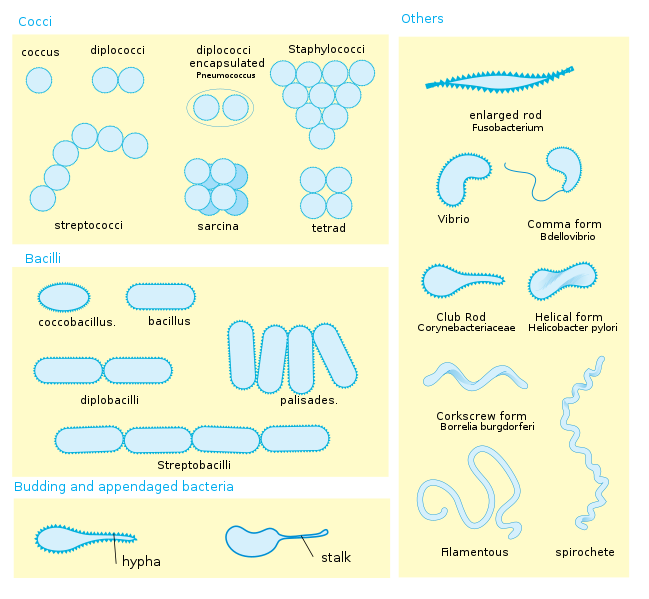
What are arrangements? (diplo-, strepto-, tetrads, sarcinae, staphylococci, palisade)
Arrangements are how bacteria are shaped and arranged in groups.
* Diplo- means they occur as a pair
* Strepto- means they are in a chain
* Tetrads are in groups of four
* Sarcinae are in groups of eight (3D like)
* Palisades are when bacilli are side-by-side (like a picket fence)
* Staphylococci are when cocci are in grape-like clusters.
* Diplo- means they occur as a pair
* Strepto- means they are in a chain
* Tetrads are in groups of four
* Sarcinae are in groups of eight (3D like)
* Palisades are when bacilli are side-by-side (like a picket fence)
* Staphylococci are when cocci are in grape-like clusters.
14
New cards
Can there be various arrangements for the same bacterium?
Yes, always
15
New cards
What conditions affect how you see arrangements?
how thick your slide is and how well it has been spread around
16
New cards
What are the stock bacteria we have that may form tetrads?
*Micrococcus luteus*
*Sporosarcina ureae*
*Kocuria rosea*
*Staphylococcus saprophyticus*
*Sporosarcina ureae*
*Kocuria rosea*
*Staphylococcus saprophyticus*
17
New cards
What are the stock bacteria that may form sarcinae?
*Sarcina aurantiaca*
*Sporosarcina ureae*
*Kocuria rosea*
*Sporosarcina ureae*
*Kocuria rosea*
18
New cards
What are the stock bacteria that may form staphylococcal arrangements?
*Staphylococcus epidermis*
*Staphylococcus saprophyticus*
*Staphylococcus saprophyticus*
19
New cards
What are the stock bacteria that may form palisade arrangements?
*Corynebacterium pseudodiphtheriticum*
20
New cards
What are the stock bacteria that may form diplococci?
*Sporosarcina ureae*
*Lactococcus lactis*
*Kocuria rosea*
*Moraxella catarrhalis*
*Lactococcus lactis*
*Kocuria rosea*
*Moraxella catarrhalis*
21
New cards
What are the stock bacteria that may form streptobacilli?
*Bacillus cereus*
*Bacillus subtilis*
*Klebsiella aerogenes*
*Bacillus subtilis*
*Klebsiella aerogenes*
22
New cards
Why is a counterstain always lighter than a primary stain?
Primary stain is usually darker and attaches to the cell walls first. Darker colors usually obscure lighter colors.
23
New cards
How do you use the following objective lens when we are observing bacteria: 10X, 20X, 40X, and 100X?
Find and focus on bacteria using the coarse and fine focus on the 10X lens. Continue to focus on the 20X and 40X lenses, then use immersion oil to fully focus on the 100X lens.
24
New cards
Name 2 things that may go wrong when you are making a negative stain
1. Putting too much dye onto the slide results in it not being able to thin out when smearing
2. Smearing it incorrectly that there is no difference between the thick and thin side
25
New cards
What is the dye used in negative staining?
Nigrosin
26
New cards
What are the stains and the procedures of Gram staining?
1. Begin with a heat-fixed emulsion
2. Cover the smear with crystal violet stain for 1 minute
3. Grasp the slide with a slider holder and gently rinse the slide with DI water
4. Cover the smear with iodine stain for 1 minute
5. Grasp the slide with a slider holder and gently rinse the slide with DI water
6. Decolorize with acetone/alcohol by allowing it to trickle down the slide until the runoff is clear. Gently rinse the slide with DI water immediately
7. Counterstain with safranin stain for 2 minutes. Rinse with DI water
8. Gently blot dry in a tablet of bibulous paper.
27
New cards
Name 4 things that may go wrong when you are Gram staining
1. If the smear is too thick, proper decolorizing will not be possible.
2. If the smear is overheated during heat fixing, the cell walls will rupture.
3. Older cultures contain more ruptured and dead cells; they may stain gram negative even if the bacteria are gram positive.
4. Excess water left on the slide will dilute reagents
28
New cards
What could have gone wrong if your Gram-positive rod looks like a Gram-negative rod?
When cells die off (too old), they don’t pick up the stain as well
29
New cards
What could have gone wrong if your Gram-negative cocci looks like a Gram-positive cocci?
Slide too thick, difficulty in decolorizing, age of the culture, etc.
30
New cards
What bacteria are endospore positive?
*Sporosarcina ureae*
*Bacillus cereus*
*Bacillus subtilis*
*Bacillus cereus*
*Bacillus subtilis*
31
New cards
What is an endospore?
A structure for the preservation of the DNA while conditions are poor
32
New cards
What does the heat do in the endospore staining process?
The heating process makes the spore coat more permeable to the stain
33
New cards
When are endospores formed?
When stressed
34
New cards
If you don't find endospores does that mean it's negative?
Not necessarily; they may not be stressed or perhaps a procedure mistake
35
New cards
Do you have to do an endospore stain to see endospores?
No, an endospore can be seen in simple and Gram stains, as a clear or empty space in a vegetative cell.
36
New cards
What are the stains used in endospore staining?
Malachite Green
Safranin
Safranin
37
New cards
What is the endospore staining procedure?
1. Begin with a heat-fixed emulsion
2. Cover the smear with a strip of bibulous paper. Apply malachite green stain. Steam for 5-10 mins. Keep the paper moist with the stain the entire time. Make sure you refill the beaker of water.
3. Grasp the slide with a slide holder. Remove the paper and dispose of it properly. Gently rinse the slide with DI water
4. Counterstain with safranin stain for 1 min. Rinse with DI water.
5. Gently blot dry in a tablet of bibulous paper.
38
New cards
What is the primary and secondary stain in endospore staining? Is there a mordant?
Primary = Malachite Green
Secondary = Safranin
The heating process acts as the mordant in this stain procedure
Secondary = Safranin
The heating process acts as the mordant in this stain procedure
39
New cards
Are there important endospore-forming bacteria?
*Clostridium difficile* causes the disease C-diff and because of its ability to produce endospores it is not readily killed by disinfectants, and can easily be transmitted in healthcare facilities
40
New cards
What is acid-fast staining?
In the Acid-Fast stain, a very concentrated stain solution of kinyoun carbolfuchsin (hot pink) is used in order to penetrate and stain the cell walls
41
New cards
Why is AF staining important?
This stain is a valuable diagnostic tool for finding tuberculosis and nocardial diseases in patient specimens
42
New cards
What AF orgs do we have?
One; *Mycobacterium smegmatis*
43
New cards
What is the AF procedure?
1. Begin with a heat-fixed emulsion
2. Apply kinyoun carbolfuchsin stain for 5-10 mins
3. Grasp the slide with a slide holder. Gently rinse the slide with DI water.
4. Continue holding the slide with a slide holder. Decolorize with acid-alcohol until the runoff is clear. Gently rinse the slide with DI water
5. Counterstain with methylene blue stain for 1 min. Rinse with DI water
6. Gently blot dry in a tablet of bibulous paper
44
New cards
What is the most important step in AF?
The decolorization step
45
New cards
What do a positive and negative AF look like?
The bacteria will be purple if positive (i.e. *Mycobacterium smegmatis*) and blue if negative
46
New cards
What can go wrong with the AF procedure?
the organism doesn't retain the stain or it fails to stain properly
47
New cards
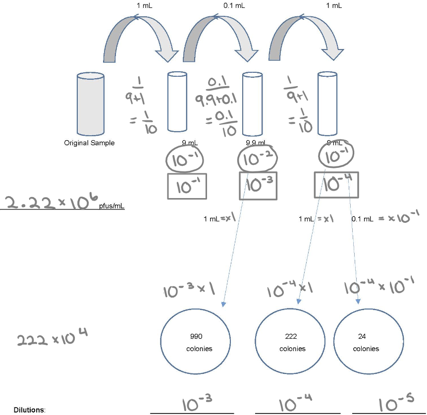
Know how to do dilution
1. Individual dilution
2. Total dilution
3. How much plating: 1.0 ml (x1) or 0.1 ml (x 10^-1)
4. (30-300) x DF