Biological Psychology
1/92
There's no tags or description
Looks like no tags are added yet.
Name | Mastery | Learn | Test | Matching | Spaced | Call with Kai |
|---|
No study sessions yet.
93 Terms
Nervous system definition?
The nervous system includes the brain, spinal cord, peripheral nerves and sensory organs such as the eye and is the primary communication system in the body.
What is the CNS?
The brain and spinal cord - it is the origin of complex commands. Brain is centre of conscious thought, and the cerebral cortex is the centre of higher thinking more developed in humans. Spinal cord is responsible for reflexes and the brain stem controls involuntary functions.
What is the PNS?
All nerves outside the CNS make it up - transmits info from outside world to CNS and from CNS to effectors.
What is PNS divided into?
What do they do?
- Somatic transmits info from sensory receptors in the sense organs to the CNS, and receives info from the CNS to instruct muscle movement.
-Autonomic transmits info to and from internal organs and is involuntary - involved in homeostasis.
What is the autonomic nervous system divided into? What do they do?
-Sympathetic is responsible for activating fight or flight (increasing heart rate etc) to prepare for stressful situations. Parasympathetic returns body to resting state by reversing any functions that were changed (decreasing heart rate etc)
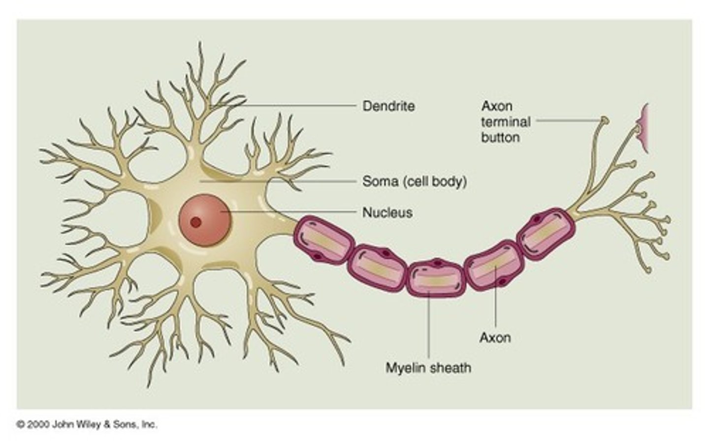
What is a neuron?
a nerve cell; the basic building block of the nervous system that process and transmit messages by chemical and electrical means.
Draw a motor neuron:
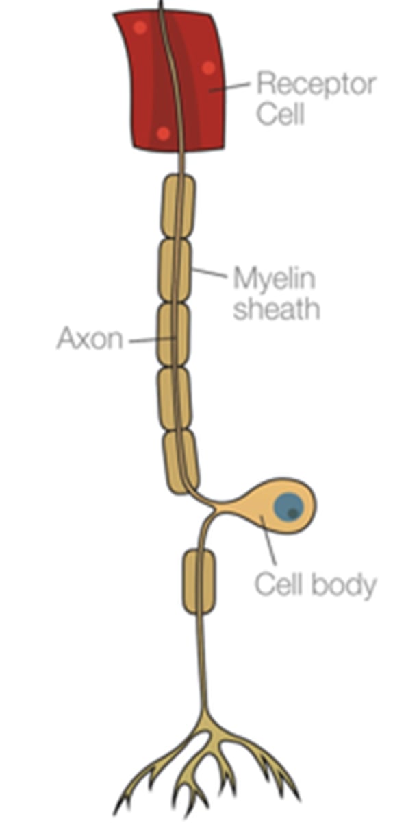
Draw a sensory neuron:
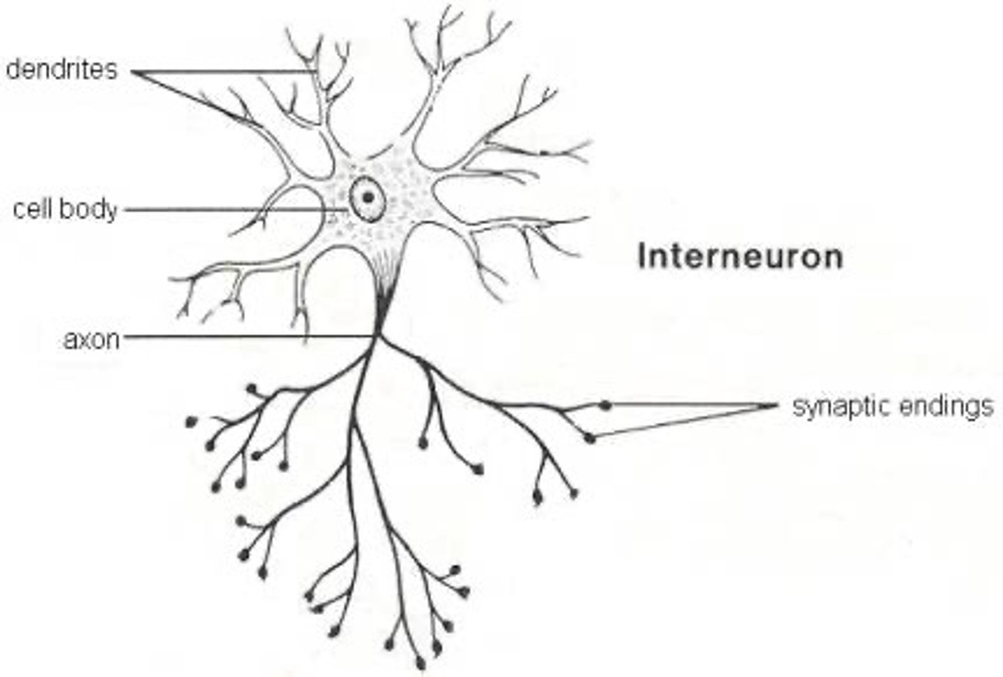
Draw a relay neuron:
Function of the sensory neuron?
picks up stimuli (receives information) from the internal or external environment, converts it to a nerve impulse, and sends the impulse to the brain or spinal cord, are unipolar so only transmit messages. Have long dendrites and short axons.
Function of the relay neuron?
connects sensory and motor neurons, or connect to other relay neurons, found in the CNS and have short dendrites and axons. They are multipolar as they send and receive many messages from different places.
Function of motor neurons?
Connect the CNS to effectors such as muscles and glands, have short dendrites and long axons.
What are the functions of the axon, myelin sheath , and dendrites?
Axon: carry impulses from cell body down the length of the neuron, the myelin sheath is an insulating layer protecting the axon and speeds up the impulse (has gaps which speeds it up), dendrites carry impulses from neighbouring neurons to cell body.
How is action potential created?
When in resting state the neuron is negatively charged compared to the outside. When activated by a stimulus the inside of the cell becomes positively charged for a spilt second which creates an action potential which creates an electrical impulse.
Draw a diagram of a synaptic cleft with the presynaptic neuron and post synaptic neuron:
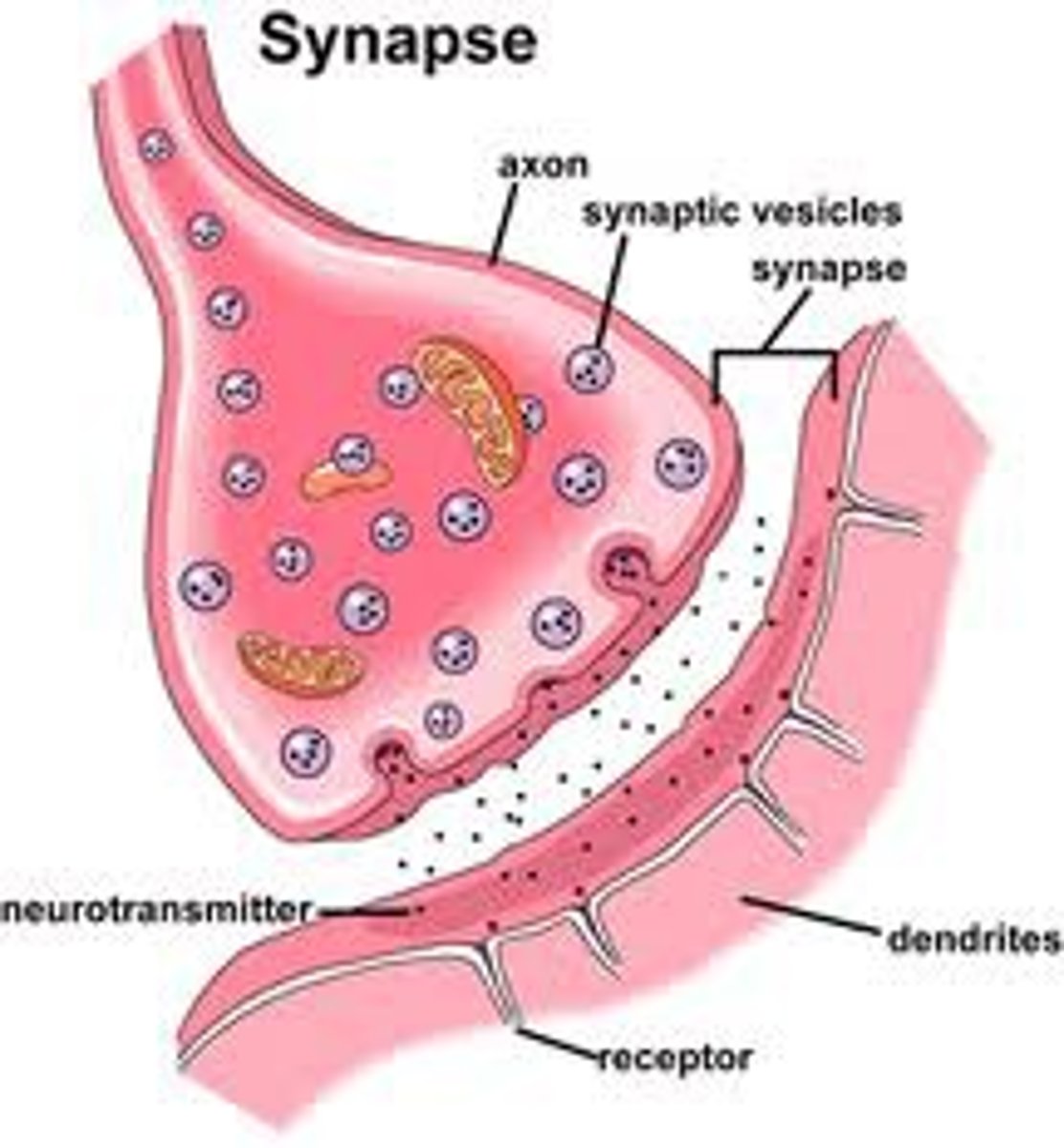
What is synaptic transmission?
- Process by which nerve impulse passes across synaptic gap from presynaptic neuron to postsynaptic neuron - neurons communicate via chemical messages.
How does the electrical impulse begin synaptic transmission?
Triggers the release of neurotransmitters from synaptic vesicles - they release contents via exocytosis.
How are neurotransmitters released across the cleft and detected, and how does the impulse get passed along?
They are released into the synaptic fluid, where the adjacent neuron must quickly take up the neurotransmitter - does this by binding to specialised surface receptors (post synaptic receptors). They are converted to an electrical impulse to travel down. Each neurotransmitter has a specific shape that binds to a specific receptor, and there are several dozens of types.
What is the process of re-uptake?
Transmission is terminated at synapse via re-uptake, as neurotransmitters are taken up via transport proteins back into the pre-synaptic neuron, where they are stored and made available for later release. How quickly the neurotransmitter is taken up determines how prolonged the effect of the neurotransmitter is - quicker reuptake leads to a shorter effect. Some drugs alter this like SSRIs.
What is excitation?
Where an excitatory neurotransmitter increases likelihood of a new action potential forming in the post synaptic cell. When detected by receptors electrical charge becomes more positive via Na+ ions entering the cell (depolarisation)
What is inhibition?
Where neurotransmitters decrease the likelihood of a new action potential forming in the post synaptic cell. When detected they hyperpolarise the neuron, eg making it more negative by K+ ions leaving the cell.
What is summation?
Combined effect of all neurotransmitters on the post synaptic neuron, and if the threshold is reached a new action potential forms
What is the endocrine system?
One of the body’s major communication systems that instructs glands to release hormones directly into the bloodstream (acts more slowly), and hormones are carried to target organs.
What are hormones?
Chemical substances circulating in the blood and only affect target organs secreted by glands
What is the pituitary gland function?
Called the master gland as it controls the release of hormones from all the other endocrine glands in the body. It also releases hormones like oxytocin and ACTH (stimulates adrenal gland).
What is the function of the thyroid gland?
Produces thyroxine which regulates metabolism and also affects heart cells to increase heart rate.
What is the overall function of the adrenal gland?
Involved in fight or flight responses
What are the functions of the adrenal cortex and medulla?
Medulla releases adrenaline for fight or flight which increases heart rate etc.
Cortex releases cortisol which contributes to long term stress.
Functions of the testes and ovaries?
Release sexual hormones to develop secondary sexual characteristics. Ovaries release oestrogen and progesterone, testes release testosterone.
Hypothalamus function?
Region of the brain controlling homeostasis, and links the nervous system with the endocrine system.
Pancreas function?
Maintains blood sugar levels by releasing insulin and glucagon
Where is the fight or flight response generated from?
The ANS, specifically the sympathetic branch, as the normal resting state (parasympathetic) is changed to arousal.
Describe the process of the fight or flight response:
Situation is appraised as stressful
Hypothalamus is alerted, which recognises the stress is acute so activates the Sympathetic Adrenomedullary pathway (SAM)
This activates the sympathetic branch of the ANS, which stimulates the adrenal medulla, part of the adrenal glands
This secretes adrenaline and noradrenaline into the blood, which prepares the body for fight or flight and brings about bodily changes like increased heart rate, decreased salivation and digestion, dilated pupils etc.
What happens after the threat has passed?
The parasympathetic branch returns the body to its resting state - its actions are antagonistic to the sympathetic system and is known as the rest and digest response.
What did Gall begin to develop in the 1790s?
The principles of phrenology - he thought that bumps on the skull could be linked to personality. This lacked scientific rigor, but it contribute to neurology.
What is localisation of function?
The theory that different areas of the brain are responsible for different behaviours, processes or activities.
What is laterisation?
The idea that some functions are only found in either the left or right hemisphere. As a general rule, activity on the left side of the body is controlled by the right hemisphere and vice versa.
What is the outer layer of the brain called?
Cerebral cortex - 3mm thick and separates us from other species as it is more developed. Described as grey matter due to cell body location.
What hemisphere usually controls language?
The left in 97% of right handed people, but in left handed people 19% have it on the right.
What did Broca discover in the 1880s?
The first language area in the left hemisphere to be discovered, called Broca’s area. He had a patient who could understand speech but could only say ‘Tan’, and after his death the patient was shown to have a lesion in an are of the brain, so this area was theorised to be responsible for speech production. Broca’s aphasia is where speech production is lost but comprehension is intact.
What did Wernicke discover?
The second language area, called Wernicke’s area. He was studying the opposite to Broca - patients could speak well but not understand speech of others. After an autopsy he found damage to an area on the left at the top of the temporal lobe. The loss of comprehension but intact speech is Wernicke’s aphasia.
What is the holistic theory of the brain?
The idea that all parts were involved in processing of thought and action.
What are the 3 main parts of the brain?
The cerebellum - comprised of small lobes and involved in motor movement coordination and basic learning/ memory. Brain stem - controls automatic actions like breathing and heart rate, links brain to spinal cord. Cerebrum has two hemispheres, left and right.
Label a diagram of the brain:
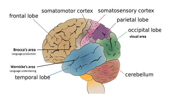
Where is the motor area and what is its function?
Back of the frontal lobe, and controls voluntary movement in the opposite side of the body
Where is the somatosensory area, and what is its function?
Front of both parietal lobes separated by a valley called the central sulcus. It is where sensory info from the skin is represented. Amount of somatosensory area devoted to a body part denotes its sensitivity.
where is the visual area and what is its function?
In the occipital lobe at the back of the brain, and the right visual field goes to left hemisphere.
Where is the auditory area, what is the function?
Temporal lobes, analyses speech based info. Damage to a part like Wernicke’s area can impact speech comprehension.
Function of the four lobes?
Frontal: motor behaviour, expressive language,higher level cognitive processes, orientation to person, place, and time.
Parietal: somatosensory cortex
Occipital: visual cortex - interpretation of visual information
Temporal: auditory cortex, receptive language, memory, emotion
What does the limbic system consist of?
Hippocampus - memory, transfer from short to long term.
Hypothalamus - controls the autonomic nervous system, homeostasis, emotions to physical responses.
Amygdala - mediates aggressive and defensive behaviour
What evidence of localisation do brain scans provide?
Petersen et al used scans to show how Wernicke's area was used during a listening task, and Broca's area during a reading task. A study of long term memory by Tulving et al revealed that semantic and episodic memories reside in different parts of the prefrontal cortex.
What evidence of localisation does neurosurgical evidence provide?
The first early attempts of surgically removing parts of the brain was the lobotomy- brutal and imprecise method of severing connections in the frontal lobe. It is still used very sparingly today in extreme OCD and depression cases. A study conducted by Dougherty et al looked at people who had an operation for OCD, and after the operation over 40% either had a partial or successful response, suggesting symptoms for mental disorders are localised.
How did the case study of Phineas Gage show localisation?
While working on the a railroad, Gage dropped his tamping iron on the rock, causing the explosive to ignite and the pole went through his left cheek, behind his left eye and took out some of his brain (frontal lobe). He survived but his personality changed, and became rude and quick tempered. Shoes how the frontal lobe may be responsible for regulating mood
How did Lashleys work provide against localisation of higher cognitive processes?
Lashley's work suggests that higher cognitive processes are not localised, but are distributed in a more holistic way. Lashley removed parts of the cortex in rats in a maze, and no area was proven to be more important than any other area in terms of ability to learn the maze. Learning appeared to need every part of the cortex, and suggests that learning is too complex to be localised.
How is plasticity an argument against localisation?
When the brain gets damaged the rest of the brain appears able to reorganise itself in an attempt to recover the lost function. This does not happen every time, but does suggest other parts are able to perform the same function, eg in strokes.
What is neuroplasticity?
Describes the brains tendency to change and adapt as a result of experience and new learning. Up to the 1960s it was thought to only do this in infancy, however it does this all the time although it is most prevalent in babies.
What is an example to show the neuroplasticity of a baby's brain?
Sometimes a baby has severe damage to one hemisphere, and if this hemisphere is removed soon after birth, as an adult they will experience few or no cognitive impairment, which shows how plasticity can allow for functions located in one hemisphere to be moved to the other.
What is synaptic pruning?
Where synaptic connections not used regularly will die
What research did Maguire et al do into plasticity?
Studied the brains of London taxi drivers and found significantly more grey matter in the posterior hippocampus than in a control group. London cabbies have to take a test called ‘the Knowledge’ - requires recall of city streets and possible routes. Longer they had been in the job, the more significant the change was, shows that learning experiences alter the brain structure.
What were the findings of Draganski et al?
They imaged the brains of medical students three months before and after their final exams. Learning induced changes were seen in the posterior hippocampus and parietal cortex as a result of the exam.
What is functional recovery?
A form of plasticity. Following damage through trauma, the brain’s ability to redistribute or transfer functions usually performed by a damaged area to another undamaged area. Healthy areas take over functions of the damaged areas.
What do neuroscientists suggest about the speed of functional recovery after trauma?
The process can occur quickly after the injury (spontaneous recovery) then slow down in the weeks and months after, and the individual may require rehabilitative treatment.
What happens to the brain during recovery?
It is able to organise and rewire itself by forming new synaptic connections close to the area of damage. Neural pathways are unmasked to enable functioning. Axonal sprouting occurs - growth of new nerve endings which connect with other undamaged nerve cells to form new neuronal pathways. Reformation of blood vessels also occurs. Homologous areas on the other side of the brain are also recruited.
Does neuroplasticity decrease with age?
Yes - reorganisation is more extensive in younger brains as it is constantly adapting to new experiences. However it is still present - in a study on golf training, after 40 hours there were changes to the motor cortex in the novice golfers compared to a control.
How has understanding of neural plasticity contributed to neurorehabilitation?
Following injury, recovery can slow down, meaning therapies must be employed, that include movement and electrical stimulation to counter deficits in cog function. Understanding of the brain’s processes help provide optimal treatment after injury to aid the process.
How does neuroplasticity sometimes have negative consequences?
Prolonged drug use has been shown to result in poorer cog functioning, and increased dementia risk in later life. 60-80% of amputees experience phantom limb syndrome - they experience sensations of the limb that are usually unpleasant. Thought that this is due to cortical reorganisation in the somatosensory area.
What support for neuroplasticity do animal studies provide?
Early evidence for plasticity was derived from animal studies, for example Hubel and Wiesel sewed one eye of a kitten shut and analysed cortical responses. The visual cortex associated with the shut eye was not idle, but continued to process info from the open eye.
What is cognitive reserve?
The concept that people who are more educated may have a better ability to recover after a brain injury - this was discovered by Schneider et al. It was found that 2/5 of patients who achieved disability free recovery had spent more than 16 years in education, compared to 1/10 patients who had less than 12 years in education.
What is the corpus callosum and its role in epilepsy?
A band of nerve fibres located deep in the brain that connects the two hemispheres, and contributes to the spread of seizures from one hemisphere to another.
How does severing the corpus callosum help epilepsy?
It prevents the spread of seizures from one side to another, and makes the seizures less severe. Split brain surgery is used as a last resort if medications have not worked.
What were the aims of Sperry’s research?
He wanted to investigate hemisphere function in split brain patients using a quasi experiment (naturally occuring IV). All 11 patients did not have the main communication line between the two hemispheres, so Sperry could see the extent to which certain hemispheres were specialised for certain functions.
What was the general procedure for the split brain research?
An image or word could be projected to a patients right visual field (processed by the left) or vice versa. In a ‘normal’ brain the corpus callosum would immediately share the info across the two hemispheres to give a complete picture, however in the split brain patients this would not happen.
What were the findings from putting an image in the patients right/ left visual field?
Right: Patient could easily describe what was seen, however on the left they could not and reported nothing was there. This is because language is usually located on the left hemisphere, so in the left visual field the image was processed on the right where there are no language centres.
What were the findings when patients were asked to identify an object presented in the left visual field by touch?
They were able to select a matching object when presented with a choice using their left hand. The patient was not able to verbally identify it or why they had chosen it, but the right hemisphere was able to understand the object and pick the correct one.
What would happen when two words were presented simultaneously on either side of the visual field (eg key on the left and ring on the right?
They would write key with their left hand and say the word ring. The right hemisphere is usually superior in drawing tasks.
How was it shown that the right hemisphere was dominant in face recognition?
When asked to match a face from a series of other faces, the picture processed by the right hemisphere would be selected, while the left hemisphere would be ignored. When a face made up of 2 halves of a face was presented - one half to each hemisphere - the left hemisphere dominated in terms of verbal description while the right dominated in terms of selecting a matching picture.
How did Sperry's brain study demonstrate lateralised brain functions?
It has shown that the left hemisphere is more geared towards analytic and verbal tasks (analyser) but the right is more adept at performing spatial tasks and music (synthesiser)
What are some strengths of Sperry's method?
Used highly specialised and standardised procedures, and the process of presenting the image to one visual field at a time successfully could demonstrate hemispheric lateralisation. This allowed him to vary aspects of the procedure so only one hemisphere was receiving info at a time, so the procedure was well controlled.
What is an issue with generalisation in Sperry's procedure?
There were only 11 patients who took part in all variations of the procedure, and they all had a history of seizures. This may have caused unique changes in the brain anyway that influenced the findings. Some of the patients also experienced more disconnection than others. The control group also did not have epilepsy which may have been inappropriate.
What is one limitation of Sperry’s work in terms of overstating differences in functions of hemipsheres?
It may have contributed to the overemphasis and oversimplification of functional distinction between the left and right hemispheres. Neuroscientists would argue the actual difference is much less clear cut than, for example verbal left and non verbal right, because there is constant communication between the 2 hemispheres while performing tasks. So his work could have led to some damaging misconceptions.
What debate did Sperry’s work prompt?
Some theorists have later argued that there are 2 brains living inside us as they are so functionally different that they could be a form of duality in the brain. However, others say that the 2 sides are highly integrated and both involved in many tasks.
How does fMRI work?
Detects changes in blood oxygenation levels and flow that occur as a result of neural activity in specific parts of the brain. When a brain area is more active it consumes more oxygen , so blood flow is directed to this area of the brain. fMRI produces 3D images that are involved in a particular mental process, and responses to simple stimuli can be looked at.
Strengths of fMRIs?
It is non invasive and does not rely on radiation, and risk free. It also has a higher spatial resolution than some other techniques , which provides a clear picture of how brain activity is localised.
Weaknesses of fMRIs?
Very expensive, and also has a poor temporal resolution - can be a lag time of up to 5 seconds of the image being formed and the initial neuron firing (due to blood flow being slower), this can make it hard to see exactly what kind of brain activity is being represented on screen.
How do EEGs work, and what do they stand for?
Electroencephalogram measures brain electrical activity via electrodes that are fixed to a persons scalp - scan recording represents the brainwaves generated from the actions of millions of neurons to provide an account overall of brain activity. Often used a diagnostic tool, as arrhythmic patterns of activity may indicate neurological abnormalities.
Strengths of EEGs?
Very helpful for diagnosing conditions like epilepsy which can show up as random bursts of activity, and also has helped with understanding stages of sleep. Relatively cheap, and high temporal resolution so it can detect brain activity at a resolution of a single millisecond.
Cons of EEGs?
Very low spatial resolution - info received is very generalised, so is not useful for pinpointing the exact source of neural activity. Researchers cannot distinguish between activities originating in different but adjacent locations.
How do ERPs work?
EEGs in their raw form are a very overly general measure of brain activity. However within the data are contained all the neural responses associated with specific sensory, cognitive and motor events that may be useful to researchers. Using a statistical averaging technique, all extraneous brain activity can be filtered out leaving only the response to a certain stimulus . What remains is the event related potential - type of brainwave triggered by particular events. There are many forms of ERP that can be linked to different cog processes.
Strengths of ERPs?
They bring more specificity to the measurement of neural processes than EEG data. They have excellent temporal resolution compared to fMRIs, so they can used widely in measurements of cog functions, and different ERPs can be linked to different processes.
Weaknesses of ERPs?
Lack of standardisation in ERP methodology between different research studies so findings are difficult to confirm. It is also very difficult to remove all background and extraneous material from the EEG data.
How do post mortem examinations work?
Involves the analysis of a person’s brain after death. Their brain is looked at in detail, and if they had a psychological disorder involving unusual mental behaviours, brain damage is looked for so that the likely cause of the behaviour can be established. The brain may be compared with a ‘normal’ control to see the extent of the difference.
Strengths of post mortems?
Vital in providing a foundation for early understanding of key brain processes. Eg Broca and Wernicke both used post mortems to establish links between language and brain structure. They improve medical knowledge and help generate further hypothesise.
Weaknesses of post mortems?
Causation is the main issue - sometimes brain damage may be found that is assumed to be the cause of the behaviour, however, this could be an unrelated trauma that did not bring about the behaviour. Also, there is an ethical issue with patients not giving informed consent for their brains to be used, but them getting used anyway.