⚡ Contraction + Fiber Types
1/15
There's no tags or description
Looks like no tags are added yet.
Name | Mastery | Learn | Test | Matching | Spaced | Call with Kai |
|---|
No analytics yet
Send a link to your students to track their progress
16 Terms
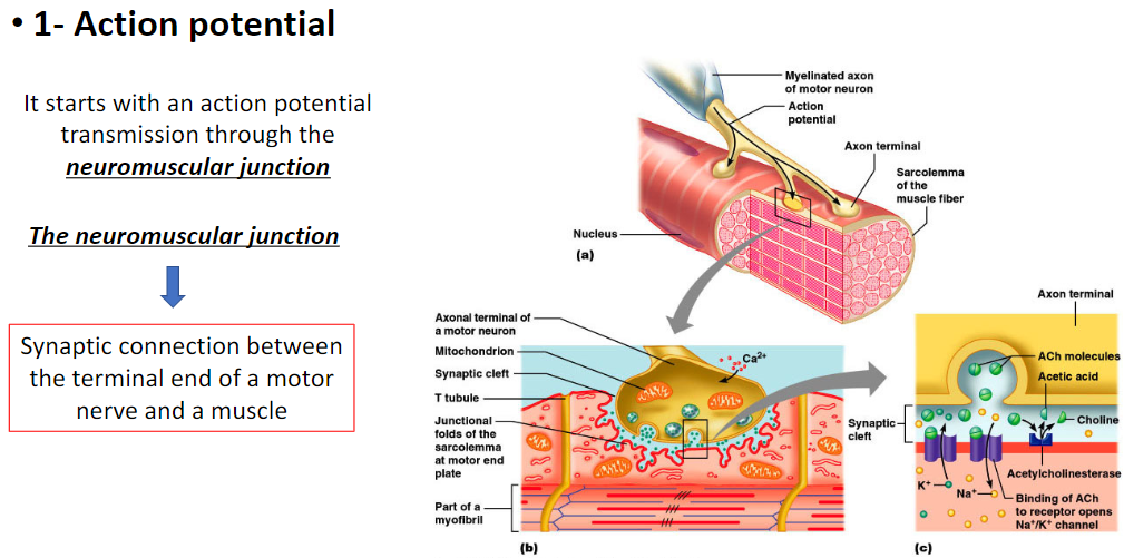
Muscle Contraction – Action Potential
Neuromuscular Junction
Synaptic connection between the terminal end of a motor nerve and a muscle fiber
Action Potential
Muscle contraction begins with an action potential
Signal is transmitted through the neuromuscular junction to the muscle fiber
Key Point
Neuromuscular junction converts nerve signals into muscle activation)
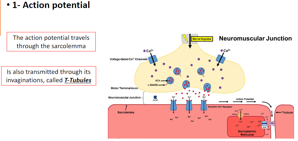
Muscle Contraction – Action Potential Transmission
Sarcolemma
Action potential travels along the muscle cell membrane (sarcolemma)
T-Tubules
Action potential is also transmitted through invaginations of the sarcolemma called T-tubules
Allows signal to reach deep into the muscle fiber
Key Point
T-tubules ensure the action potential rapidly reaches the interior of the muscle fiber for coordinated contraction)
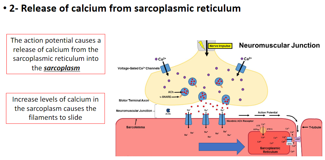
Muscle Contraction – Calcium Release
Sarcoplasmic Reticulum (SR)
Action potential triggers release of calcium (Ca²⁺) from the SR into the sarcoplasm (cytoplasm of skeletal muscle cell)
Effect on Filaments
Increased Ca²⁺ levels in the sarcoplasm cause actin and myosin filaments to slide
Key Point
Calcium release from SR is essential for initiating filament sliding and muscle contraction)
Muscle Contraction – Thin Filament Activation
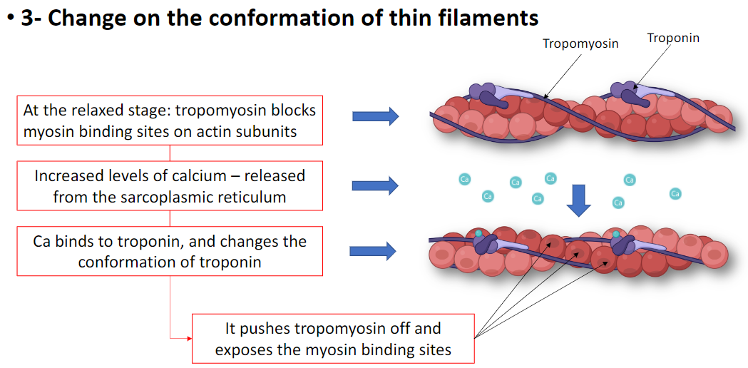
Relaxed Stage
Tropomyosin blocks myosin binding sites on actin subunits
Calcium Effect
Ca²⁺ released from the sarcoplasmic reticulum binds to troponin
Changes troponin conformation
Pushes tropomyosin off myosin binding sites
Key Point
Calcium binding to troponin exposes actin sites for myosin attachment, enabling contraction)
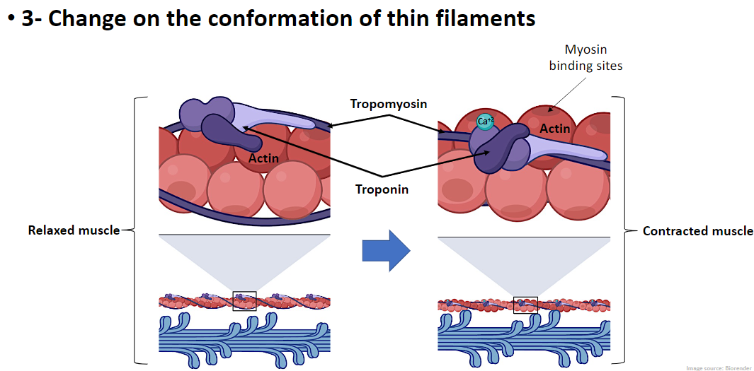
Muscle Contraction – Thin Filament Conformation
Relaxed Muscle
Tropomyosin covers myosin binding sites on actin
Myosin cannot bind, muscle remains relaxed
Contracted Muscle
Ca²⁺ binds to troponin
Troponin changes conformation and pulls tropomyosin away
Tropomyosin moves, exposing myosin binding sites on actin
Myosin heads attach, initiating contraction
Key Point
Calcium-induced conformational change in thin filaments allows myosin–actin interaction for muscle contraction)
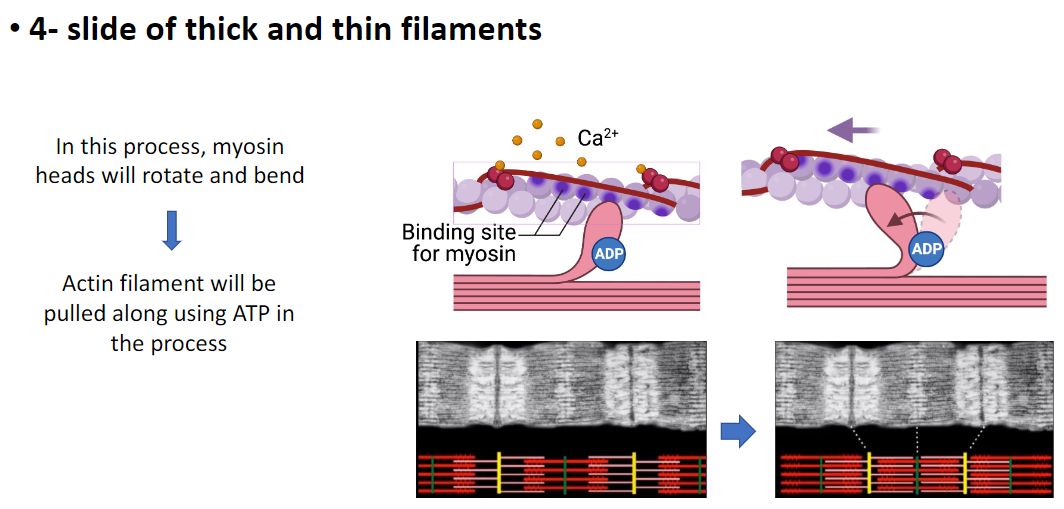
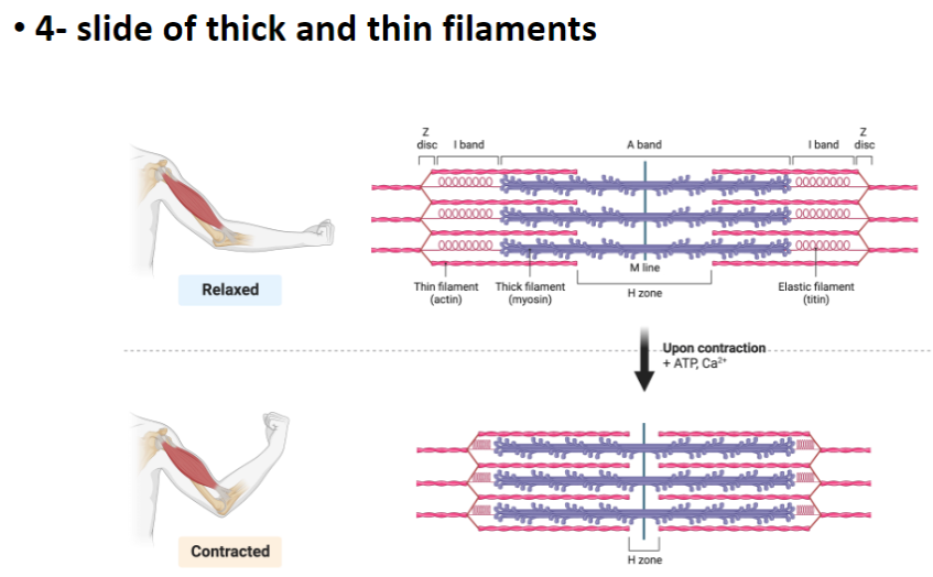
Muscle Contraction – Filament Sliding
Myosin Action
Myosin heads rotate and bend during contraction
Attach to actin filaments and pull them
ATP Role
ATP provides the energy for myosin movement and filament sliding
Effect on Filaments
Thin (actin) and thick (myosin) filaments slide past each other
Shortens the sarcomere, generating muscle contraction
Key Point
ATP-powered myosin movement causes actin–myosin sliding, which shortens the sarcomere and contracts the muscle)
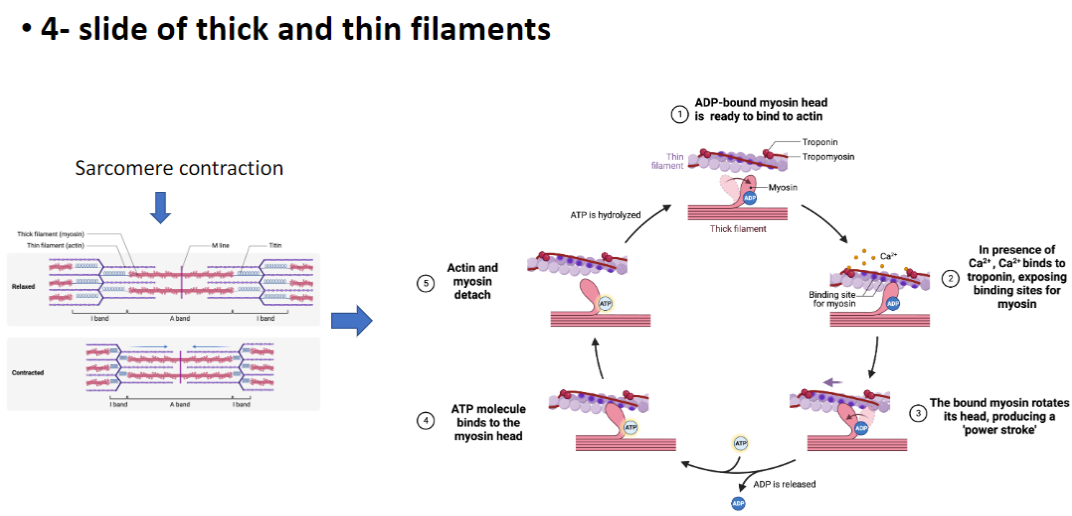
Muscle Contraction – Sarcomere Sliding
Relaxed Sarcomere
Actin (thin filaments) and myosin (thick filaments) are not bound
Tropomyosin blocks myosin binding sites on actin
Activation
Ca²⁺ binds to troponin
Troponin changes conformation, moves tropomyosin, exposing myosin binding sites
Myosin Binding and Power Stroke
ADP-bound myosin heads attach to actin
Myosin head rotates, producing a power stroke
Thin filaments are pulled past thick filaments, shortening the sarcomere
ATP Role
ADP is released and ATP binds to myosin head, causing detachment from actin
ATP hydrolysis re-cocks the myosin head for the next power stroke
Key Point
Cycle of Ca²⁺ binding, myosin attachment, power stroke, and ATP hydrolysis drives sarcomere shortening and muscle contraction)
Muscle Contraction – H Zone
H Zone
Central region of the A band containing only thick filaments (myosin)
Shortens during contraction as filaments slide
ATP and Ca²⁺ Role
Ca²⁺ exposes myosin binding sites on actin by binding to troponin which moves tropomyosin
ATP powers myosin head movement and detachment
Key Point
H zone shortens, titin maintains alignment, and ATP + Ca²⁺ enable contraction)
Muscle Contraction – Stepwise Process (Structural Hierarchy & ATP Role)
1 – Action Potential Initiation
Signal starts at motor neuron
Transmitted through the neuromuscular junction to the muscle fiber (cell)
2 – Action Potential Propagation
Travels along the sarcolemma (muscle membrane)
Also transmitted deep into the fiber via T-tubules
Ensures signal reaches interior of muscle fiber
3 – Calcium Release from Sarcoplasmic Reticulum (SR)
Action potential triggers Ca²⁺ release from SR into the sarcoplasm (cytoplasm of muscle cell)
Inside the muscle fiber surrounding the myofibrils
4 – Thin Filament Activation
Ca²⁺ binds troponin on the thin filament (actin)
Troponin changes conformation, moves tropomyosin
Exposes myosin binding sites on actin
5 – Cross-Bridge Formation
ADP-bound myosin heads on thick filament attach to exposed actin sites
Inside the sarcomere, thick and thin filaments begin interaction
6 – Power Stroke
Myosin heads rotate and bend, pulling actin filaments toward sarcomere center
Shortens the sarcomere, A band remains constant, H zone and I band shorten
7 – ATP Role in Detachment
ATP binds myosin head, causing it to detach from actin
ATP hydrolysis re-cocks the myosin head into a ready position (ADP + Pi-bound)
Cycle can repeat as long as Ca²⁺ and ATP are present
8 – Sarcomere Shortening
Multiple myofibrils within the muscle fiber shorten
Muscle fiber contracts, generating force
9 – Relaxation
Ca²⁺ pumped back into SR
Troponin/tropomyosin re-block myosin binding sites
Sarcomere returns to resting length, H zone and I band lengthen
Key Point
Muscle contraction is a coordinated process:
Signal (T-tubules) → Ca²⁺ release (SR) → thin filament activation → myosin cross-bridge cycling → sarcomere shortening
ATP controls myosin attachment/detachment and resets the head, enabling repeated cycles of contraction)
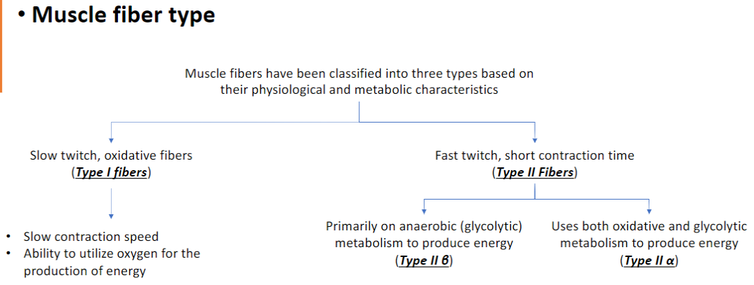
Muscle Fiber Types – Classification and Characteristics
Type I – Slow Twitch, Oxidative Fibers
Contract slowly
Use oxygen for energy production (aerobic metabolism)
Resistant to fatigue, suited for endurance activities
Type II – Fast Twitch Fibers
Type II β – Fast Glycolytic
Contract quickly
Rely primarily on anaerobic (glycolytic) metabolism for energy
Fatigue rapidly, suited for short, intense bursts
Type II α – Fast Oxidative-Glycolytic
Use both aerobic and anaerobic metabolism
Intermediate speed and fatigue resistance
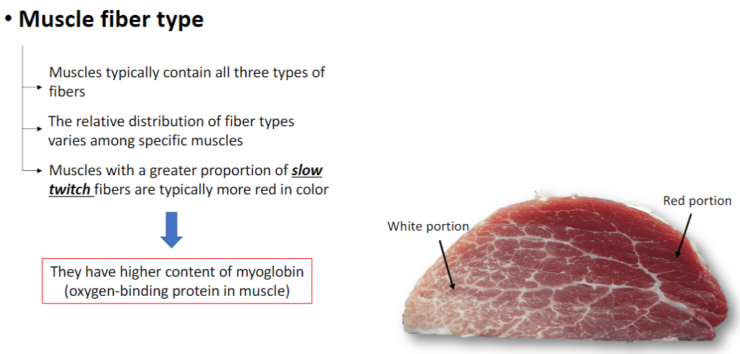
Muscle Fiber Distribution – Characteristics
Mixed Fiber Composition
Most muscles contain all three fiber types
Relative distribution varies among specific muscles
Slow Twitch Fibers (Type I)
Muscles with more slow twitch fibers appear red
High myoglobin content (oxygen-binding protein)
Suited for endurance and continuous activity
Fast Twitch Fibers (Type II)
Muscles with more fast twitch fibers appear white
Lower myoglobin content
Suited for short, intense bursts of activity
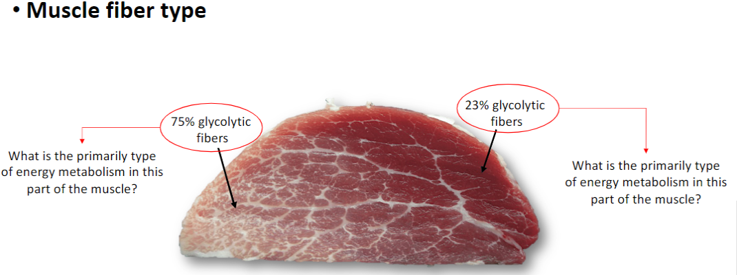
Muscle Fiber Energy Metabolism – Glycolytic Proportion
23% Glycolytic Fibers (77% Oxidative)
Majority are oxidative fibers (Type I, slow twitch)
Primary energy metabolism is aerobic (oxidative)
75% Glycolytic Fibers
Majority are glycolytic fibers (Type II, fast twitch)
Primary energy metabolism is anaerobic (glycolytic)
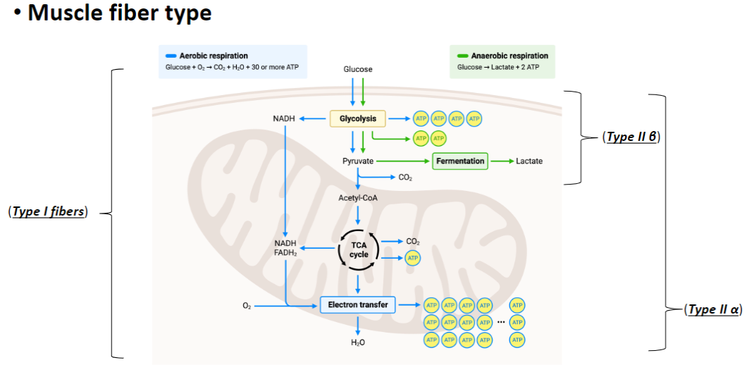
Muscle Fiber Energy Metabolism – Aerobic vs Anaerobic
Type I Fibers – Slow Twitch, Oxidative
Use aerobic respiration
Glucose + O₂ → CO₂ + H₂O + ~30 ATP
Pathway: Glycolysis → Pyruvate → Acetyl-CoA → TCA cycle → Electron Transport Chain
High ATP yield, fatigue-resistant
Type IIa Fibers – Fast Oxidative-Glycolytic
Use both aerobic and anaerobic metabolism
Intermediate ATP yield
Can sustain moderate force and activity
Type IIb Fibers – Fast Glycolytic
Use anaerobic respiration
Glucose → Lactate + 2 ATP
Pathway: Glycolysis → Fermentation to Lactate
Rapid energy production, fatigues quickly
Key Point
Fiber type determines energy pathway:
Type I → aerobic (needs O2), oxidation of glucose
Type IIa → mixed metabolism, aerobic oxidation of glucose / anaerobic fermentation
Type IIb → anaerobic fermentation to lactate
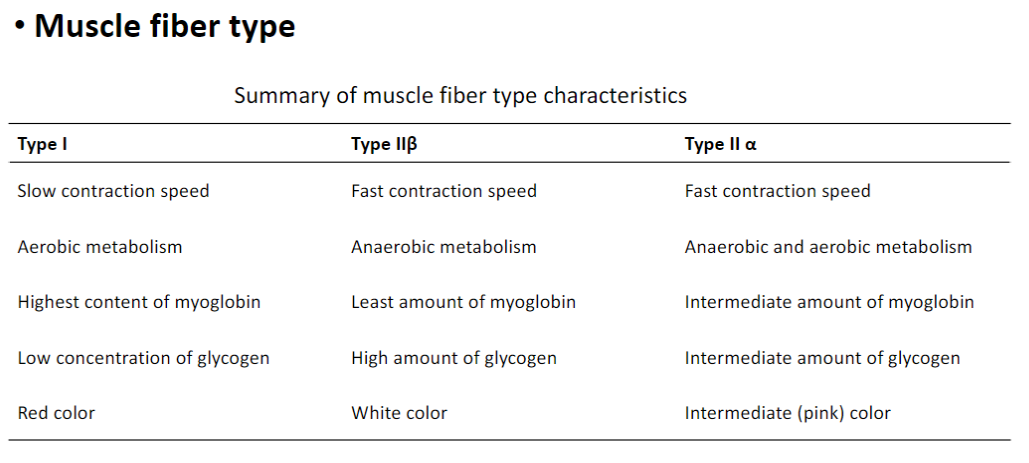
Muscle Fiber Types – Summary
Type I (Slow Twitch)
Slow contraction speed
Uses aerobic metabolism
Highest myoglobin content
Low glycogen concentration (since glycogen is used to make the glucose)
Red color
Type IIβ (Fast Glycolytic)
Fast contraction speed
Uses anaerobic metabolism
Least myoglobin
High glycogen content
White color
Type IIα (Fast Oxidative-Glycolytic)
Fast contraction speed
Uses both aerobic and anaerobic metabolism
Intermediate myoglobin
Intermediate glycogen
Intermediate (pink) color