Cell structure , Microscopy , Origins of life , Cell Specialization | Quizlet
1/295
There's no tags or description
Looks like no tags are added yet.
Name | Mastery | Learn | Test | Matching | Spaced | Call with Kai |
|---|
No study sessions yet.
296 Terms
What is a cell
The basic unit of all living things
cell theorey
1. Every organism is composed of one or more cells
2. The cell is the smallest unit possessing properties of life
3. All cells arise from pre-existing cells
4. Therfore, if we find a new sliving thing, we can deduce that it is also made of cells
Three types of cells
Prokaryotes: Bacteria Archea
Eukaryotes
Why aren't "functions of life" the same as "life itself"?
They describe processes required to maintain life, not what life fundamentally is.
What is a key difference between living and non-living things and like what is the meaning of this difference
Living things use energy to keep themselves in a highly ordered state. A structured, organized condition that living things actively maintain. For example, Damage must be repaired and there must be periodic regeneration through reproduction. The ability to maintain a highly ordered state must be inherited when a living thing reproduces. Non-living things may have some of these properties, but not all of them.
Describe the types of evidence that help decide that a cell is alive
• Individual cells use energy to maintain a highly ordered state.
• Some cells in a multicellular organism may stop doing this. These cells are clearly dead, for example, hair cells or cells in the outer layers of skin.
• Cells can divide to produce more cells.
• Cells can be taken from the body and cultured. For example, HeLa cells have been kept in cultures since 1951.
Why are viruses not considered living?
Viruses are NOT cells—(remember that cells are the smallest things that can be considered alive)—viruses do not have organelles, they do not have a metabolism, and they cannot reproduce outside of a host cell. They are not self susustaining
Why is the cell the smallest unit of life?
Within a multicellular organism, individual cells also have all the properties of life, but subcellular components do not
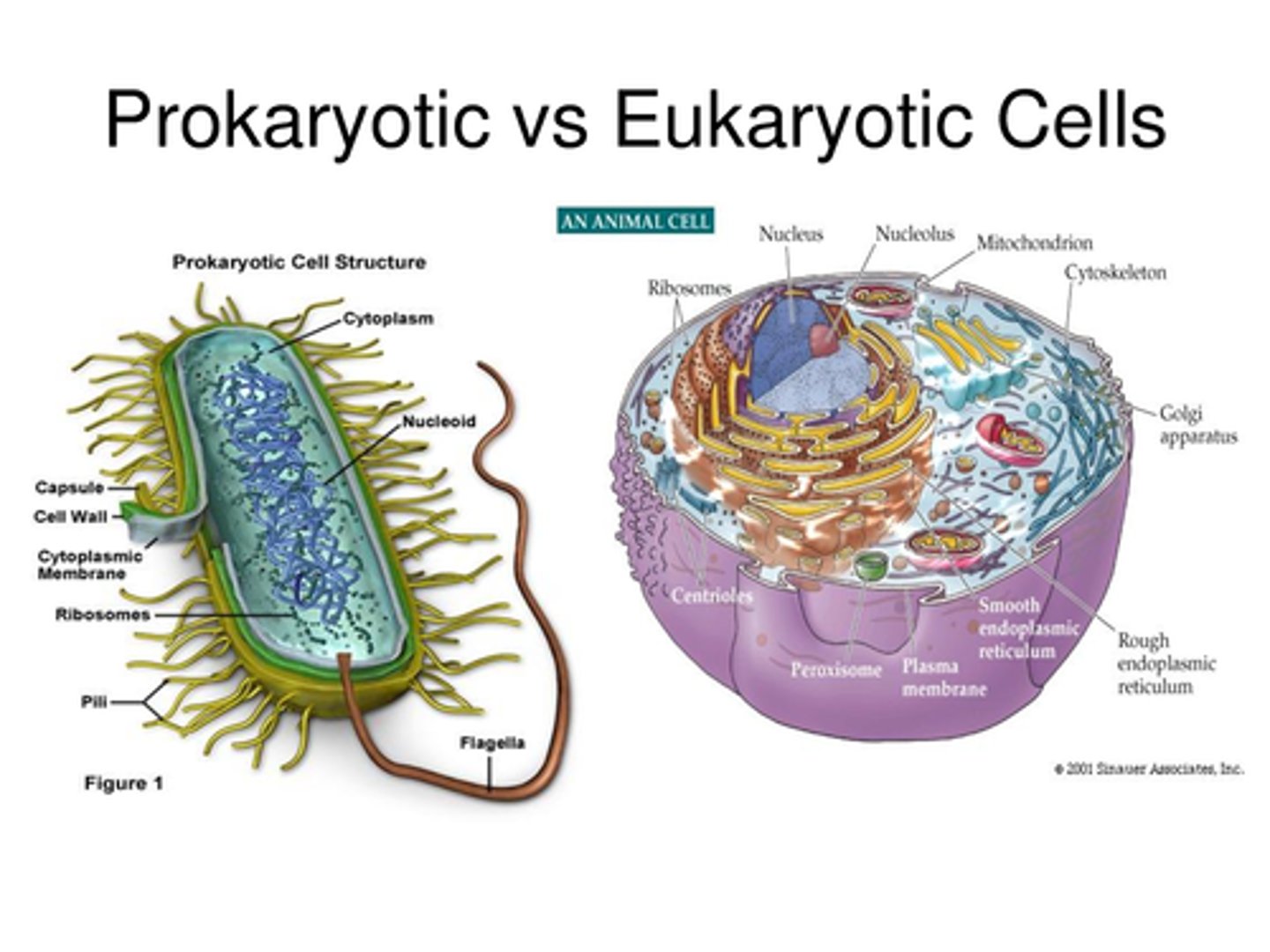
What are prokaryotes?
Bacteria and Archaea
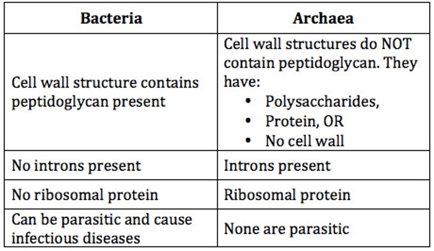
What are archaea
A single celled organism that is prokaryotic and lives in extreme environments.
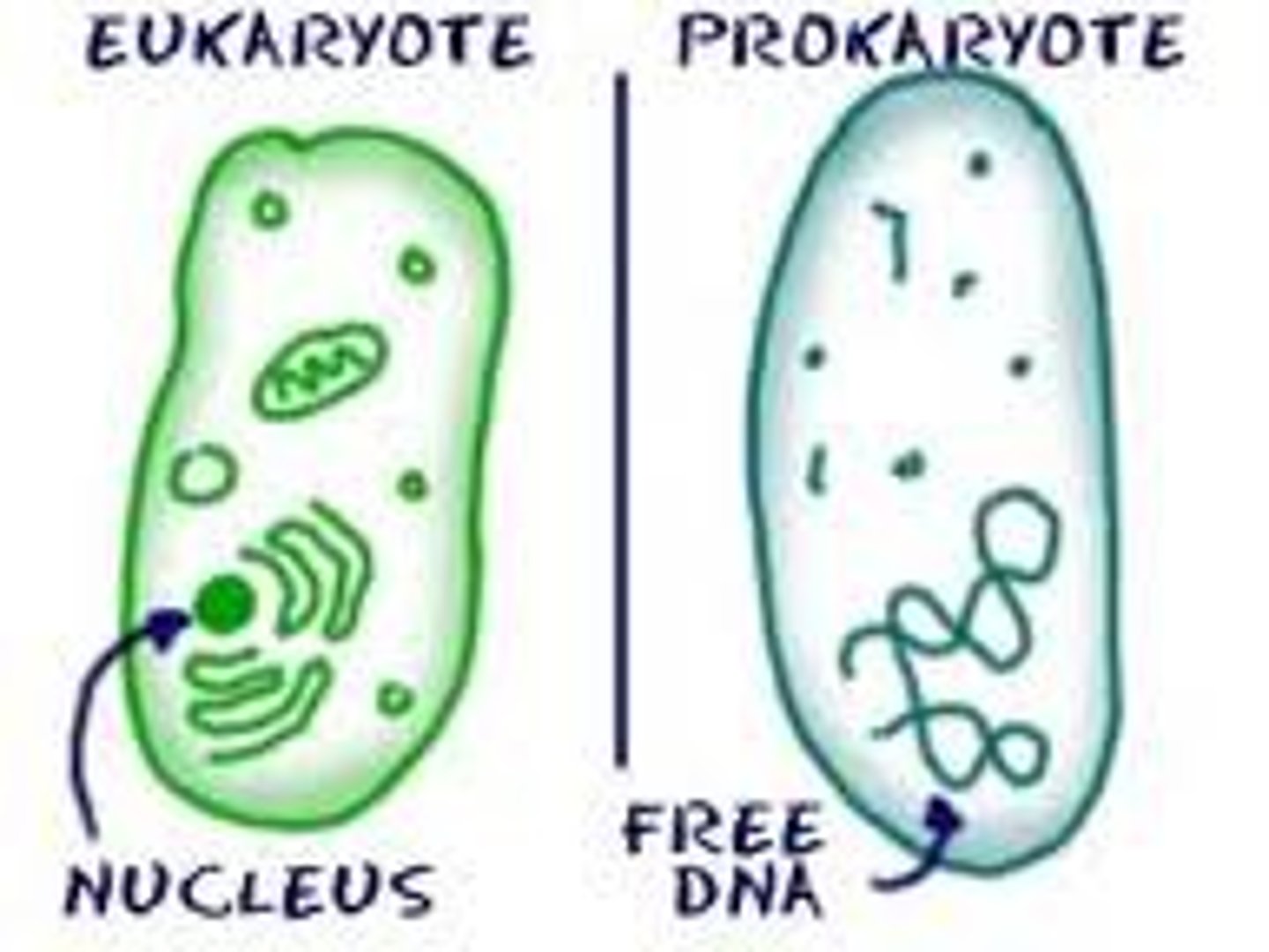
WHat is the key feature of prokaryotes
no nucleus
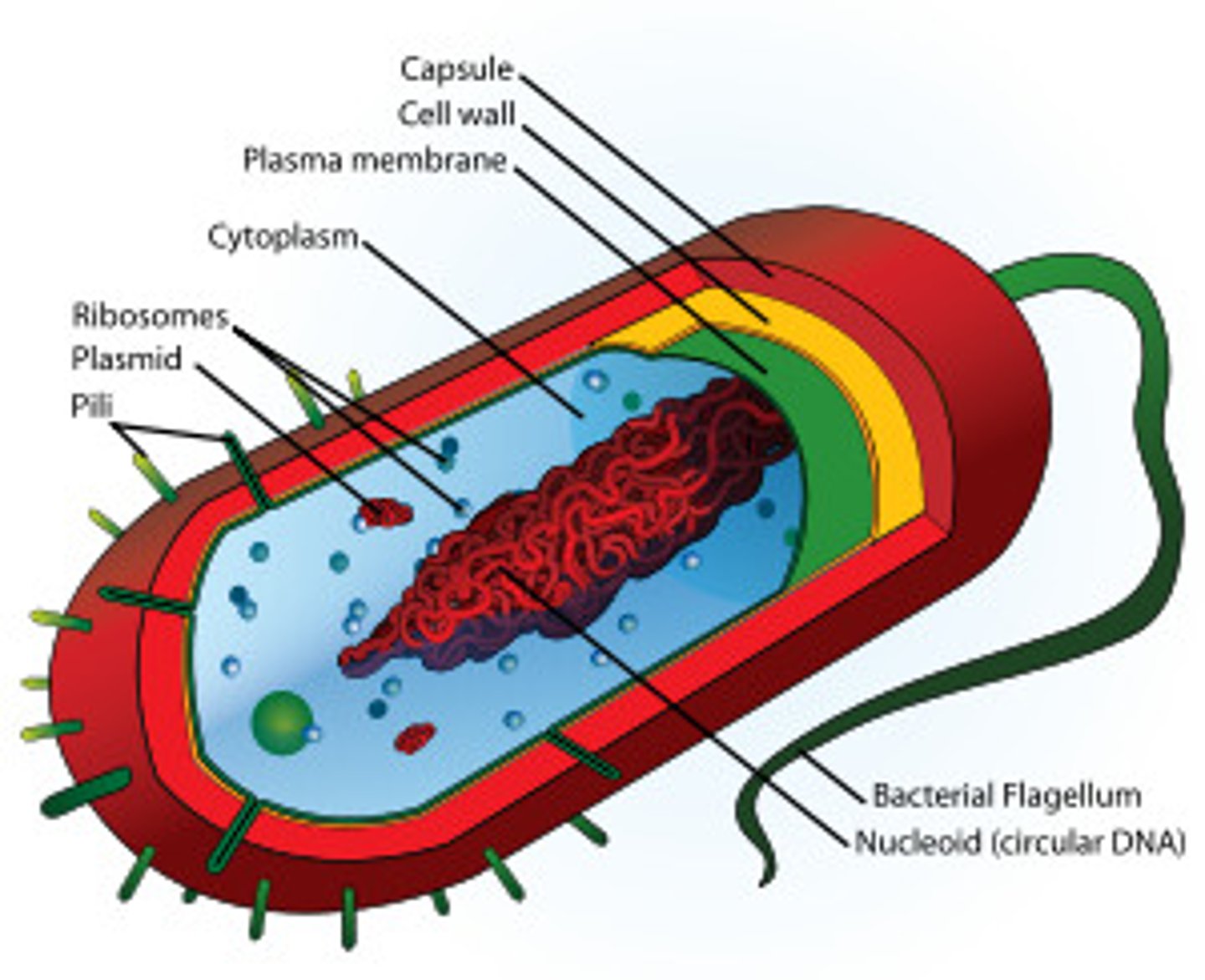
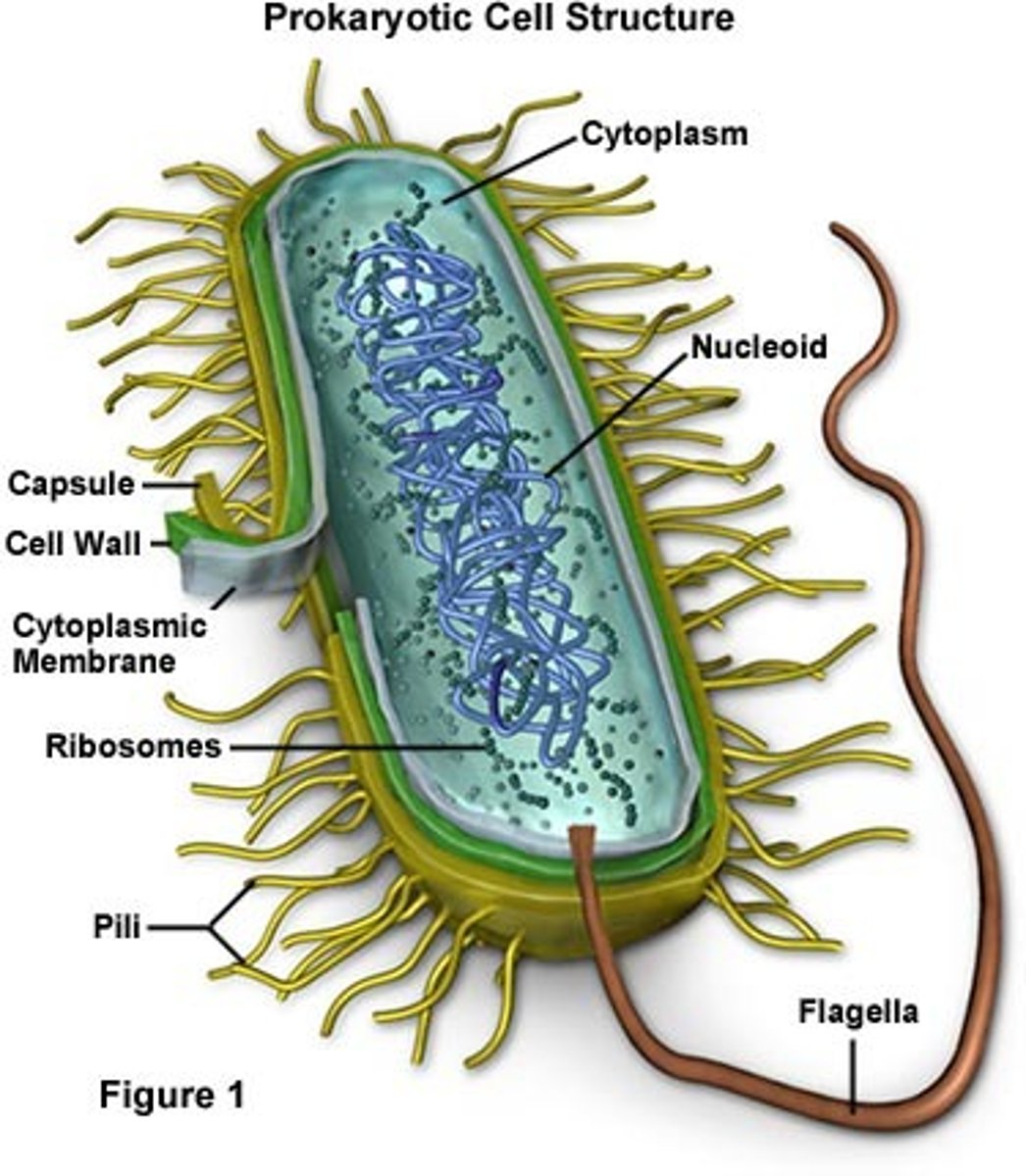
explain like the enclosure of a prokaryotic cell
Theres the membrane plus the cell wall which is made out of peptidoglycan
What is the role of the cell wall
This structure is thicker and stronger than the membrane. It protects the cell, maintains its shape and supports the plasma membrane to prevent it from bursting
What do prokaryotic cells have instead of a nucleus
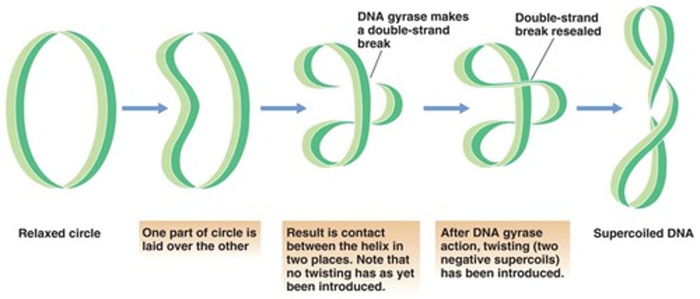
they have a nucleoid region in which there is a single chromosome of dna (a dna molecule) which is CIRCULAR and naked meaning that it is NAWT associated with proteins
Naked vs not naked DNA
not naked is like wrapred around proteins called hstones
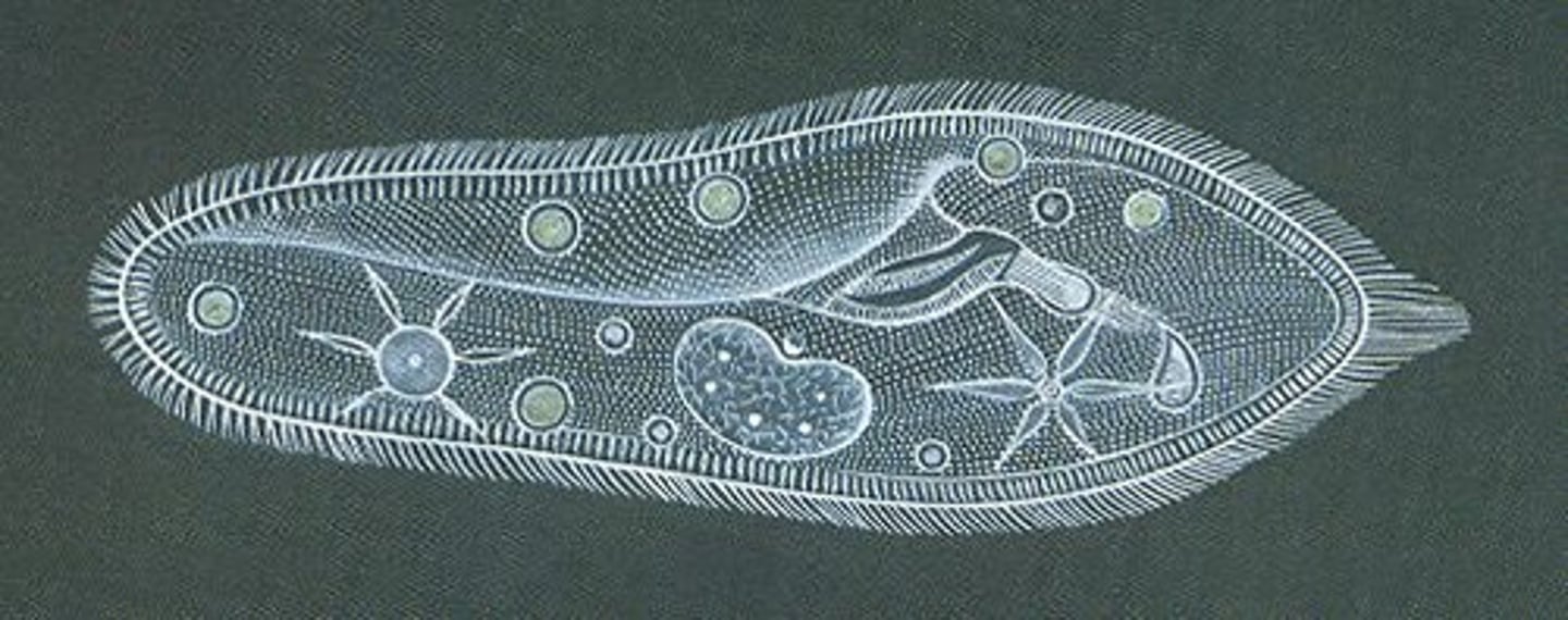
Describe the inside of a prokaryotic cell
its all just cytoplasm, no compartments seperated by membranes, lots of ribosomes.
Describe the ribosomes in prokaryotic cells
They are smaller, being only 70S
The S indicates how fast they sink to the bottom to become sediment during centifugation. Because they have a relitivley smaller number, they sink slower and thus are smaller
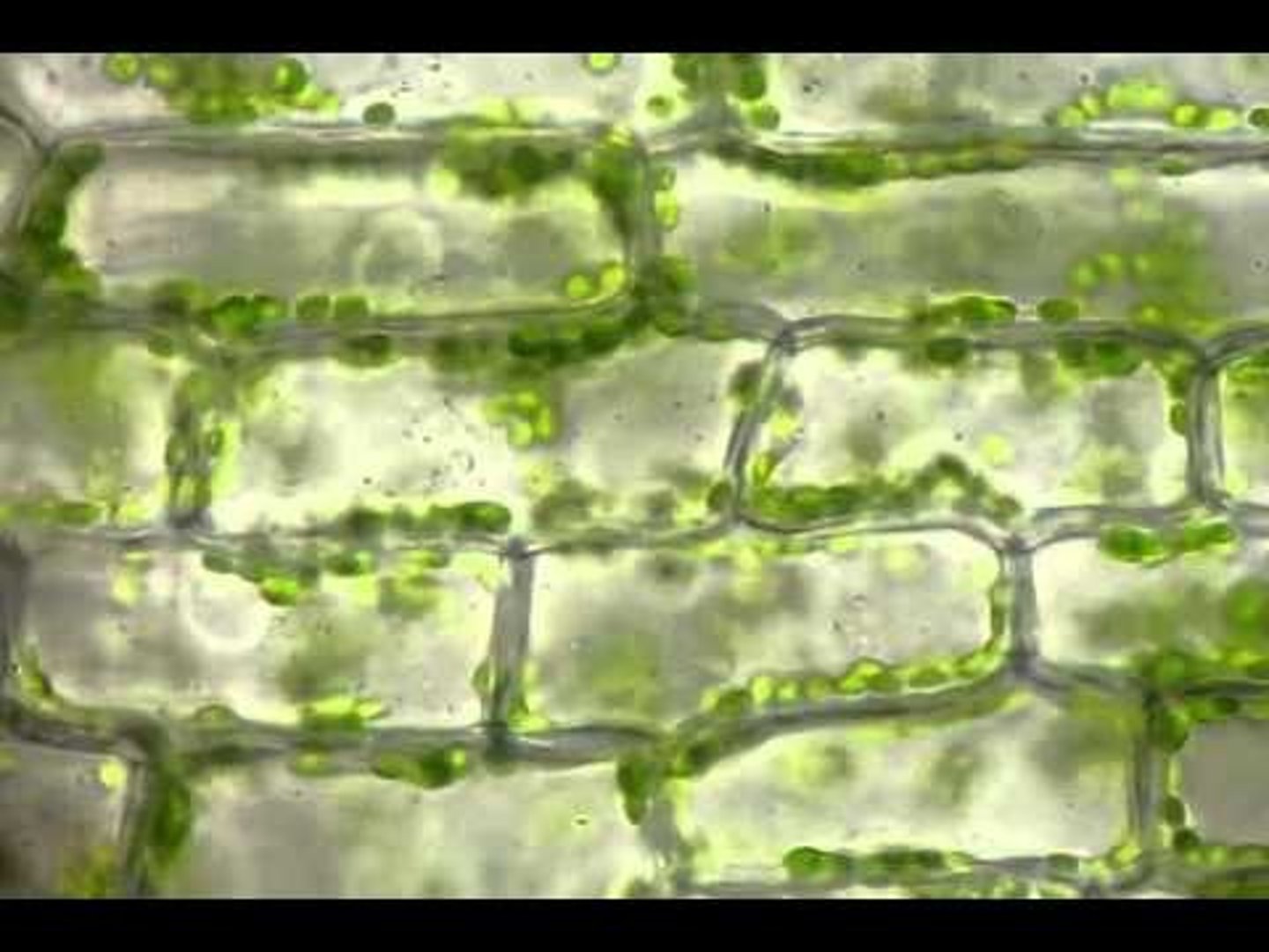
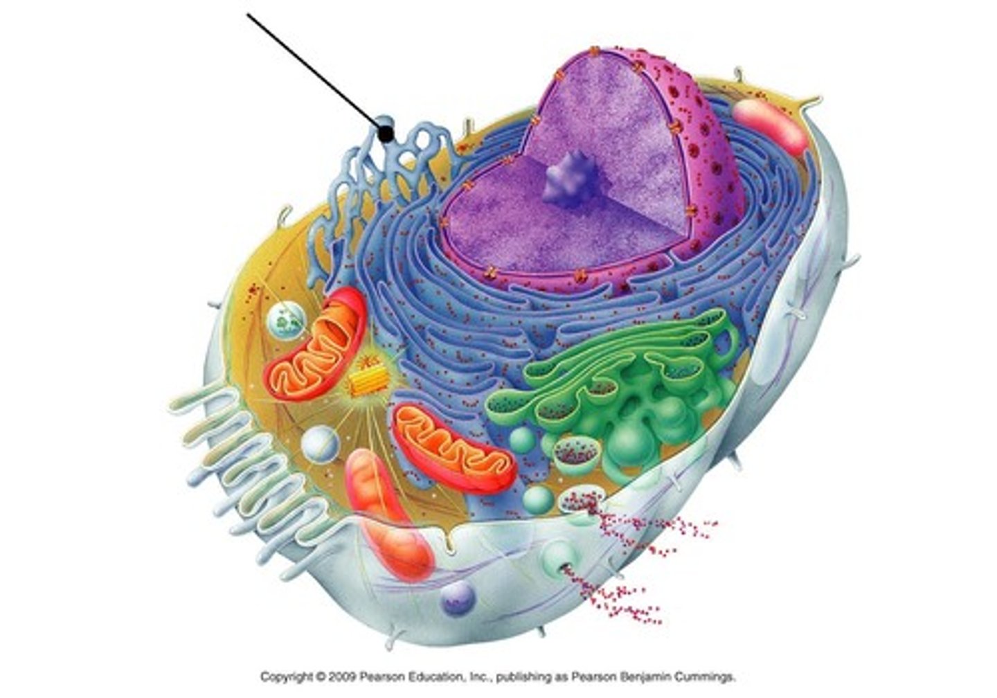
explain the enclosurw and basic inside of eukaryotic cells
Plasma mebrane, some have a cell wall, on the inside its is compartmentalized
4 distinguishing features of eukaryotic cells
-compartmentalized
-nucleus
-80s tibosome
- Mitochondria
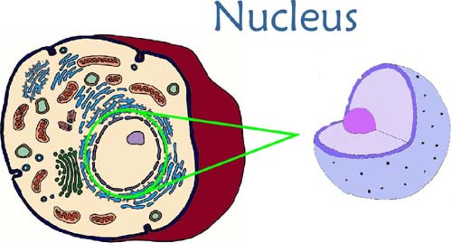
Nucleus (eukaryotic cells) structure and role
This compartment holds the cell's chromosomes. The nucleus has a double membrane with pores through it
Cell chromosomes (euk)
Each chromosome consists of one long DNA molecule attached to proteins, except when a cell is preparing to divide and the DNA is replicated. The DNA molecules are linear rather than circular. The proteins are histones, arranged in globular groups like small beads, with the DNA wound around the outside.
Ribsomes in eukaryotic cells
There are some structural differences to those in prokaryotes and they are larger in size, meaning they sink to the bottom to become sediment during centrifugation, making them have a Svedberg unit of 80 (80S)
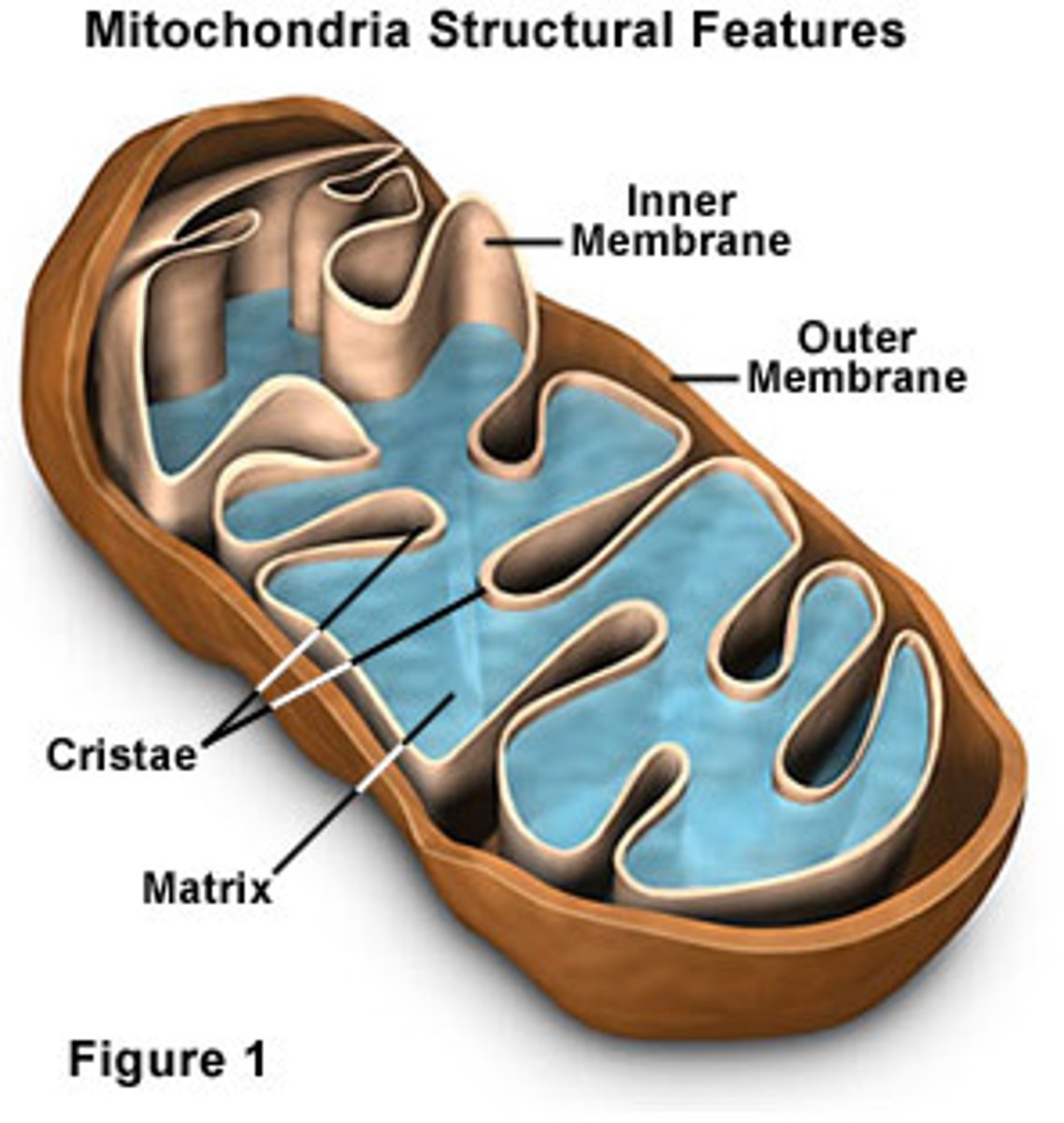
Mitochondria form and function
Surounded by double membrane, the inner membrane is folded inwards to increase surface area
Carry out aerobic respiration , so in eukaryotes, only cells that DONT respire aerobically DONT have a mitochondria
What is an organelle?
A tiny cell structure that carries out a specific function within the cell
Name the organelles in eukaryotes with no membrane
80S ribosomes
Name the organelles in eukaryotes with one membrane
rough endoplasmic reticulum (ER), smooth ER, Golgi apparatus, lysosomes, vesicles, vacuoles,
Name the organelles in eukaryotes with two membranes
nucleus, mitochondria, chloroplasts.
Name the cell structures in cells that are NOT organnels and their function
cell wall—outside the plasma membrane so outside the boundary of the cell (extracellular),
cytoplasm—has diverse rather than specific functions,
cytoskeleton—a structure that extends through the cytoplasm and is not discrete.
What developments in science allowed scientists to research the functions of various organelles
Ultracentrifuges, which can rotate at more than 40,000 revolutions per minute (rpm), were invented in the 1920s, and used for cell fractionation from the 1930s onwards. Organelles can be released from the tissue by homogenization and then can be separated into different types because they settle to the base of the centrifuge tube (sediment) at different speeds.
advantages of compartmentalizing in cells
• Enzymes and substrates for a particular process can be much more concentrated than if they were spread throughout the cytoplasm.
• Substances that could cause damage to the cell can be kept inside the membrane of an organelle. For example, the digestive enzymes of a lysosome could digest and kill a cell, if they were not safely stored inside the lysosome membrane.
• Conditions such as pH can be maintained at an ideal level for a particular process, which may be different from the levels needed for other processes in a cell.
• Organelles with their contents can be moved around within the cell.
• There is a larger area of membrane available for processes that happen within or across membranes.
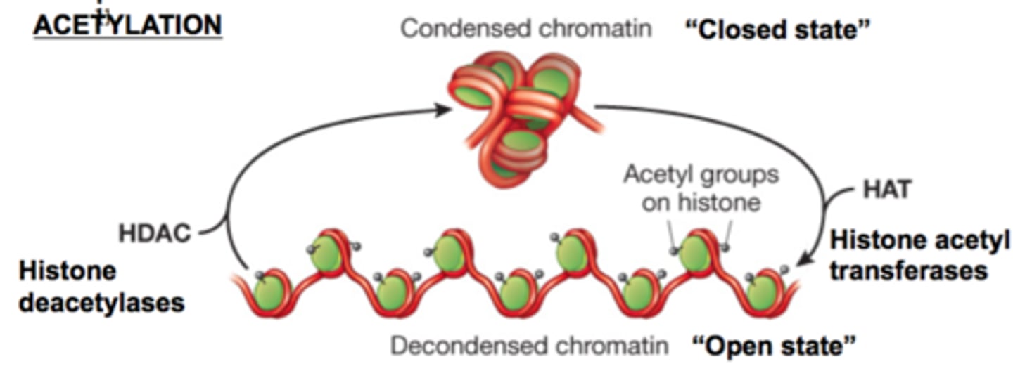
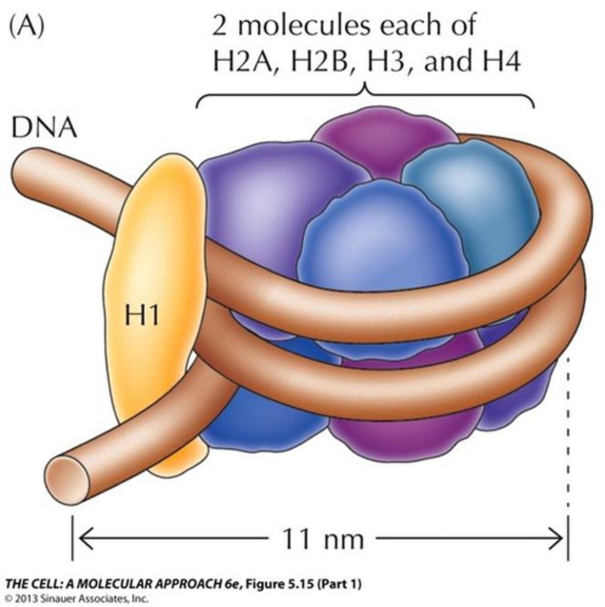
What is the nucleosome core made of?
Eight histone proteins: two copies each of four different histone types, forming a disc-shaped core.
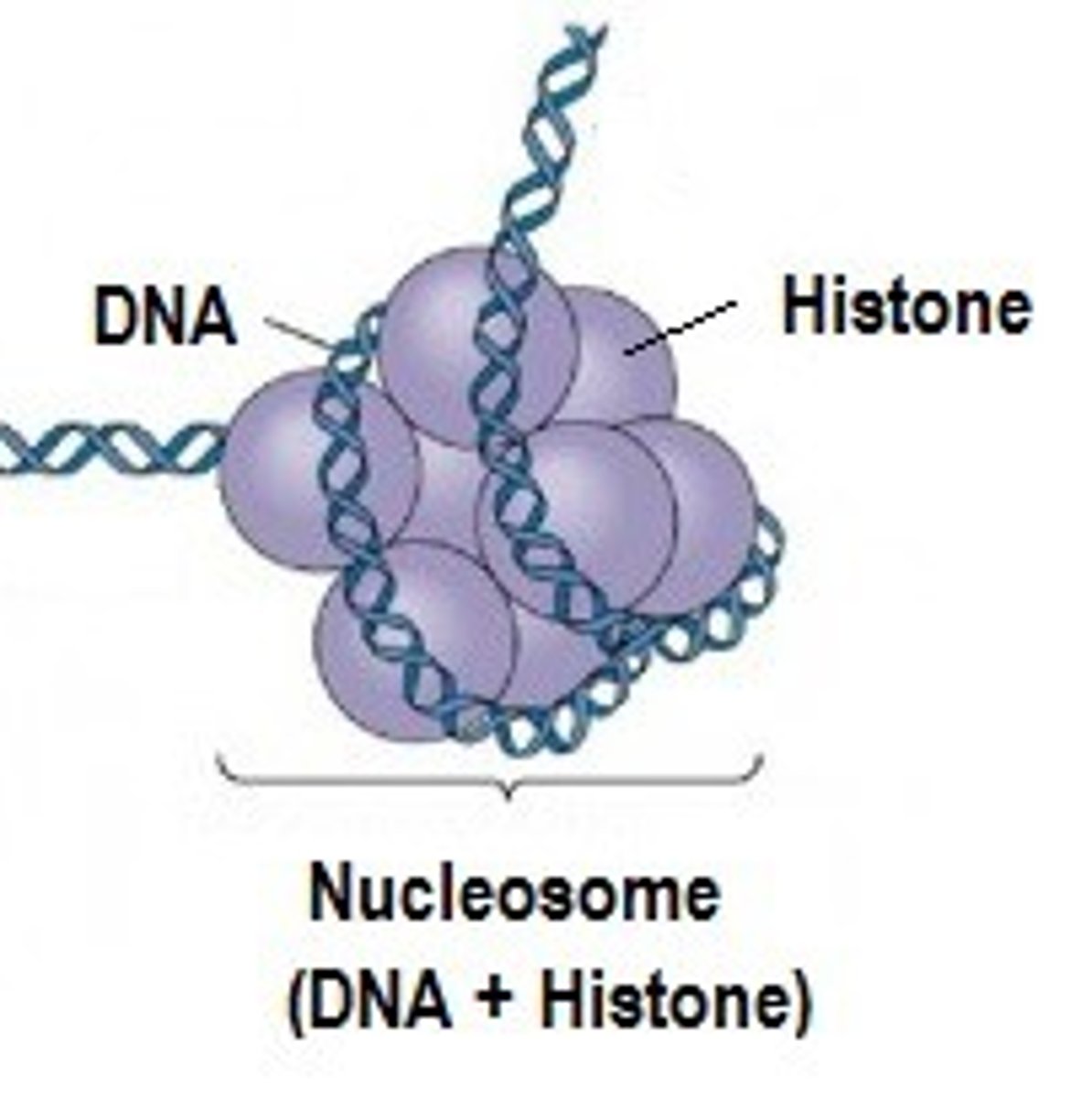
How is DNA arranged around the nucleosome core?
The DNA is wound about twice around the histone core
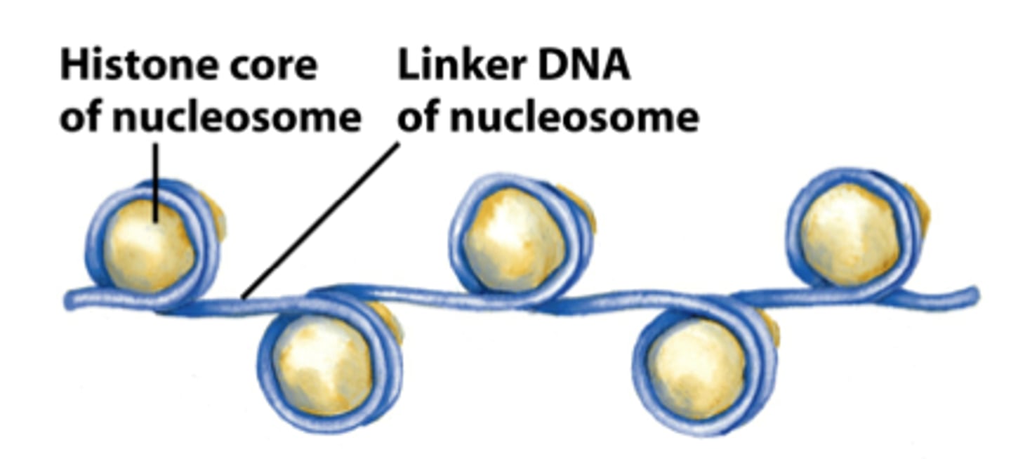
What is histone H1, and what else is found between nucleosomes?
H1 helps reinforce DNA binding to the nucleosome and may aid chromosome packaging before division; linker DNA connects adjacent nucleosomes.
Explain the role of the cytoplasm
Metabolic reactions take place in the cytoplasm, including the reactions that release energy by respiration. Enzymes in the cytoplasm catalyse these reactions.
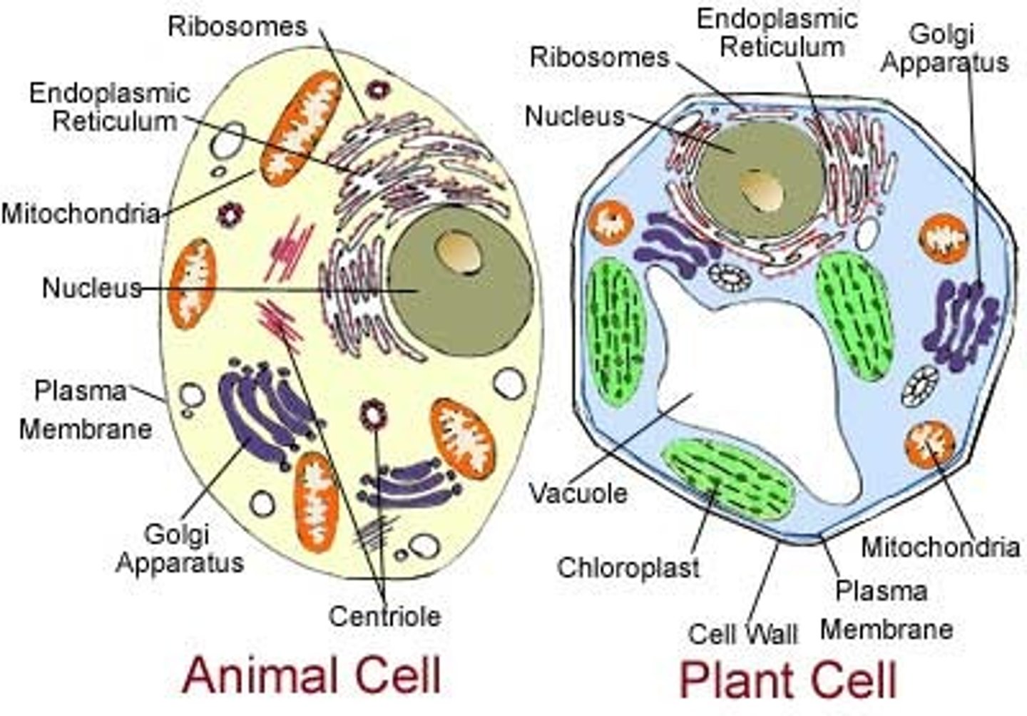
Describe the structure and role of plastids and compare them in plants, animals, and fungi
A family of organelles with two outer membranes and internal membrane sacs
Animals and fungi- none
Plants- Plant cells have varied types such as chloroplasts (for photosynthesis) and amyloplasts (to store starch).
Describe the structure and role of cell walls and compare them in plants, animals, and fungi
Animals and fungi- none
Plants- Cell wall A rigid layer outside the plasma membrane to strengthen and protect the cell. Cells of fungi and plants have walls, composed of chitin in fungi and cellulose in plants
Describe the structure and role of vacuoles and compare them in plants, animals, and fungi
A fexible fluid-flled compartment surrounded by a single membrane
Animals- wo types of small temporary vacuole occur in some animal cells but not plant or fungal cells: contractile vacuoles that expel excess water by exocytosis and food vacuoles that digest food or pathogens taken in by endocytosis.
Plants and fungi- There is often a large permanent vacuole in cells of fungi and plants, used for storage of substances and pressurizing the cell
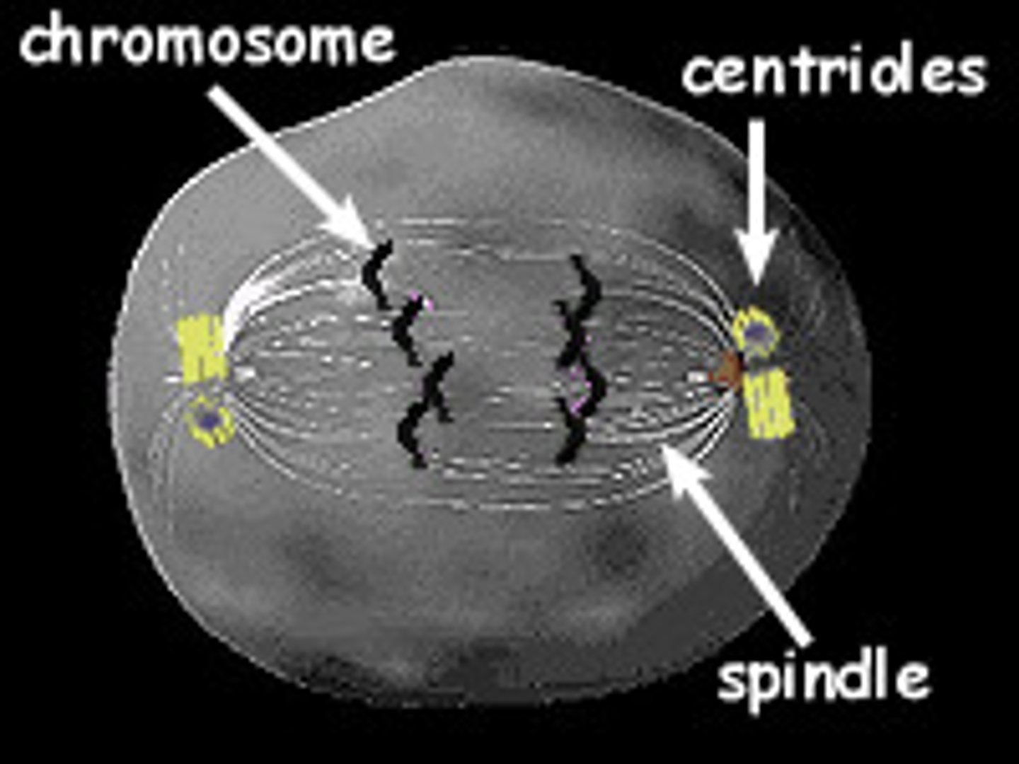
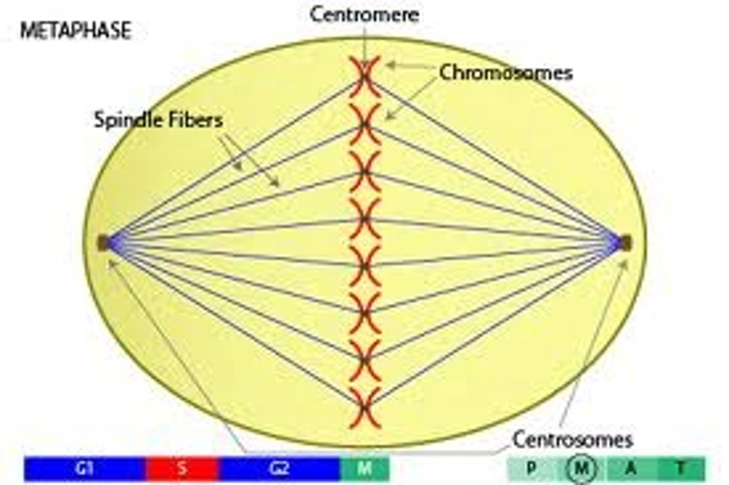
Describe the structure and role of centrioles and compare them in plants, animals, and fungi
Cylindrical organelles that organise the assembly of structures composed of microtubules
Animals- they form the core of centrosomes that organize assembly of a spindle of microtubules during mitosis and meiosis and the 9 + 2 microtubules in cilia and flagella
Plants and fungi- Absent, except in fungi and plants with swimming male gametes, which have a centriole at the base of the flagellum
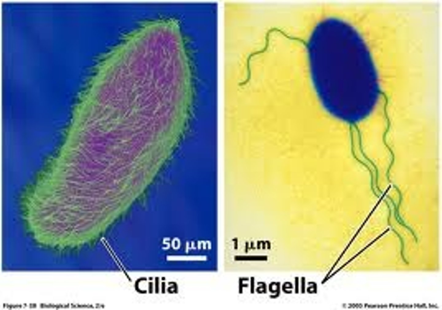
Describe the structure and role of Undulipodia and compare them in plants, animals, and fungi
Cilia and flagella are whip-like structures with a 9+2 arrangement of microtubules inside and plasma membrane on the outside. They protrude from the cell and generate movement by a beating action
Animals- Some types of animal cell have many cilia, which are small and move fluids adjacent to the cell. Male gametes (sperm) in animals have a single flagellum (tail), which is much longer than cilia and causes the sperm to move.
Plant and fungus -cells have no cilia. Some plants, including ferns and mosses, have motile male gametes with a flagellum, but conifers, flowering plants and almost all fungi do not.
Example of anucleate cells
Mature red blood cells in mammals
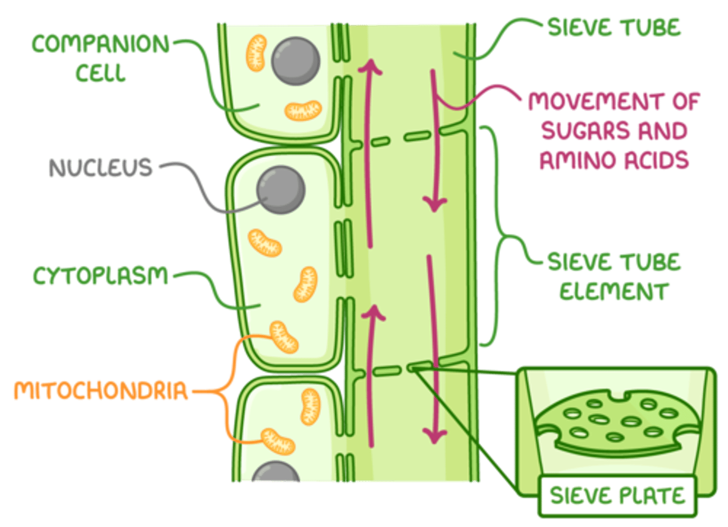
Phloem sieve tube elements
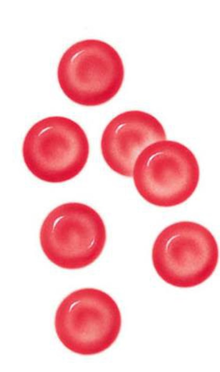
Describe the nucleus in mature red blood cells
In mammals, these cells do not have a nucleus. At a late stage in their development in bone marrow, the nucleus is moved to the edge of the cytoplasm and the small part of the cell containing it is pinched o and destroyed by a phagocyte. Removal of the nucleus makes red blood cells smaller and more exible, but they cannot repair themselves if they are damaged. For this reason, they have a lifespan of only 100 to 120 days.
Describe the nucleus in phloem sieve tube elements
Phloem sieve tube elements are the subunits of the tubes that transport sugar-containing sap in plants. They initially have a nucleus but it breaks down, so sap can flow more easily. However, they are supplied with proteins by adjacent companion cells, which have a nucleus and rough ER.
Examples of multinucleate cells
Skeletal muscle
Aseotate fungi
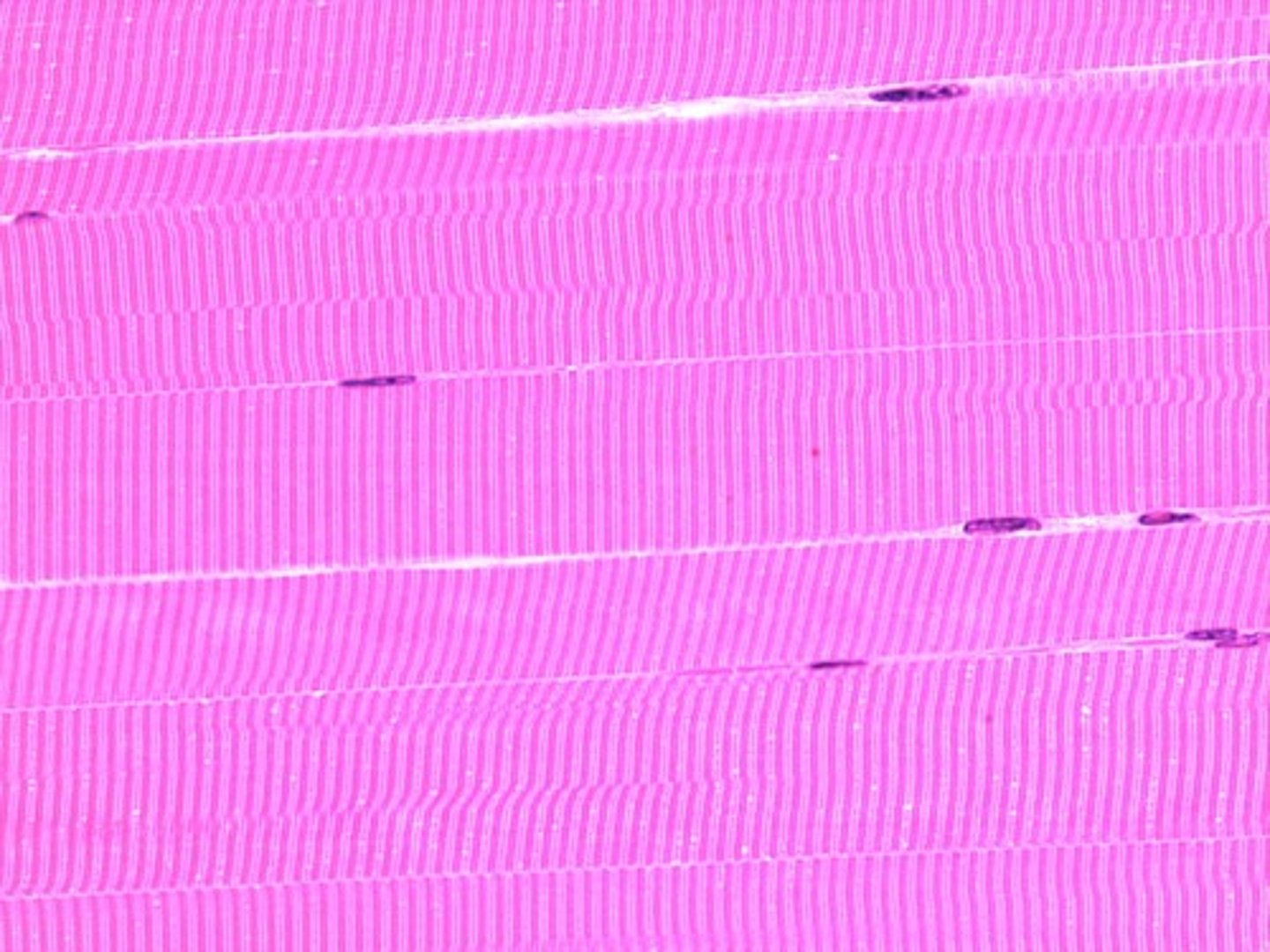
Describe the nucleus of skeletal muscle
Skeletal muscle is made up of muscle fibres. Each fibre is enclosed inside a plasma membrane like a cell, but is 300 or more mm long (so much larger) and contains hundreds of nuclei. They have striated appearance due to regular arrays of protein filaments used in muscle contraction.
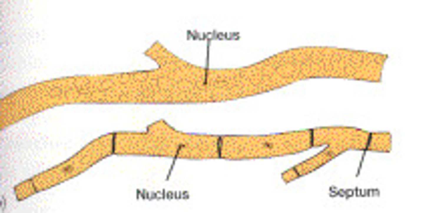
Describe the nucleus of aseptate fungi
Aseptate fungi consist of thread-like structures called hyphae. These hyphae are not divided up into subunits containing a single nucleus. Instead, there are long undivided sections of hypha which contain many nuclei.
All nucleus info (euk)
The nuclear membrane is double and has pores through it.
The nucleus contains the chromosomes, consisting of DNA associated with histone proteins.
Uncoiled chromosomes are spread through the nucleus in the areas that appear pale and grainy.
The small areas that are more densely stained, mostly around the edge of the nucleus, contain parts of chromsomes that have remained coiled up (condensed).
The nucleus is where DNA is replicated and transcribed to form mRNA, which is exported via the nuclear pores to the cytoplasm.
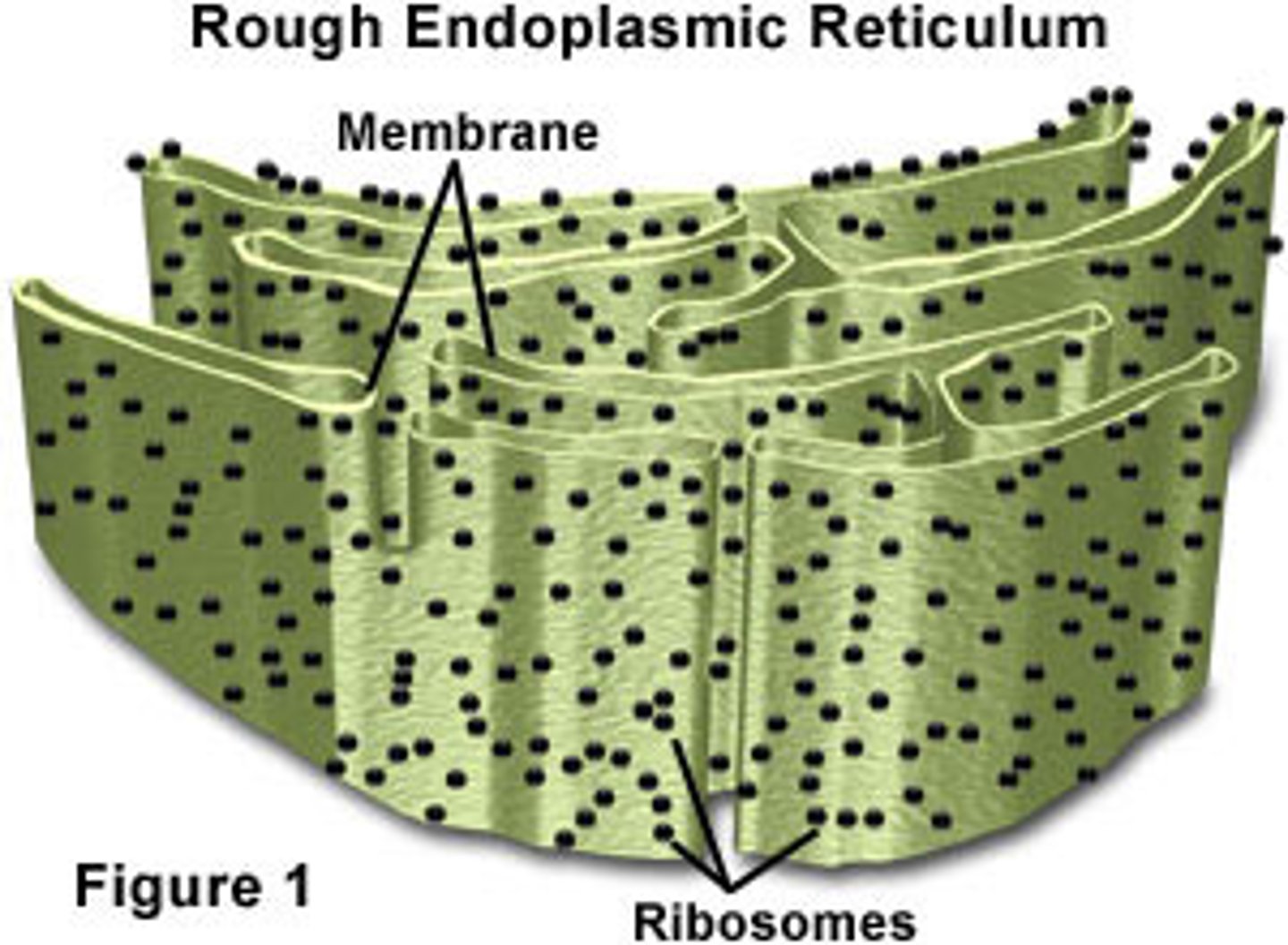
All RER info (euk)
The rER consists of flattened membrane sacs, called cisternae.
Ribosomes are attached to the outside of these cisternae.
They are larger than in prokaryotes and are classied as 80S.
The main function of the rER is to synthesize protein for secretion from the cell.
Protein synthesized by the ribosomes of the rER passes into its cisternae. It is then carried by vesicles, which bud o and are moved to the Golgi apparatus.
all SER info (euk)
Smooth endoplasmic reticulum consists of a branched network of tubular membranes.
In electron micrographs, it appears as circles or ovals of membrane. The membrane is smooth because there are no ribosomes attached.
Smooth ER has a variety of functions. It is used to synthesize lipids, phospholipids and steroids.
A special type of smooth ER stores calcium ions in muscle when it is relaxed.
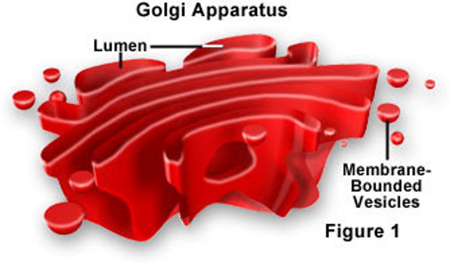
All golgi info (euk)
This organelle consists of flattened membrane sacs called cisternae (as in rER).
However, these cisternae are not as long, are often curved, do not have attached ribosomes and have many vesicles nearby.
The Golgi apparatus processes proteins brought in vesicles from the rER.
Most of these proteins are then carried in vesicles to the plasma membrane for secretion.
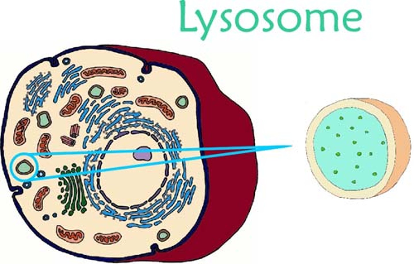
All lysosome info (euk)
These are approximately spherical with a single membrane.
They are formed from Golgi vesicles. They contain high concentrations of protein, which makes them densely staining in electron micrographs.
They contain digestive enzymes, which can be used to break down ingested food in vesicles.
These enzymes can also break down organelles or even whole cells.
All mitochondrion info (euk)
A double membrane surrounds mitochondria.
The inner membrane is invaginated to form structures called cristae.
The fluid inside is called the matrix. The shape of mitochondria is variable but usually spherical or ovoid.
They produce ATP for the cell by aerobic cell respiration. Fat is digested here if it is being used as an energy source in the cell.

All free ribosomes info (euk)
These appear as dark granules in the cytoplasm and are not surrounded by a membrane.
They have the same size as ribosomes attached to the rER—about 20nm in diameter (known as 80S).
Free ribosomes synthesize protein, releasing it to work in the cytoplasm, as enzymes or in other ways.
Ribosomes are constructed in a region of the nucleus called the nucleolus.
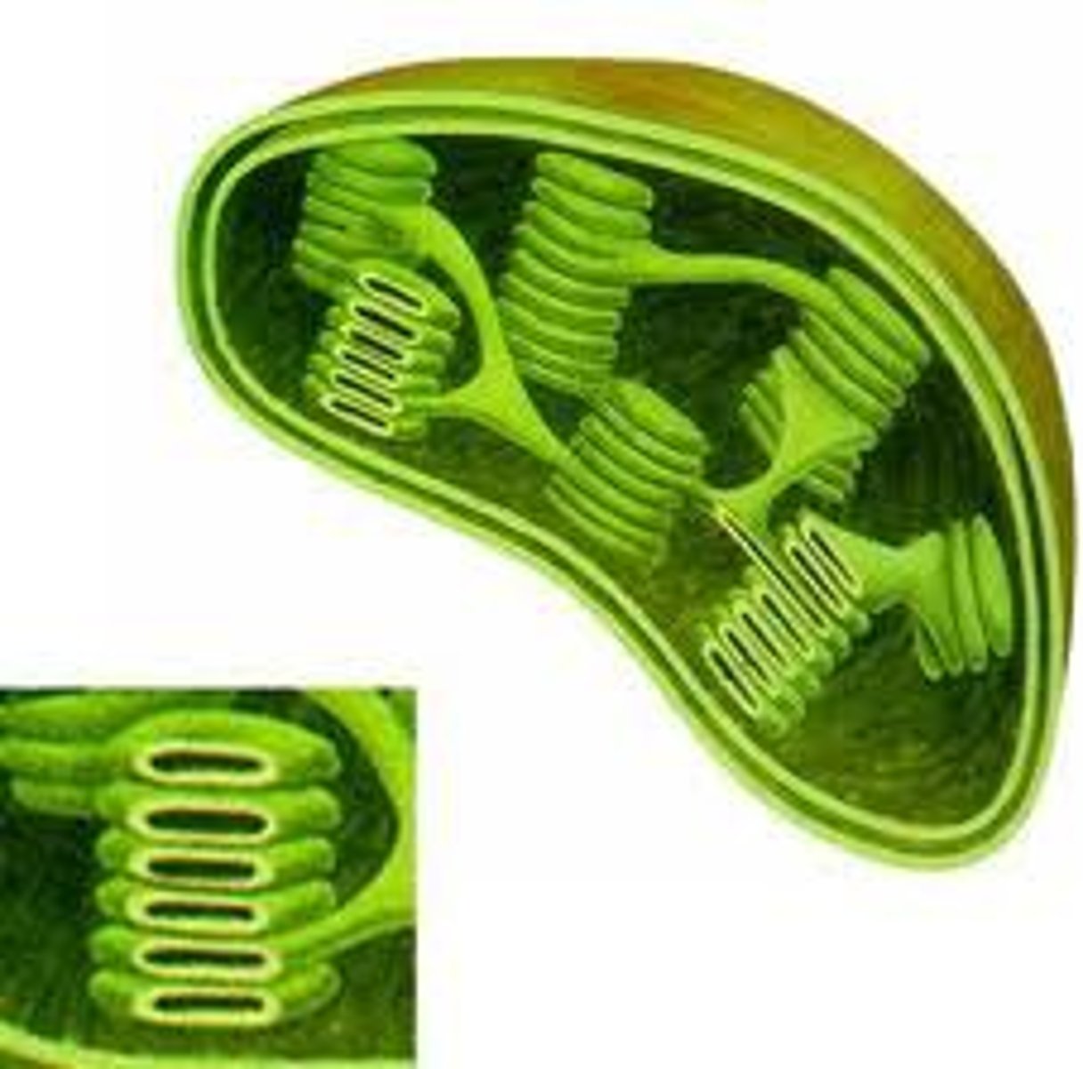
all chloroplast info (euk)
A double membrane surrounds the chloroplast.
Inside are stacks of thylakoids, which are flattened sacs of membrane.
The shape of chloroplasts is variable but usually spherical or ovoid.
They produce glucose and a wide variety of other organic compounds by photosynthesis.
If chloroplasts have been photosynthesizing rapidly, they may contain starch grains.

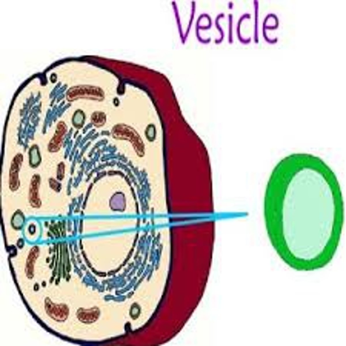
All vacuoles and vessicles info (euk)
These organelles consist of a single membrane with fluid inside.
Many plant cells have large vacuoles that occupy more than half of the cell volume.
Some animals absorb foods from outside and digest them inside vacuoles.
Some unicellular organisms use vacuoles to expel excess water.
Vesicles are very small vacuoles used to transport materials inside the cell.
All Microtubles and centrioles info ( euk)
The cytoplasm of cells contains small cylindrical fibers called microtubules.
They have a variety of roles, including moving chromosomes during cell division.
Animal cells have structures called centrioles, which consist of two groups of nine triple microtubules.
Centrioles form an anchor point for microtubules during cell division and also for microtubules inside cilia and flagella.
All cytoskeleton info (euk)
The cytoskeleton is constructed from several types of protein fibre.
Tubulin is used to make microtubules, and actin is used to make microfilaments.
These structures can easily be constructed or deconstructed,so the cytoskeleton is dynamic.
Microtubules guide the movement of components within the cell.
They help plant cells to construct cell walls.
A layer of microlaments just inside the plasma membrane helps animal cellsto maintain theirshape.
All cillia and flagella info (euk)
These are whip-like structures projecting from the cell surface.
They contain a ring of nine double microtubules plus two central ones.
Flagella are larger and usually only one is present, as in a sperm.
Cilia are smaller, and many are present.
Cilia and agella can be used for locomotion. Cilia can also be used to create a current in the fluid next to a cell.

What is the smallest size the naked eye can usually see?
About 0.1 mm (100 µm).
How much can a single convex lens typically magnify?
Up to about 20×.
Why do microscopes use two or more lenses?
Total magnification is multiplied, so much smaller structures become visible.
Ex: total magnification with a 10× eyepiece and 40× objective
400x
What does the eyepiece do?
You look through it; it provides part of the magnification (often 10×).
What does the objective lens do?
Provides the main magnification (e.g., low/medium/high power).
What is the stage for?
Holds the slide/specimen.
What do coarse and fine focus knobs do?
Coarse = big focus changes; fine = small adjustments to sharpen the image.
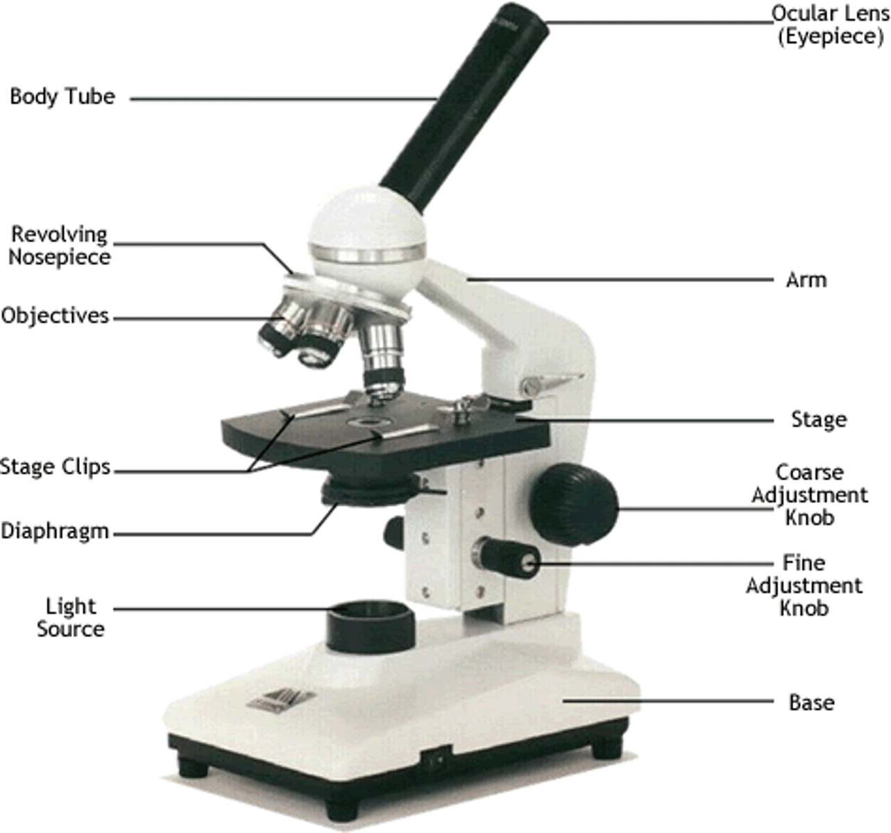
What do the condenser/light source help with?
They direct and control light through the specimen.
What is the best order for focusing?
Start on low power, focus (coarse then fine), center the specimen, then switch to higher power.
When moving to higher magnification, what should you do first?
Put the most important part of the specimen in the center of the field of view.
Why always begin with low power?
Easier to find the specimen and focus before zooming in.
What is a temporary mount?
A quick slide preparation (not permanent) for viewing cells/tissues.
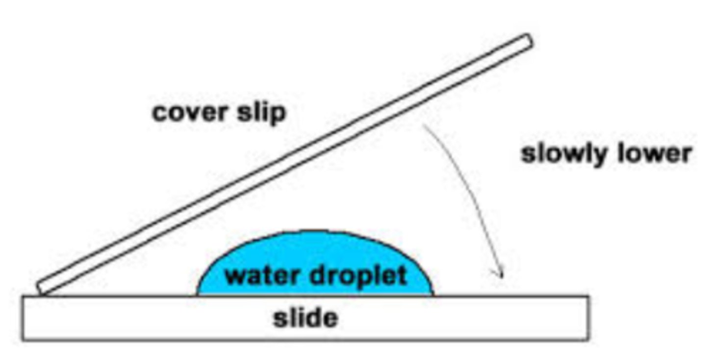
Steps to make a temporary mount (core method)?
Put specimen in a thin layer → add water or stain → lower cover slip carefully (avoid bubbles) → blot excess fluid.

Why use stains?
To make pale/transparent structures more visible.
Why can photography be better than drawings?
Photos contain real data rather than subjective interpretation.
What makes a good biological drawing?
Clear single lines, correct proportions, labels, and only key structures (not lots of shading).
What is an artefact in microscopy?
Something not naturally present, introduced by slide prep (e.g., air bubbles).
What is an eyepiece graticule?
A scale in the eyepiece used to measure structures, but it must be calibrated.
Why must a graticule be calibrated for each objective lens?
Each objective changes the scale value, so 1 unit can represent different µm at different magnifications.
Magnification formula (for drawings/micrographs)?
Magnification= actual size of specimen/ size of image
Key rule when using the magnification formula?
Use the same units for image size and actual size (convert if needed).
Unit conversions you often needed for microscopy scale
1 mm = 1000 µm; 1 µm = 10⁻⁶ m.
What is a scale bar?
A line labelled with the actual distance it represents (more reliable than stated magnification).
how to produce scale bar
To obtain the length of scale bar that should be added to the image, multiply the appropriate length that a scale bar indicates (e.g. 10 μm) by the magnification.
When were microscopes first invented?
17th century
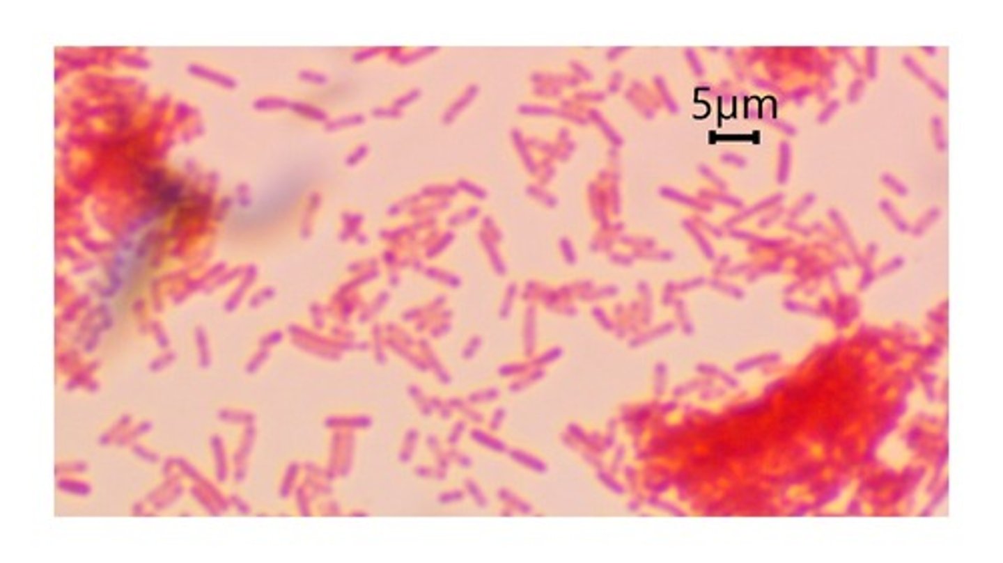
What did improved light microscopes (2nd half of the 19th century) allow scientists to discover/observe?
Bacteria and other unicellular organisms, chromosomes, and processes like mitosis, meiosis, gamete formation, and fertilization.
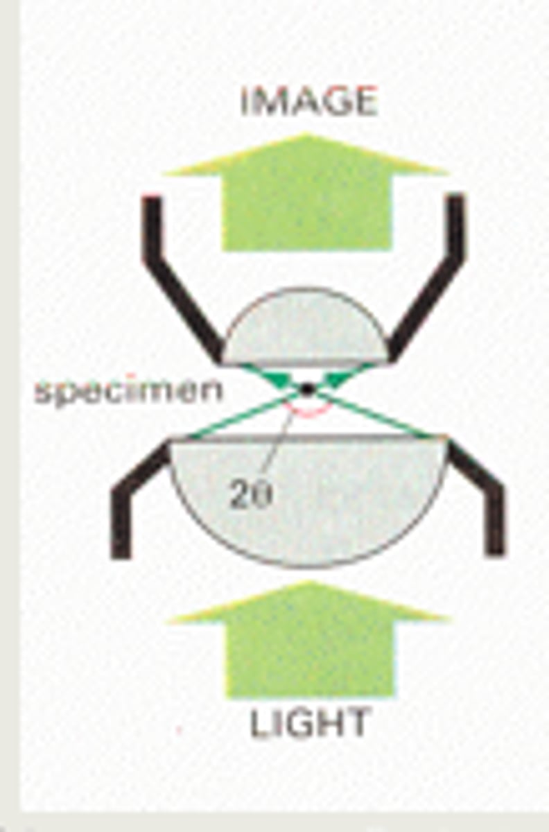
Why does increasing magnification above ~400× become difficult with light microscopes?
Because of resolution limits linked to the wavelength of visible light—close points blur together.
What is "resolution" in microscopy?
The ability to distinguish two close points as separate.
What is the approximate resolution limit of the unaided eye?
About 0.1 mm (= 100 µm = 100,000 nm).
What is the approximate resolution limit of a light microscope?
About 0.0002 mm (= 0.2 µm = 200 nm).
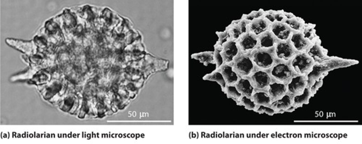
What is the approximate resolution limit of an electron microscope?
About 0.000001 mm (= 0.001 µm = 1 nm).
Why do electron microscopes have higher resolution than light microscopes?
Electrons have a much shorter wavelength than visible light.
How high can magnification go in some electron microscopes
Up to about 1,000,000×.
What is a major advantage of electron microscopes?
They reveal ultrastructure (very fine details) because of better resolution.
Give 2 disadvantages of electron microscopes.
Images are black and white (colour must be added artificially), and specimens must be in a vacuum so living cells can't be examined (electron beams can be destructive).
Why are light microscopes still widely used?
They can examine living material and produce images in colour.
Why are stains useful in microscopy?
Many cell chemicals are colourless, so stains help structures become distinguishable.
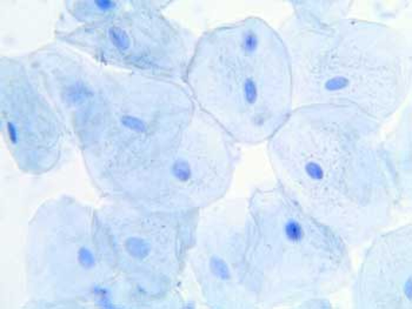
what does methylene blue stain?
It binds to DNA and RNA, staining the nucleus dark blue and cytoplasm a lighter blue.
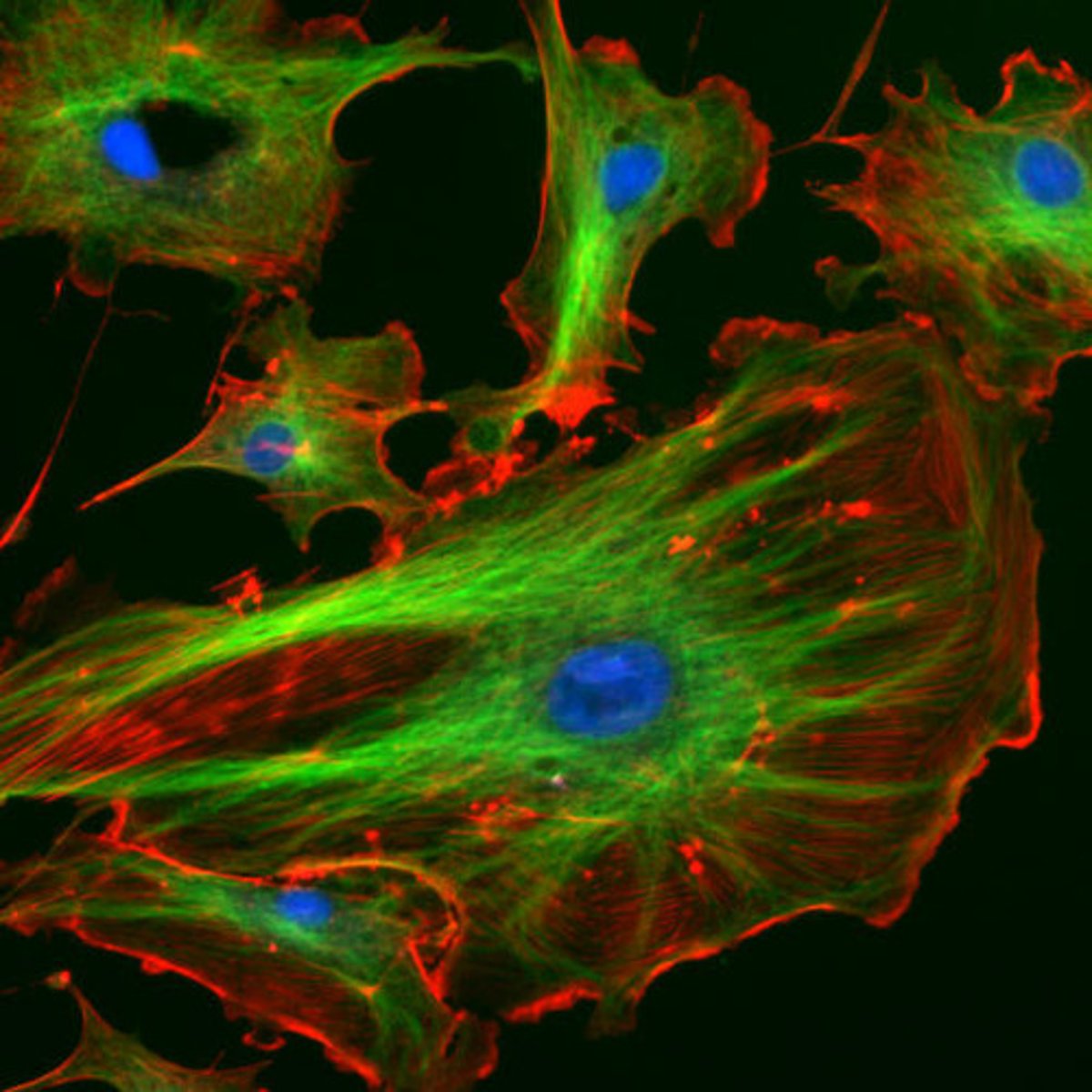
What is fluorescence?
When a substance absorbs light and re-emits it at a longer wavelength.
What special equipment is used in fluorescence microscopy to get very bright images?
An intense light source such as high-power LEDs or lasers that emit a single wavelength.
What is immunofluorescence?
A technique where antibodies bind specific antigens in cells and are linked to fluorescent markers.
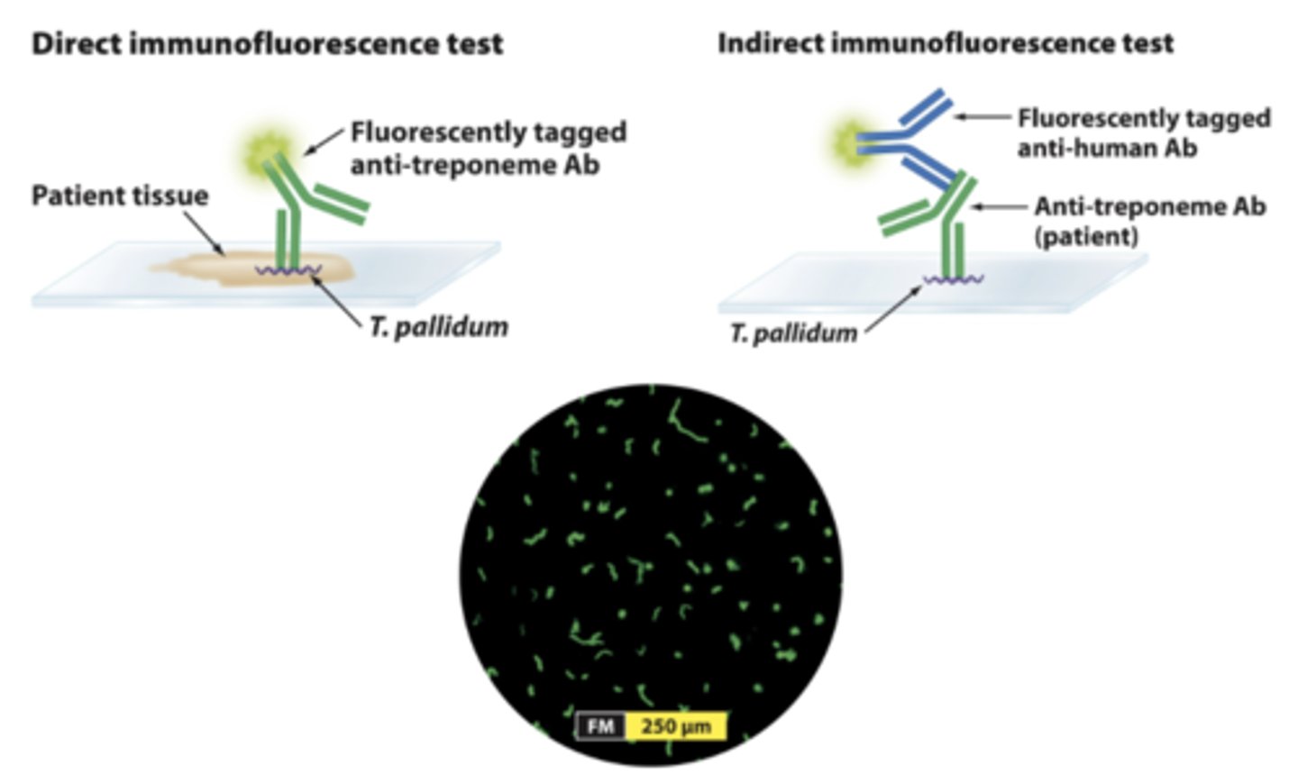
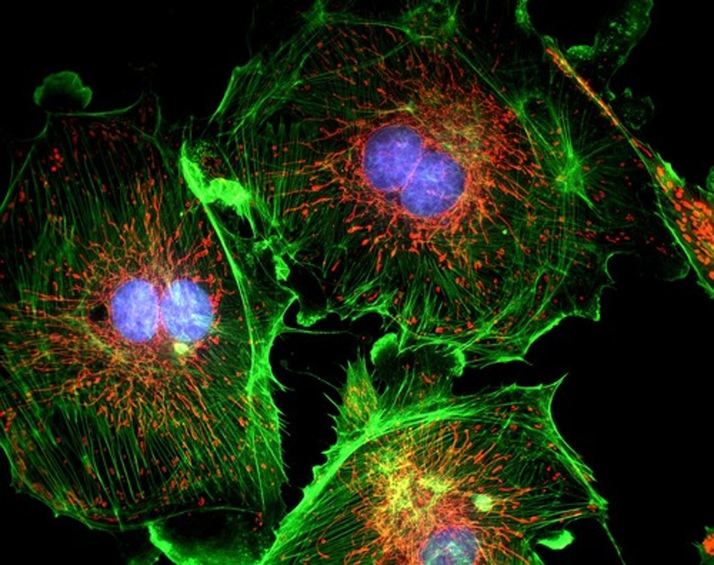
What is the advantage of using multiple fluorescent markers in immunofluorescence?
You can make multicolour images showing where different molecules are located.
What can immunofluorescence help scientists find out about a cell?
Whether a specific type of protein is being produced and where it is in the cell.