2) Trematodes - Eggs and Larvae
1/27
There's no tags or description
Looks like no tags are added yet.
Name | Mastery | Learn | Test | Matching | Spaced | Call with Kai |
|---|
No study sessions yet.
28 Terms
genus and species?
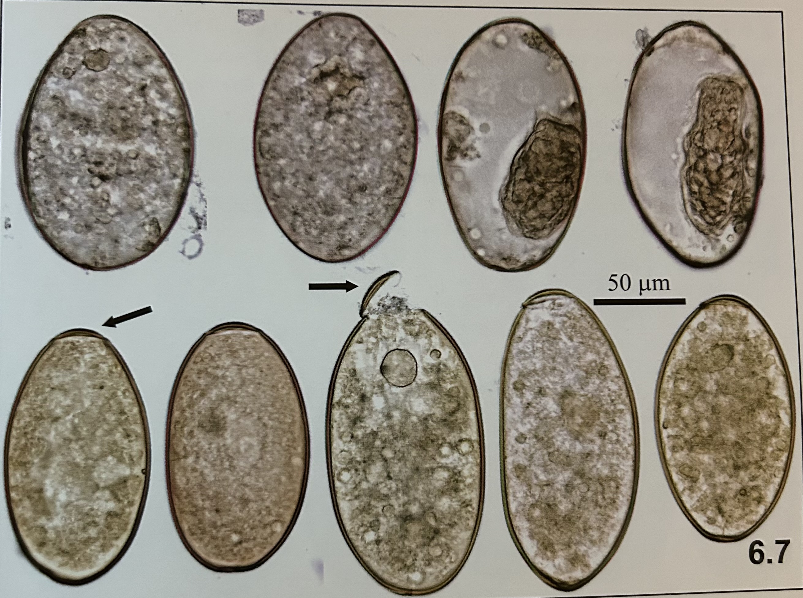
Fasciolopsis buski (egg)

structures of Fasciolopsis buski (egg)
operculum
(top row, two right = embryonated)
(bottom row = Fasciola hepatica)

developmental stage?
egg (Fasciolopsis buski)

genus and species?
Clonorchis sinensis (egg)
structures of Chlonorchis sinensis (egg)?
SMALL SIZE
(2nd and 3rd row magnified)
operculum
formed miracidiumd

developmental stage?
egg (chlonorchis sinensis)
genus and species?
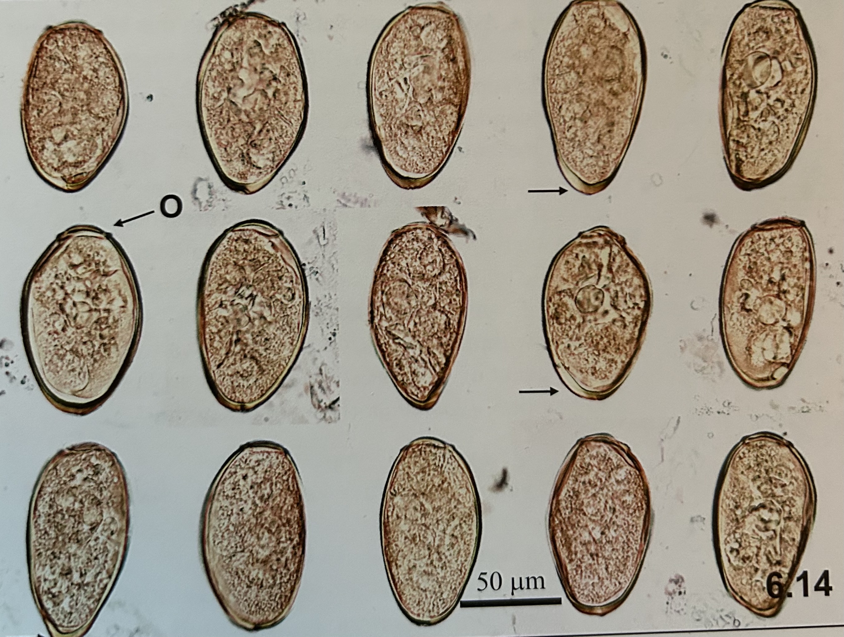
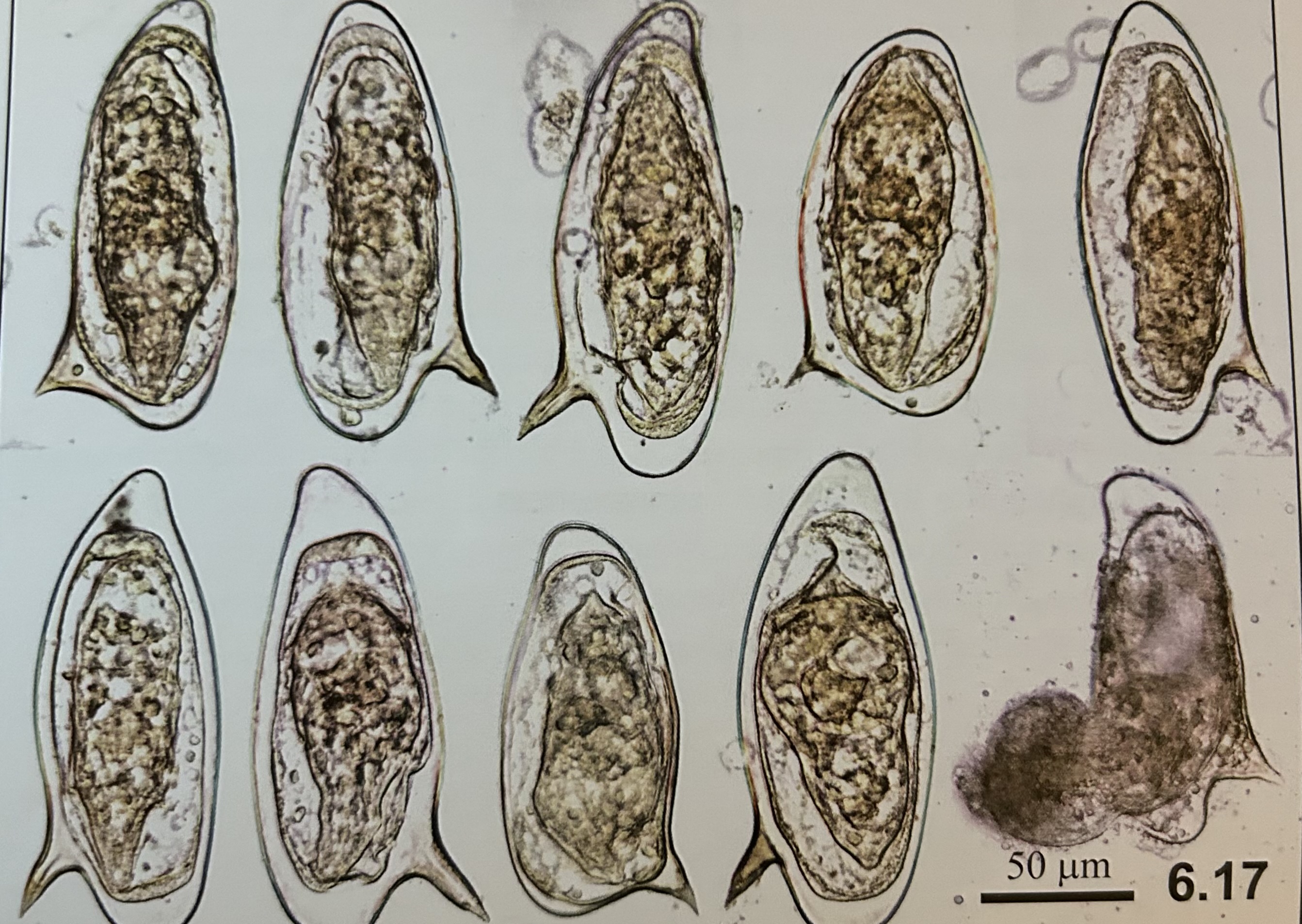
Paragonimus westernmani (egg)

structures of Paragonimus westernmani (egg)?
thick abopercular wall (arrow)
wide operculum (O) sitting in rim

developmental stage?
Paragonimus westernmani (egg)
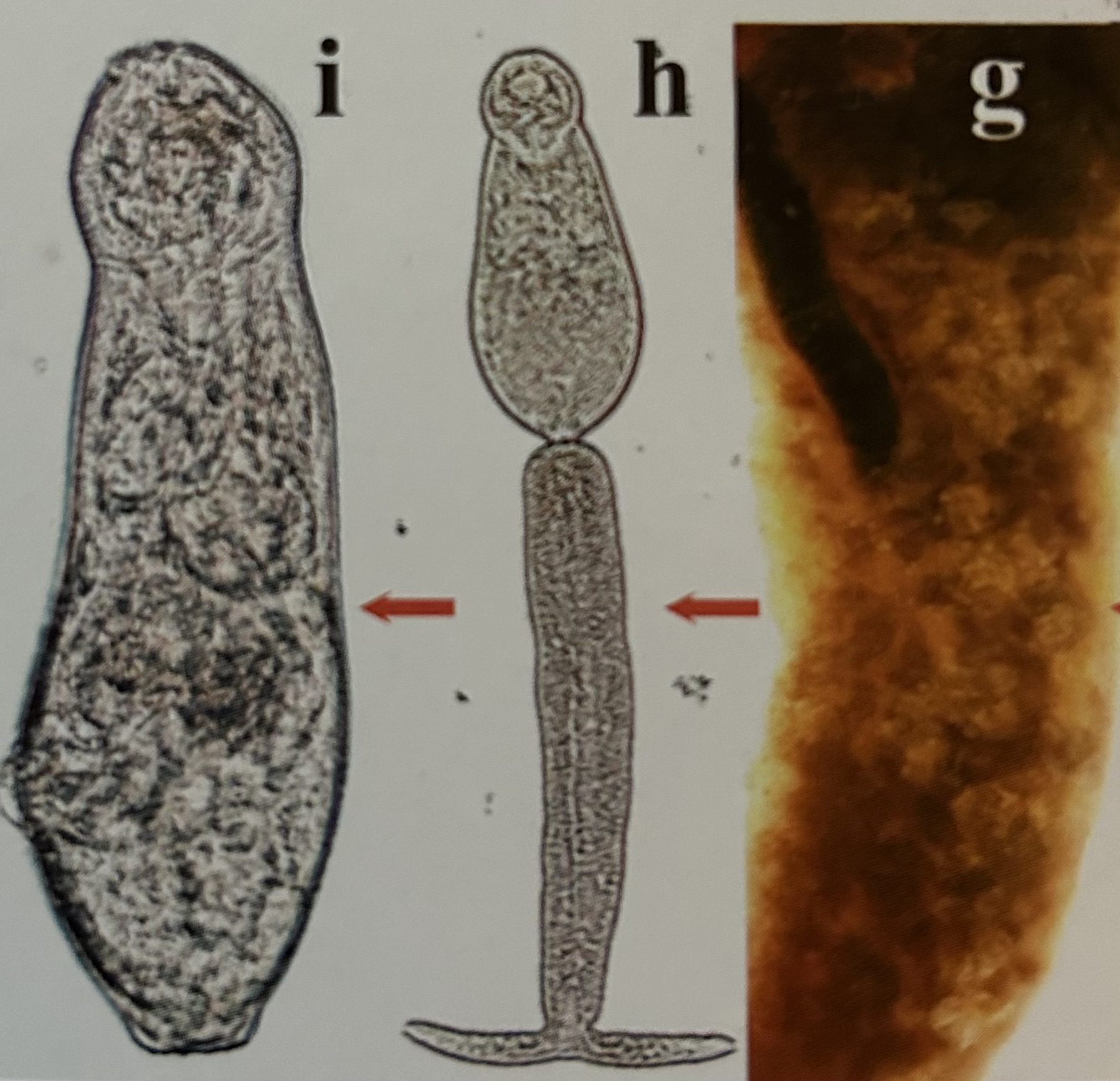
genus and species?
paragonimus westernmani (larvae)

developmental stage?
paragonimus westernmani (larvae)
genus and species?
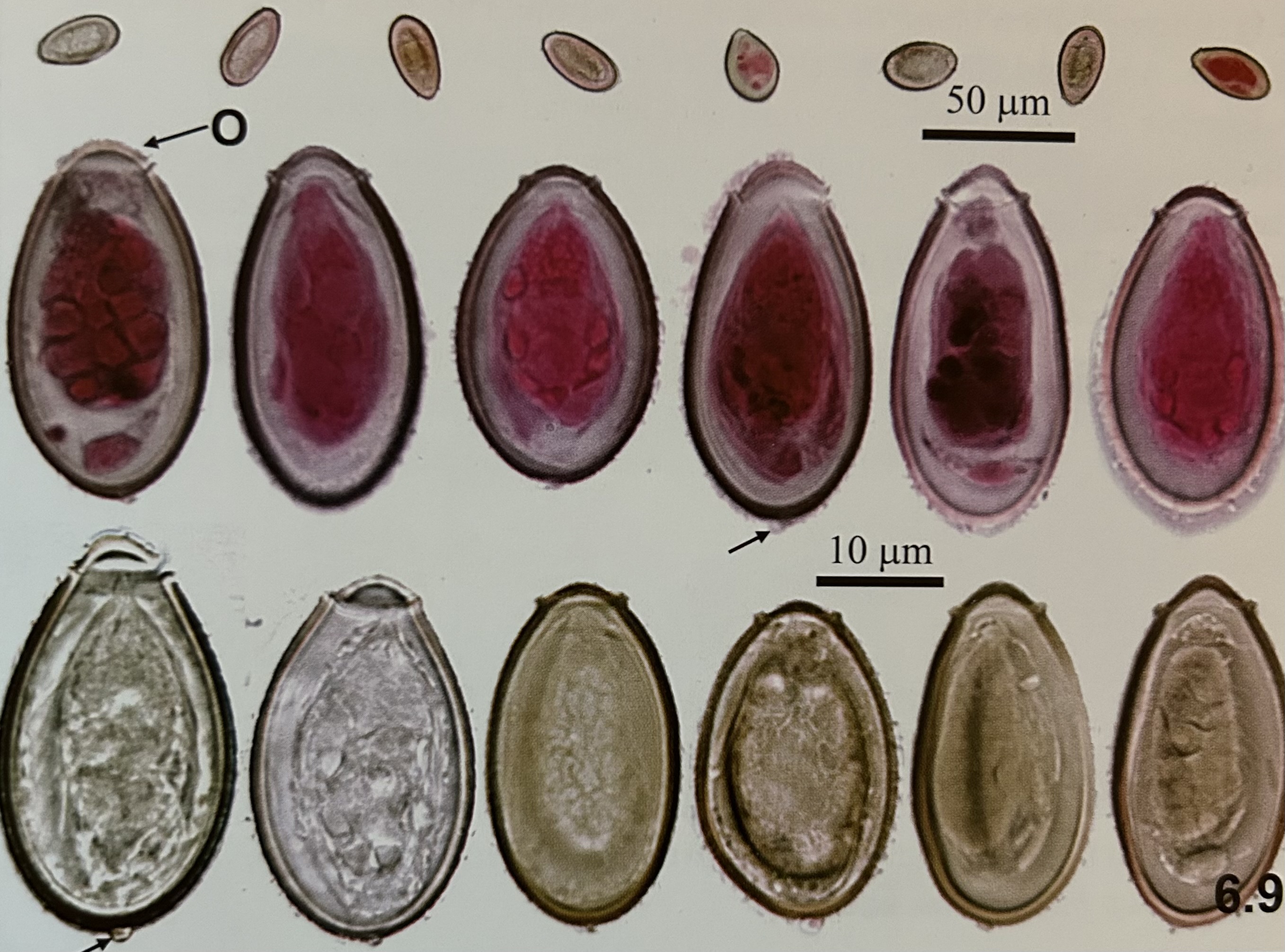
schistosoma mansoni (eggs)

structures of schistosoma mansoni (egg)?
fully formed miracidium
large lateral spine

developmental stage?
schistosoma mansoni (egg)
genus and species? (h)
Schistosoma mansoni (Cercaria)

structures of Schistosoma manosoni (cercaria)
forked tail that attaches 2 human skin
→ penetrate, called schistosomula

genus and species?
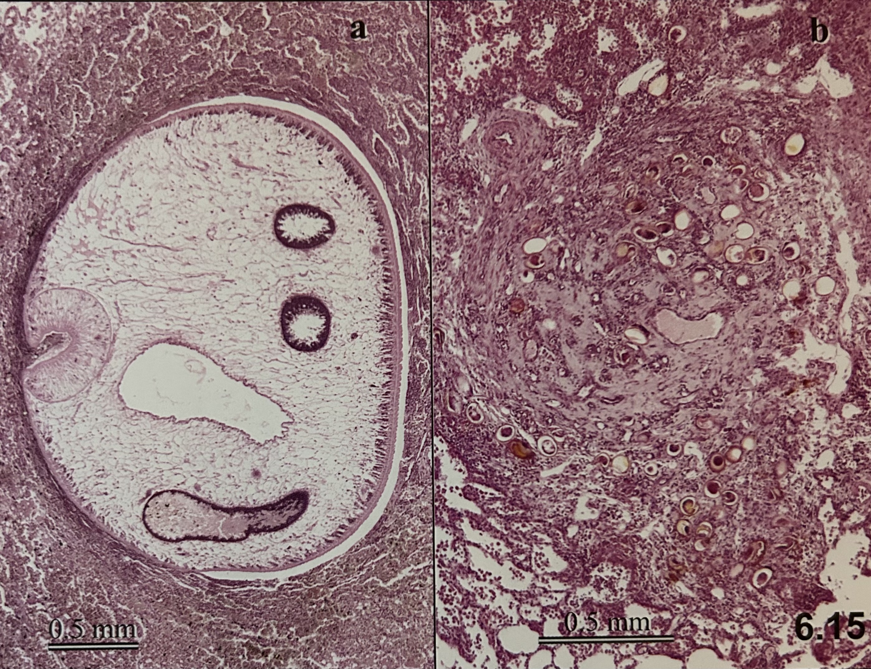
Schistosoma mansoni in liver histological section (egg)
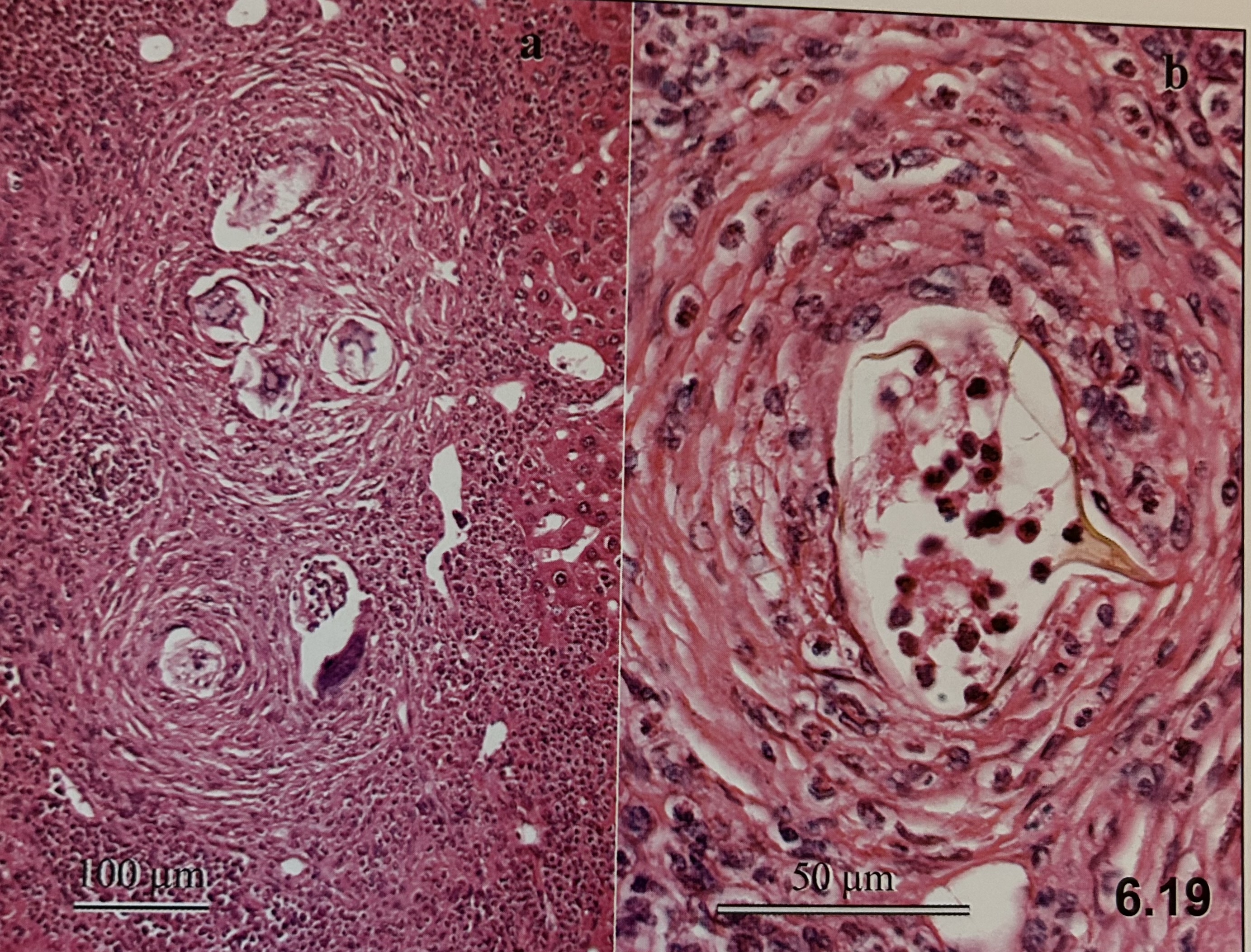
developmental stage?
Schistosoma mansoni in liver histological section (egg)

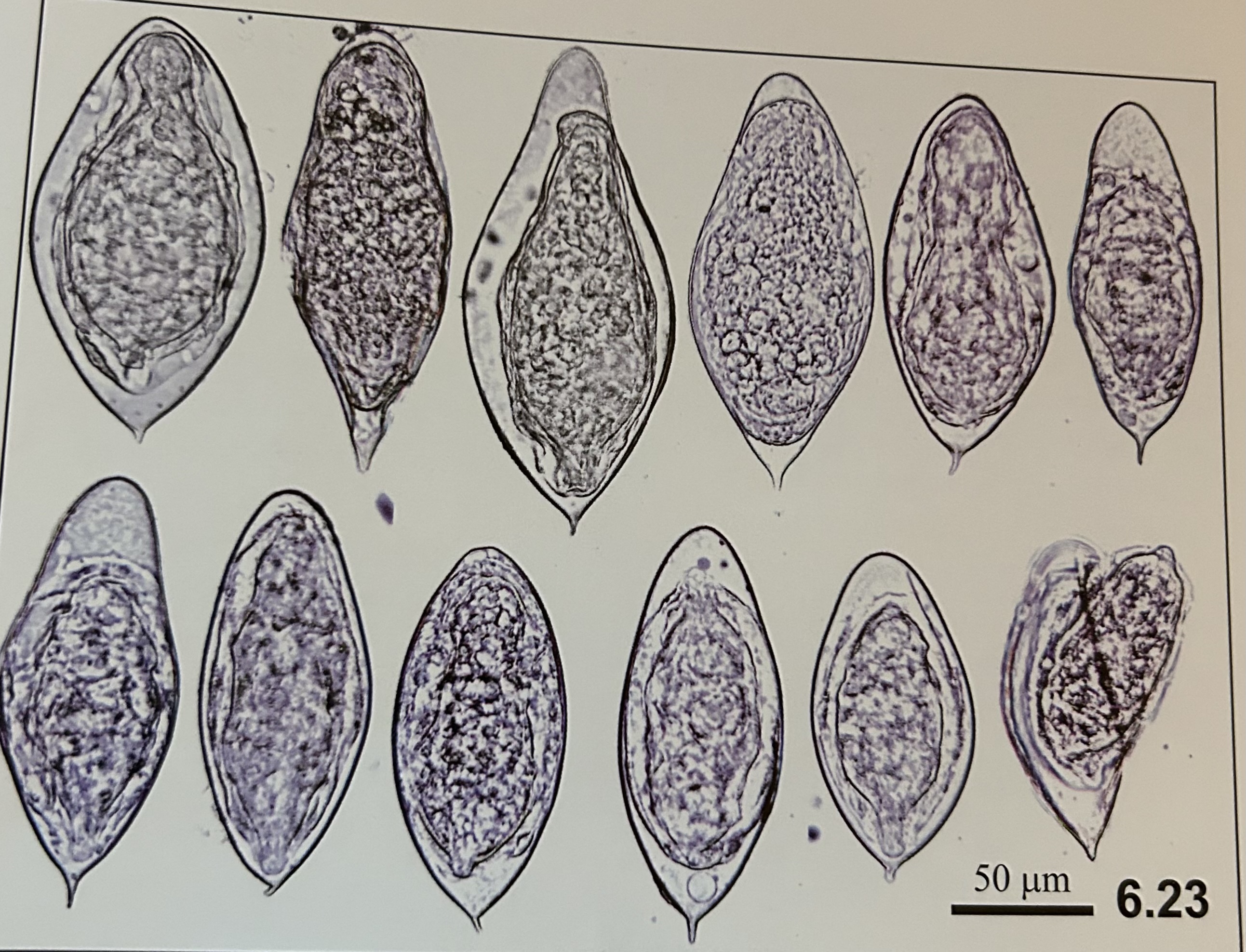
genus and species?
Schistosoma japonicum (eggs)

structures of Schistosoma japonicum (eggs)?
fully formed miracidium
minute lateral spine

developmental stage?
Schistosoma japonicum (eggs)
genus and species?
Schistosoma haematobium (eggs)

structures of Schistosoma haematobium (eggs)?
terminal spine
fully formed miracidium

developmental stage?
Schistosoma haematobium (eggs)
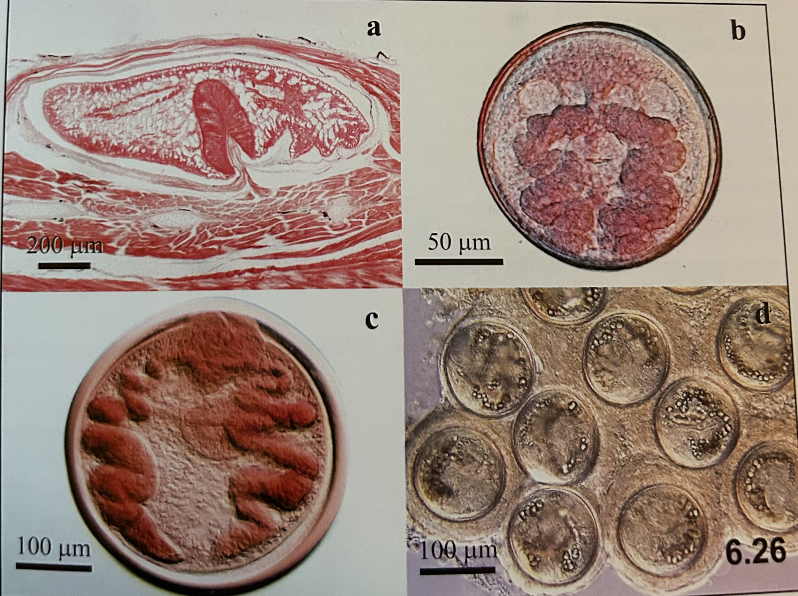
genus and species? (b and c)
b. Fasciola hepatica (metacercariae)
c. Paragonimus westernmani (metacercariae)

developmental stages?
b. Fasciola hepatica (metacercariae)
c. Paragonimus westernmani (metacercariae)

genus and species?
Fasciola hepatica (meracidium)
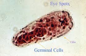
structures of Fasciola hepatica (meracidium)
eye spots
cilia
germinal cells