EXAM 2 BIO 250 (Lecture slides)
1/227
There's no tags or description
Looks like no tags are added yet.
Name | Mastery | Learn | Test | Matching | Spaced | Call with Kai |
|---|
No analytics yet
Send a link to your students to track their progress
228 Terms
Skeletal System 2 Divisions
Axial skeleton
Appendicular skeleton
Axial skeleton (Skeletal System 2 Divisions)
skull
vertebral column (spine)
thorax (ribcage)
Appendicular skeleton (Skeletal System 2 Divisions)
limbs (arms and legs)
pelvic girdle
shoulder girdle
Skeletal System Components
bones
cartilage
ligaments (bones to bones)
tendons (muscle to bones)
Skeletal System Functions
Supports body
Protects soft organs:
brain, heart, lungs, spinal cord
Attachment for skeletal muscles
Stores minerals & fats:
calcium, phosphorus, lipids
2 Types of Osseous (BONE) Tissues
Compact bone
Spongy bone
Compact bone (2 Types of Osseous (BONE) Tissues)
dense, smooth, homogeneous
Spongy bone (2 Types of Osseous (BONE) Tissues)
density varies, porous, heterogeneous
Long bones (Bones Classified by Shape)
long shaft
in limbs
Long bone examples (Bones Classified by Shape)
femur = thigh
humerus = UPPER arm
Short bones (Bones Classified by Shape)
mostly spongy
Short bone examples (Bones Classified by Shape)
carpals = wrist bones
tarsals = ankle “TALUS: singular”
sesamoid bones = INSIDE tendon
Sesamoid bone example [SHORT BONE] (Bones Classified by Shape)
patella = kneecap
Flat bones (Bones Classified by Shape)
often CURVED, NOT COMPLETELY flat
spongy bone BETWEEN 2 THIN layers of compact bones
Flat bone examples (Bones Classified by Shape)
skull
ribs
sternum = breastbone
Irregular bones (Bones Classified by Shape)
don’t fit in other categories
Irregular bone examples (Bones Classified by Shape)
vertebrae = backbones
pelvic bones
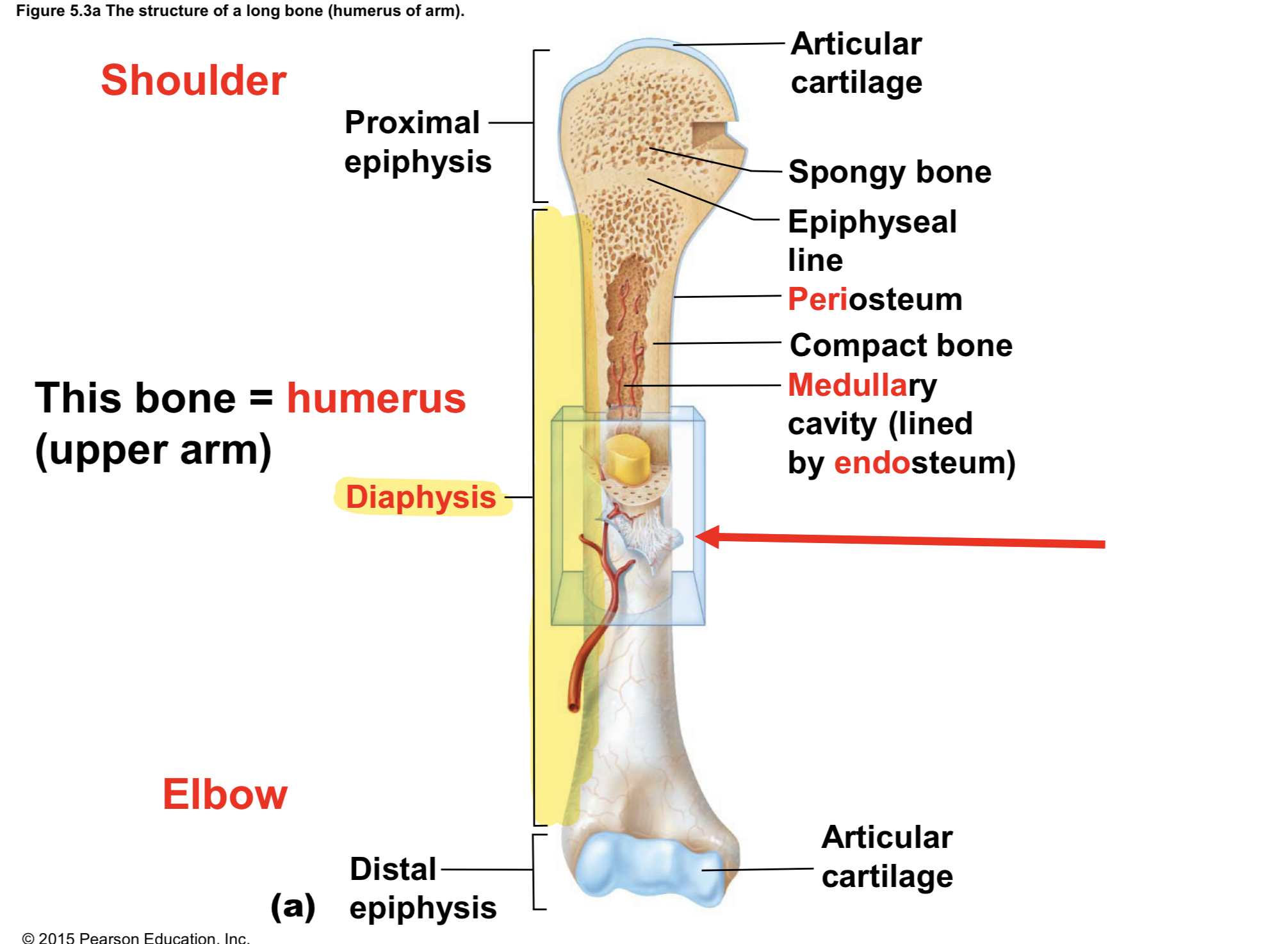
Diaphysis (Gross Anatomy of LONG Bones)
LONG shaft of the bone
MOSTLY COMPACT bone
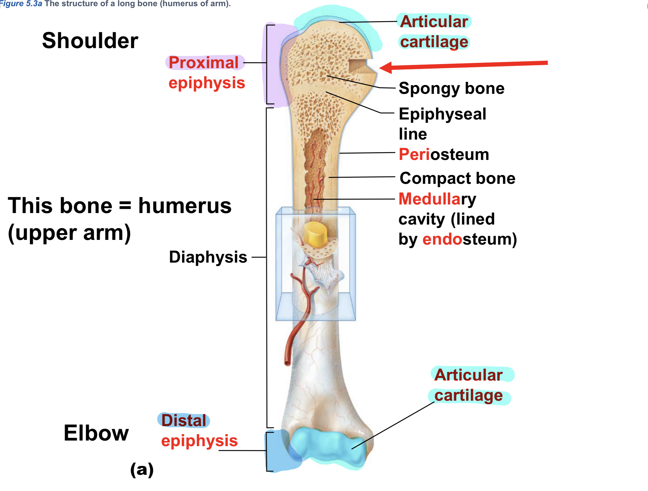
Epiphyses (Gross Anatomy of LONG Bones)
Proximal & Distal
MOSTLY SPONGY + THIN cover of COMPACT BONE
articular cartilage
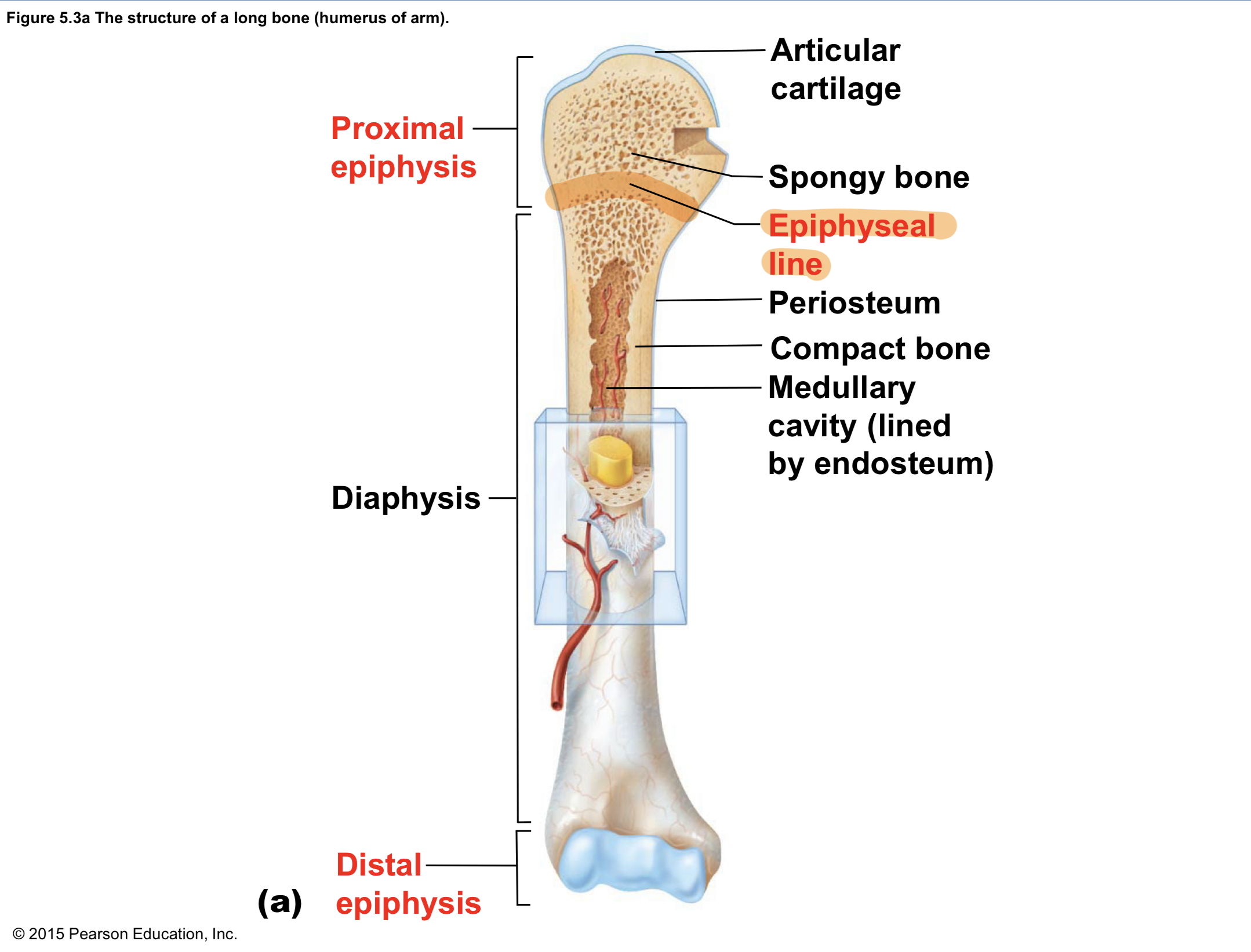
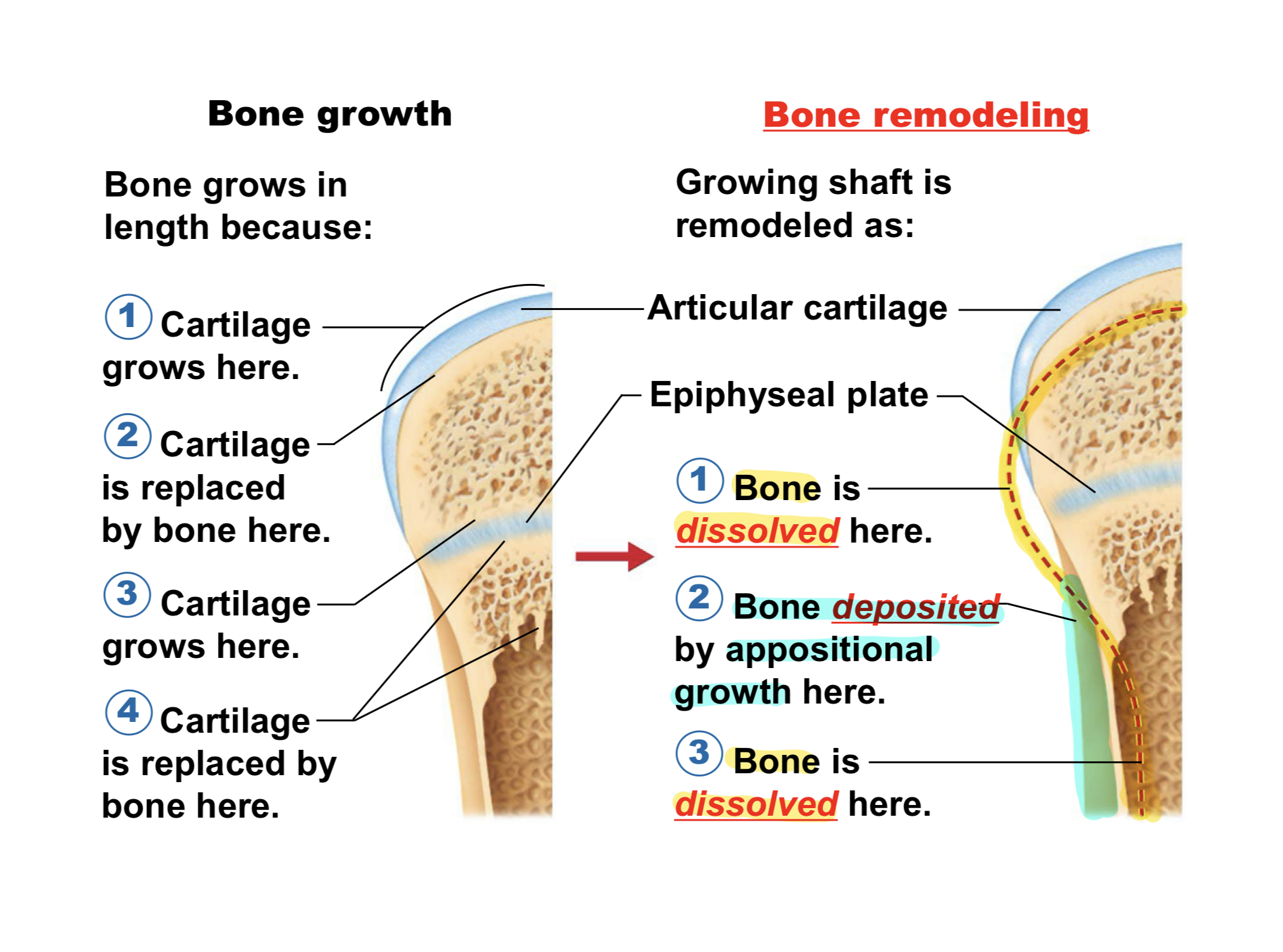
Epiphyseal PLATE (Gross Anatomy of LONG Bones)
flat plate of HYALINE cartilage in YOUNG/GROWING bone
makes long bone grow longer = growing taller!
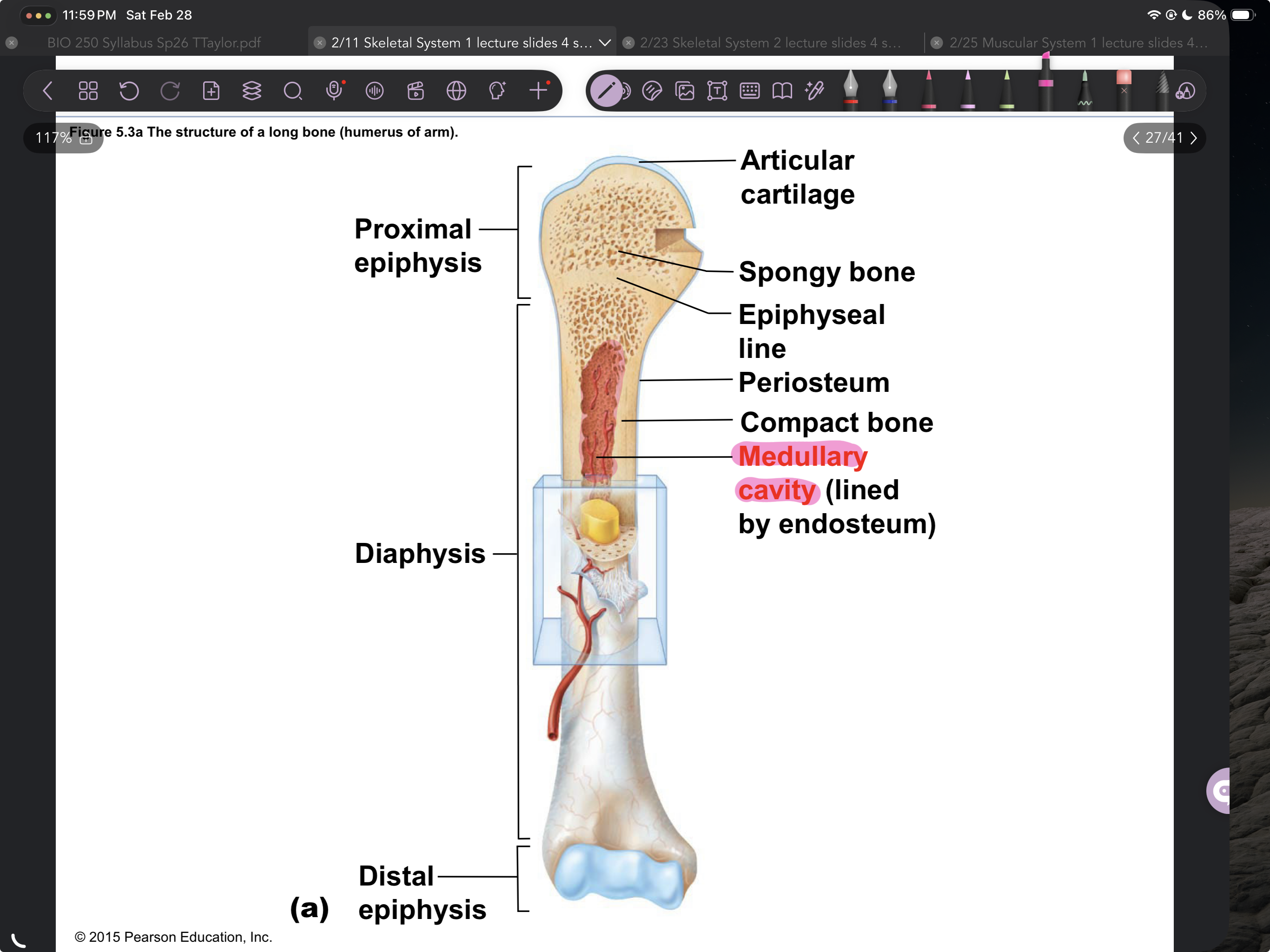
Marrow(medullary) cavity (Gross Anatomy of LONG Bones)
INSIDE diaphysis
yellow marrow in adults = mostly fat
red marrow in infants = for hematopoiesis
Bone Components
bone = CONNECTIVE TISSUE (has extracellular matrix)
HARD due to Ca salts in matrix
FLEXIBLE due to collagen fibers in matrix
OSTEOcytes (Compact Bone Histology)
BONE cells
lacunae: inside “shells” LACUNA: singular
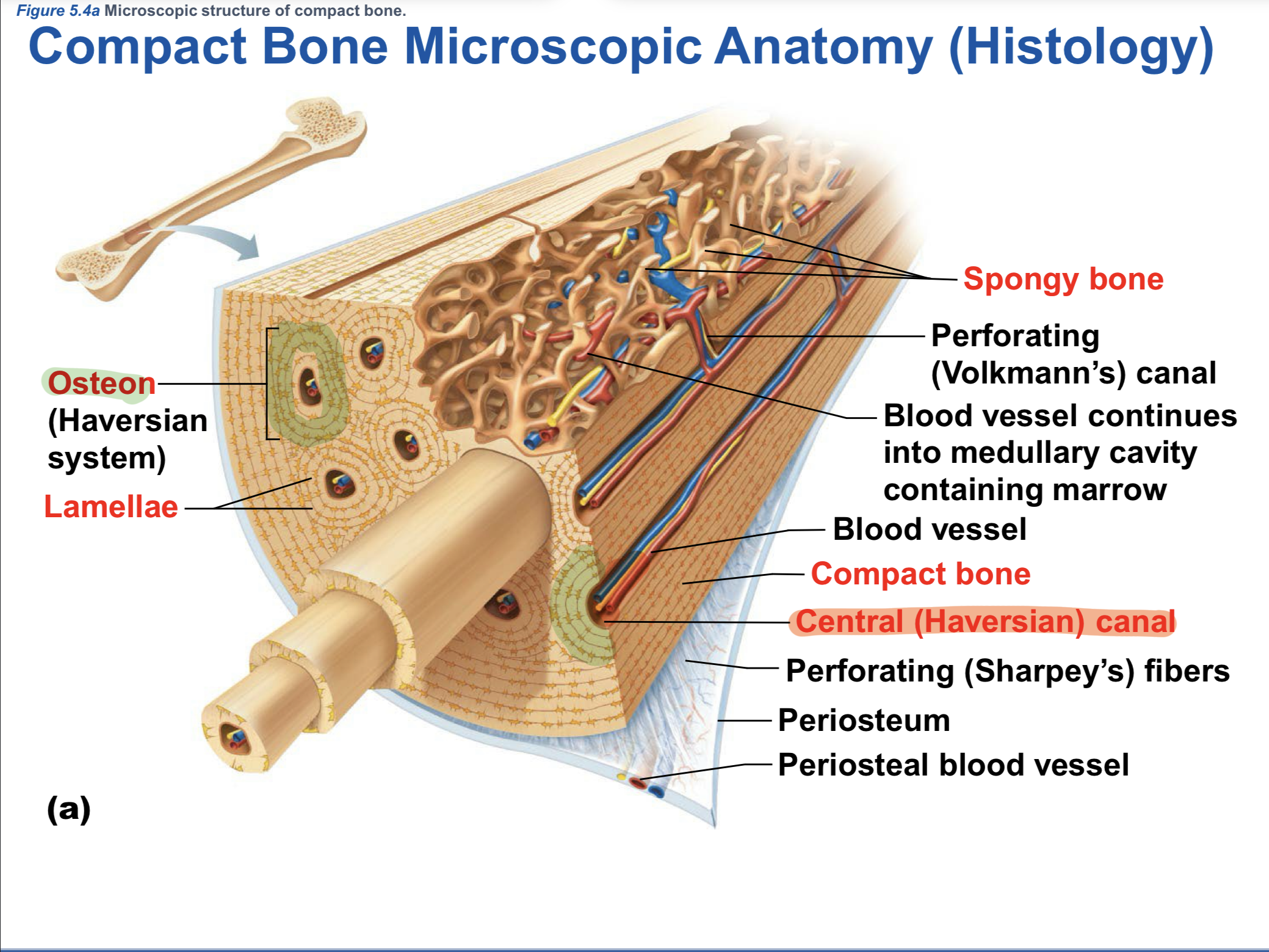
Central (Haversian) canals (Compact Bone Histology)
tube at CENTER of OSTEON
lengthwise thru bone
vessels & nerves go thru it
Canaliculi (Compact Bone Histology)
TINY CANALS, connect central canal & lacunae
Canaliculi TRANSPORT (Compact Bone Histology)
nutrients to osteocytes (glucose)
waste from osteocytes
Ossification (Bone Formation & Growth)
deposition of bony matrix in long bones BEFORE BIRTH
OsteoBLASTS [Ossification] (Bone Formation & Growth)
bone-depositing cells
swarm onto fetal skeleton made of hyaline cartilage
OsteoBLASTS digest away [Ossification] (Bone Formation & Growth)
cartilage skeleton
OsteoBLASTS deposit [Ossification] (Bone Formation & Growth)
hard bony matrix in its place
Ossification creates (Bone Formation & Growth)
medullary cavity in long bone
2 cartilages in long bones AFTER BIRTH that AREN’T REPLACED (Bone Formation & Growth)
articular cartilage
epiphyseal plate
Articular cartilage & Epiphyseal plates made of (Bone Formation & Growth)
hyaline cartilage
New cartilage grows ON (Bone Formation & Growth)
EXTERNAL faces of Articular/Epiphyseal cartilages
OsteoBLASTS digest old cartilage & deposit hard bony matrix ON (Bone Formation & Growth)
INTERNAL faces of Articular/Epiphyseal cartilages
Appositional growth (Bone Formation & Growth)
how long bones grow wider = increased diameter
controlled by human growth hormone
Epiphyseal plate cartilage CONVERTED to (Bone Formation & Growth)
bony matrix at START of ADULTHOOD
becomes epiphyseal line
growth stops
Bone Remodeling happens throughout life due to: (Bone Remodeling)
Gravity = bearing weight
Exercise = muscles pulling on bones
Ca levels in blood
Calcitonin (Bone Remodeling)
stimulus = INCREASED Ca level in blood
effector = thyroid gland
response = thyroid releases calcitonin
Calcitonin release activates (Bone Remodeling)
OsteoBLASTS: bone-depositing cells
pull out Ca ions OUT of blood
deposit Ca INTO bony matrix (bones)
OsteoBLASTS moving Ca from blood to bones leads to: (Bone Remodeling)
DECREASED Ca levels in blood = NEGATIVE FEEDBACK
Parathyroid hormone (PTH) (Bone Remodeling)
stimulus = DECREASED Ca level in blood = HYPOcalcemia
effector = parathyroid glands
response = parathyroid releases PTH
PTH release activates (Bone Remodeling)
OsteoCLASTS = bone-dissolving cells
dissolve Ca salts FROM bony matrix
release Ca ions INTO blood
OsteoCLASTS dissolving Ca from bony matrix & releasing into blood leads to: (Bone Remodeling)
INCREASED Ca levels in blood = NEGATIVE FEEDBACK
2 Types (Bone Fractures)
Simple (closed) fracture
Compound (open) fracture
Simple (closed) fracture (Bone Fractures)
doesn’t penetrate skin
Compound (open) fracture (Bone Fractures)
penetrates out of skin
Treatment of Bone Fractures
Reduction
Immobilization
Reduction (Treatment of Bone Fractures)
CLOSED reduction = bones MANUALLY placed back into position by physician
OPEN reduction = bones secured SURGICALLY
Immobilization (Treatment of Bone Fractures)
bone placed in cast/splint while it repairs
How Body Repairs Bone Fractures:
Hematoma
Fibrocartilage callus
Bony callus
Bone remodeling
Hematoma forms (How Body Repairs Bone Fractures)
blood-filled swelling/bruise
Fibrocartilage callus forms (How Body Repairs Bone Fractures)
in a couple weeks
cartilage matrix, bony matrix, & collagen fibers brought by blood to splint the break
Bony callus REPLACES (How Body Repairs Bone Fractures)
fibrocartilage callus
osteoblasts deposit bony matrix = more weeks
Bone remodeling occurs (How Body Repairs Bone Fractures)
in response to mechanical stress AFTER cast/splint removed
osteoblasts & osteoclasts do it
Tendons & ligaments are
AVASCULAR = poorly supplied with blood vessels
often need surgery, unlike bones (repair themselves)
Articulations
joints
where 2+ bones meet
Articulation Functions
hold bones in place
allow movement (most joints, NOT ALL)
Articulations are classified by:
function & structure
FUNCTIONAL Types of Joints
Synarthrosis
Amphiarthrosis
Diarthrosis
Synarthrosis (FUNCTIONAL Joints)
immobile
Amphiarthrosis (FUNCTIONAL Joints)
slightly mobile
Diarthrosis (FUNCTIONAL Joints)
freely mobile
STRUCTURAL Types of Joints
Fibrous
Cartilaginous
Synovial
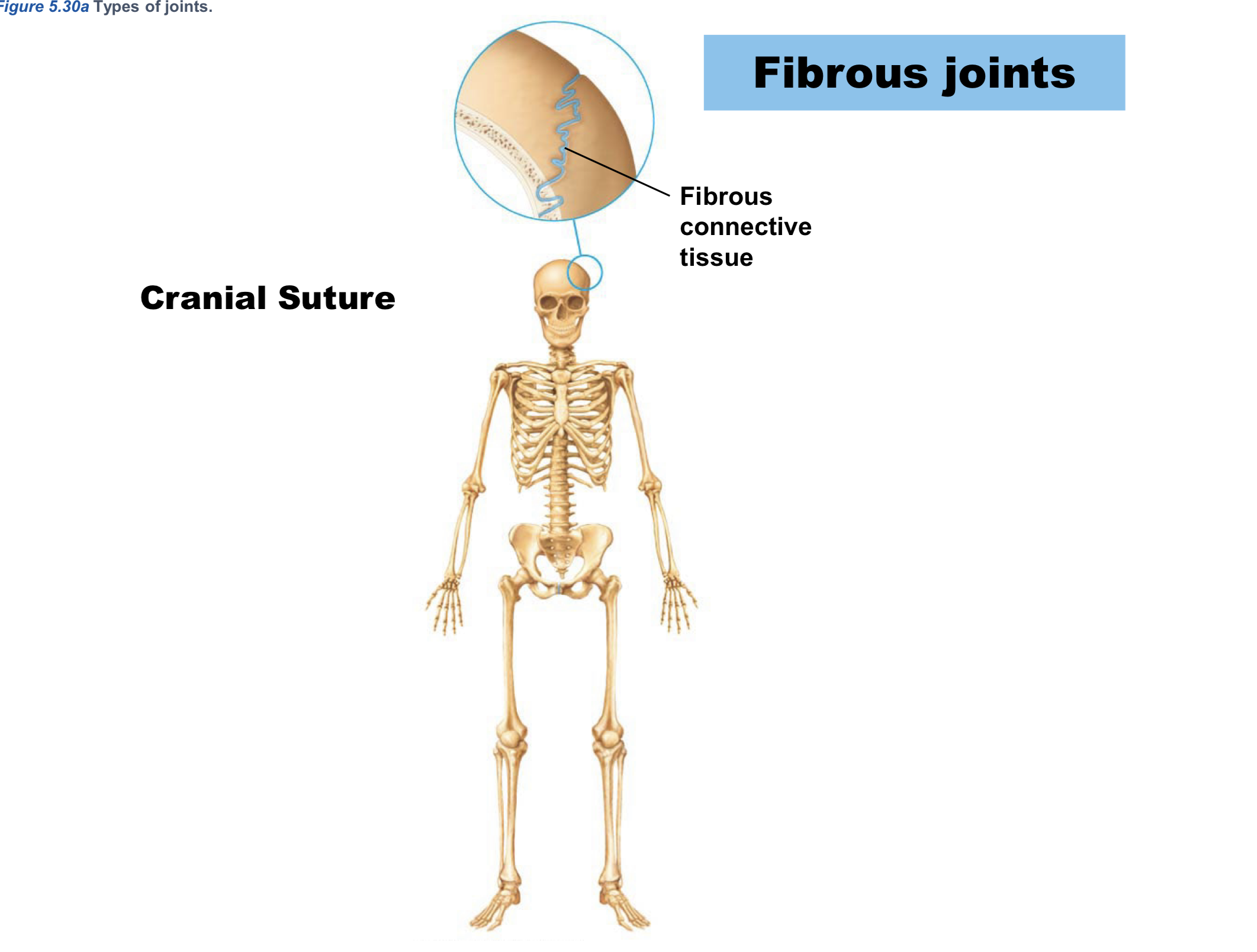
Fibrous (STRUCTURAL joints)
fibrous connective tissue between bones
usually synarthrosis
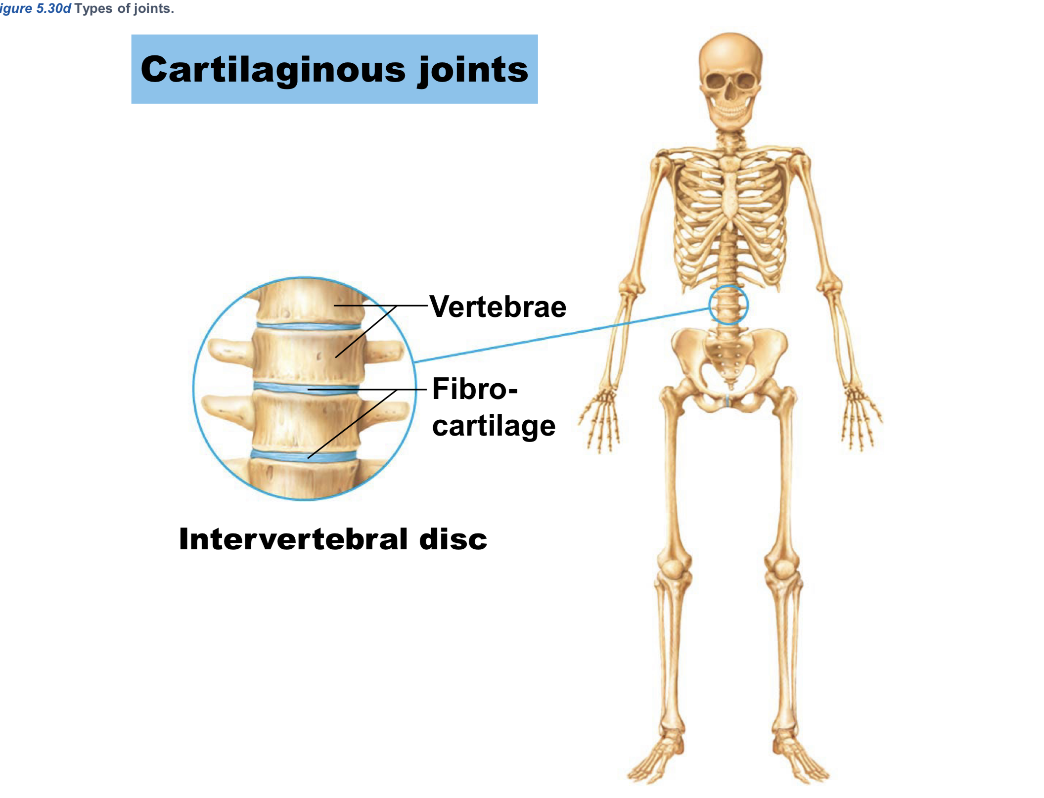
Cartilaginous (STRUCTURAL joints)
cartilage between bones
usually amphiarthrosis
Synovial (STRUCTURAL joints)
fluid-filled cavity between bones = fluid motion!
diarthrosis
Synovial Joint 4 Structures (STRUCTURAL joints)
Articular cartilage
Articular capsule
Fluid-filled cavity = synovial fluid!
Reinforcing ligaments (bone to bone)
Synovial Joint Extra Structures [reduce stress/friction]
bursae
tendon sheaths
menisci
![<p>Bursa<span style="color: red;">e</span> (Synovial Joint Extra Structures [reduce stress/friction])</p>](https://assets.knowt.com/user-attachments/73b247f3-15d3-4ccf-a9ab-e612f70f283e.png)
Bursae (Synovial Joint Extra Structures [reduce stress/friction])
flattened fibrous sacs
lined w/ synovial membrane
filled w/ synovial fluid
![<p>Tendon sheath<span style="color: red;">s </span><span style="color: rgb(255, 255, 255);">(</span>Synovial Joint Extra Structures [reduce stress/friction])</p>](https://assets.knowt.com/user-attachments/16b1939b-5b3e-4de1-a20f-051b2d58f21b.png)
Tendon sheaths (Synovial Joint Extra Structures [reduce stress/friction])
elongated bursae wrapped around tendons
tendons connect skeletal muscle to bone
Menisci (Synovial Joint Extra Structures [reduce stress/friction])
fibrocartilage discs in knee joints
MENISCUS: singular
Synovial Joint Types:
Plane
Hinge
Pivot
Condylar
Saddle
Ball & Socket
Synovial Joint Directions of Movement
uniaxial
biaxial
multiaxial
Uniaxial (Synovial Joint Directions of Movement)
movement in 1 direction
Biaxial (Synovial Joint Directions of Movement)
movement in 2 directions
Multiaxial (Synovial Joint Directions of Movement)
movement in multiple directions
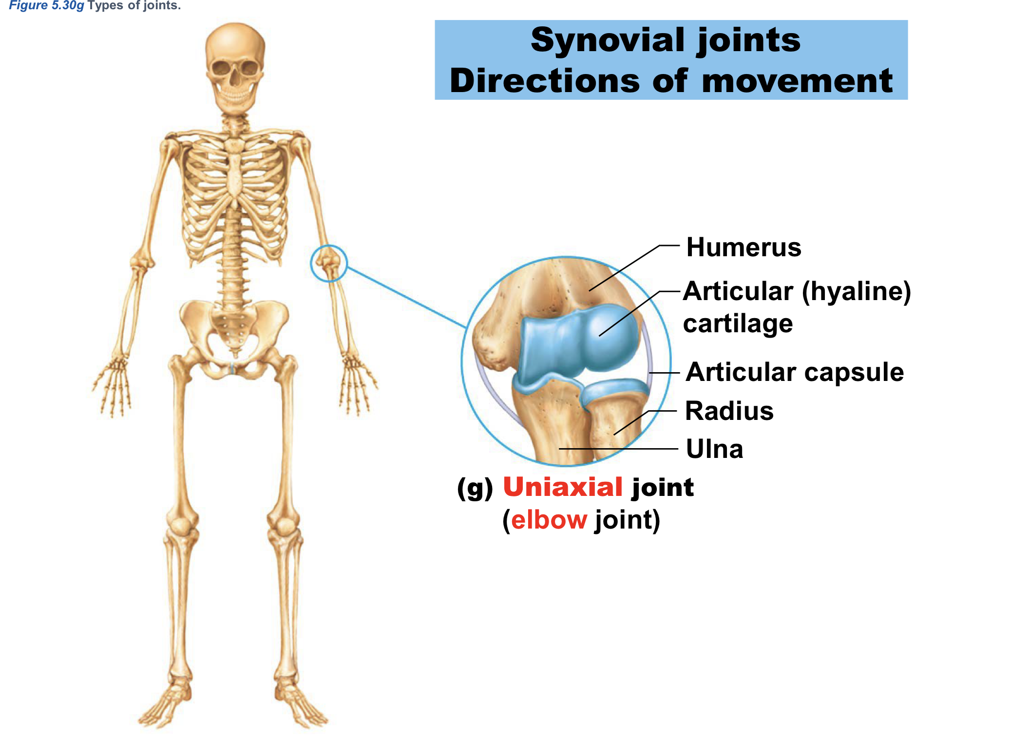
Uniaxial joint example:
elbow joint
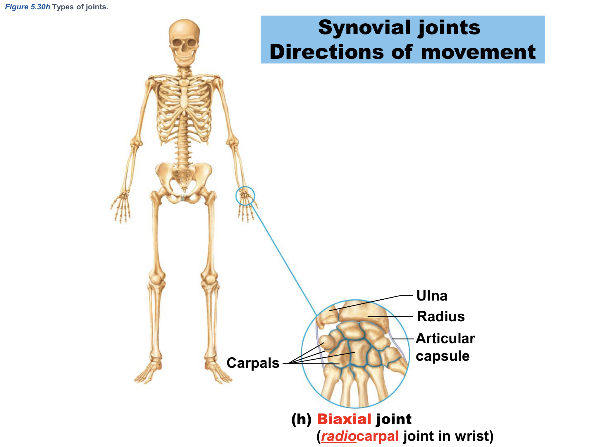
Biaxial joint example:
radoiocarpal joint in wrist
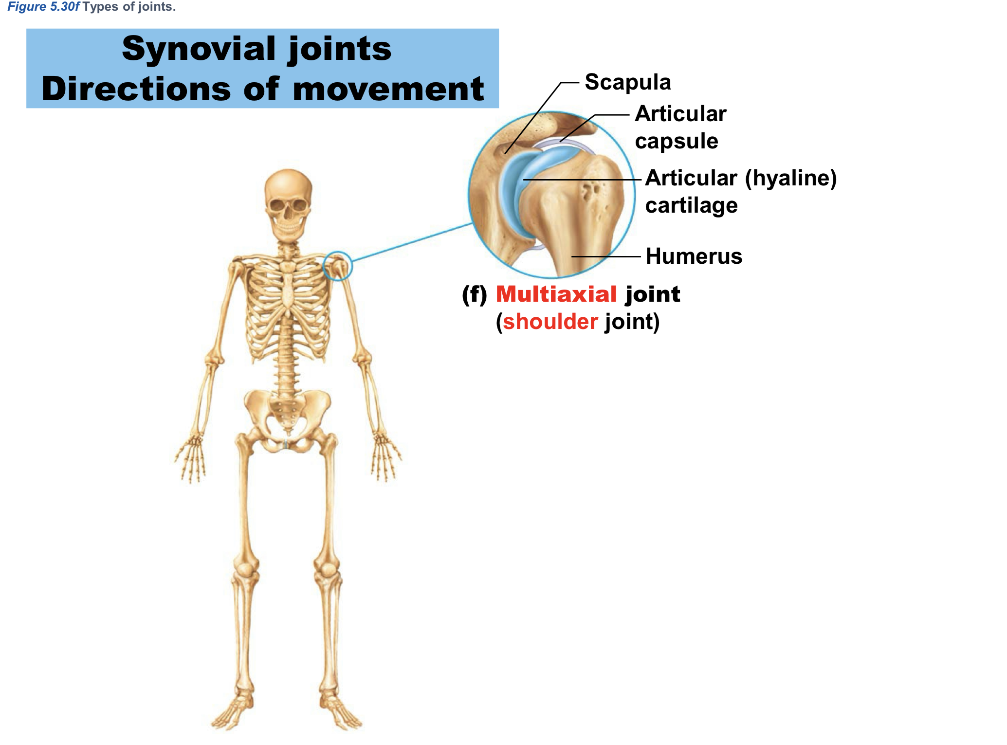
Multiaxial joint example:
shoulder joint
Terms relating to Muscles (Muscular System)
myo
mys
sarco
Cardiac Muscle Control (Muscular System)
involuntary
Cardiac Muscle Location (Muscular System)
WALLS of HEART
Cardiac Muscle Function (Muscular System)
pumps blood through blood vessels
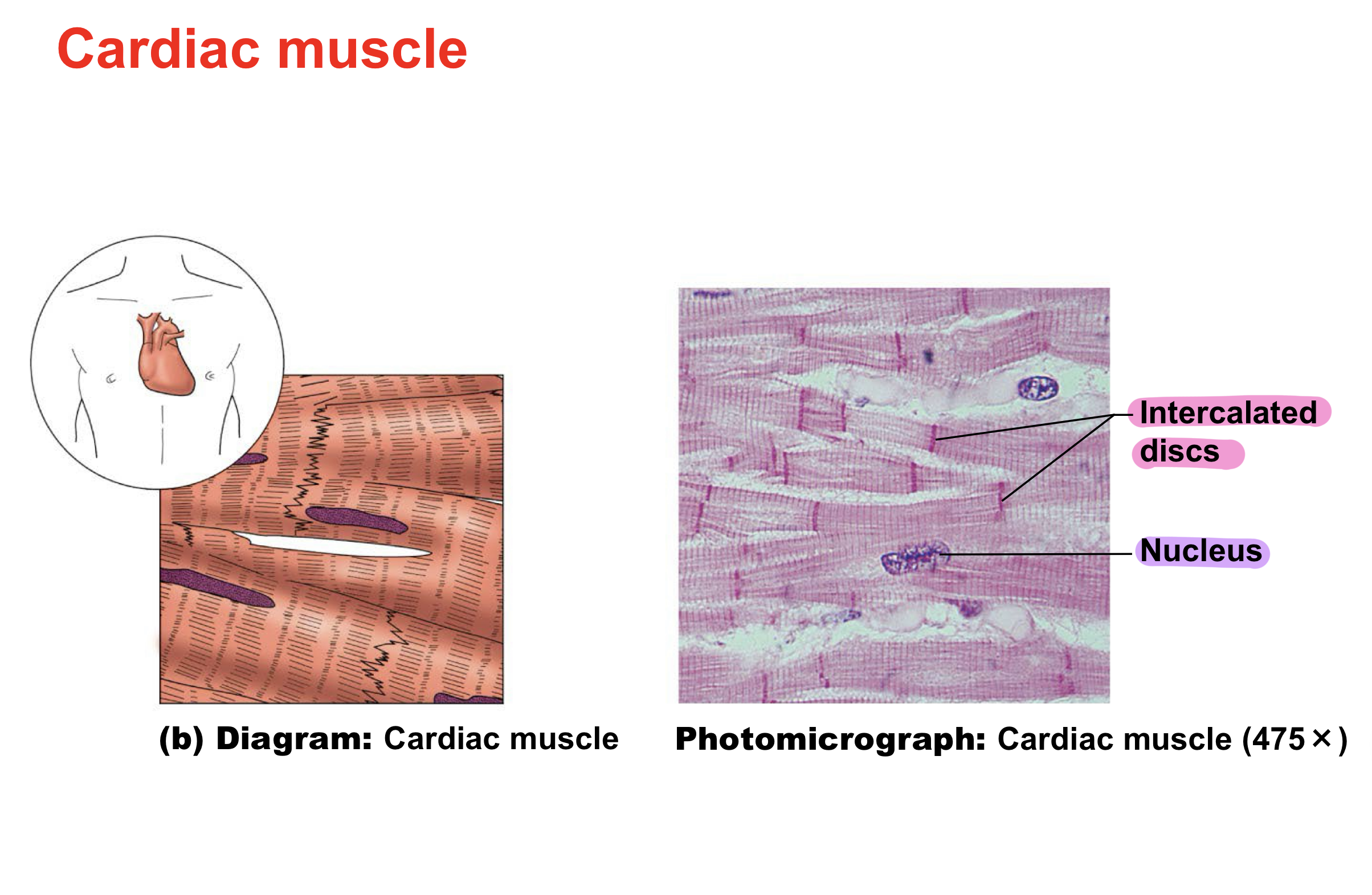
Cardiac Muscle Unique features of Fibers (cells) (Muscular System)
Intercalated discs
Striated (striped)
Short
Branched
Smooth Muscle Control (Muscular System)
involuntary
Smooth Muscle Location (Muscular System)
WALLS of hollow organs
stomach, uterus, blood vessels, urinary bladder
Smooth Muscle Function (Muscular System)
PROPELS substances through organs
food, urine, blood, sperm & egg
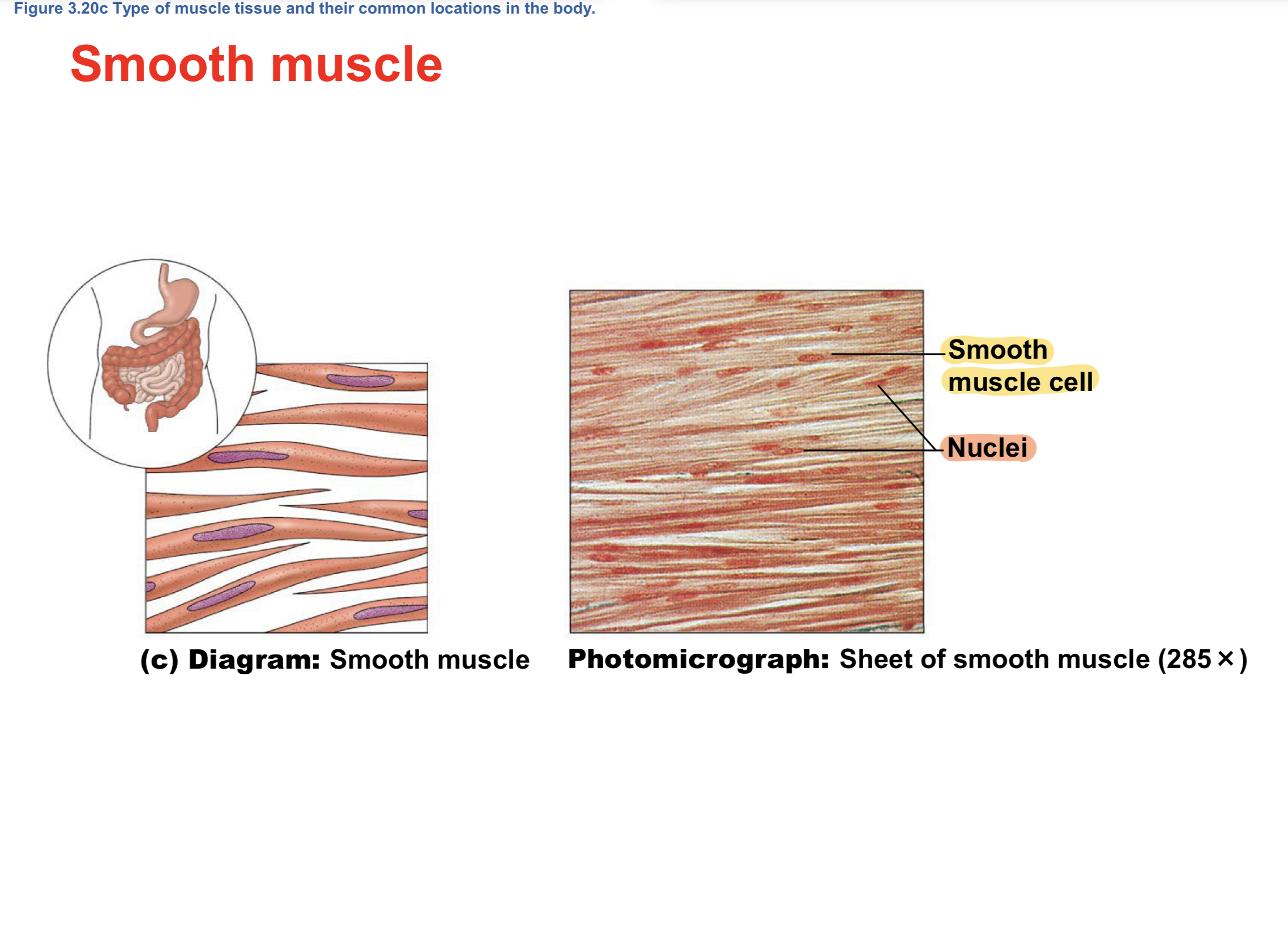
Smooth Muscle Unique features of Fibers (cells) (Muscular System)
NOT striated
Pointed at 2 ends
Skeletal Muscle Control (Muscular System)
voluntary
Skeletal Muscle Location (Muscular System)
attached to bones & skin
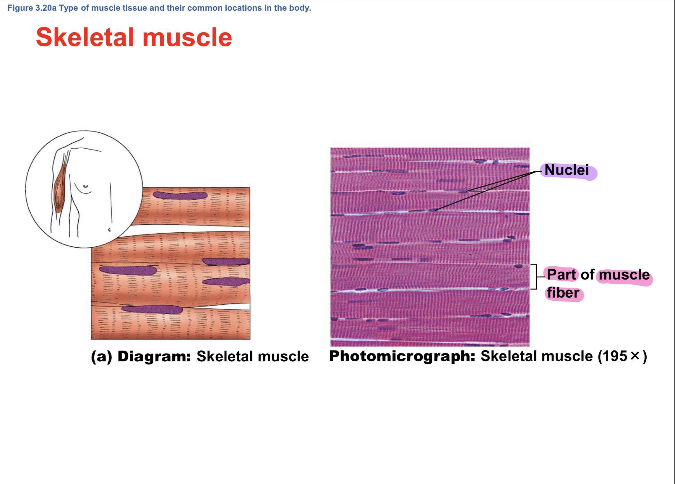
Skeletal Muscle Unique features of Fibers (cells) (Muscular System)
Multinucleate (>1 nucleus)
Striated (striped)
Long, straight cylinders
Skeletal Muscle FunctionS (Muscular System)
movement
maintain body position (posture)
stabilize joints
generate heat
facial expressions
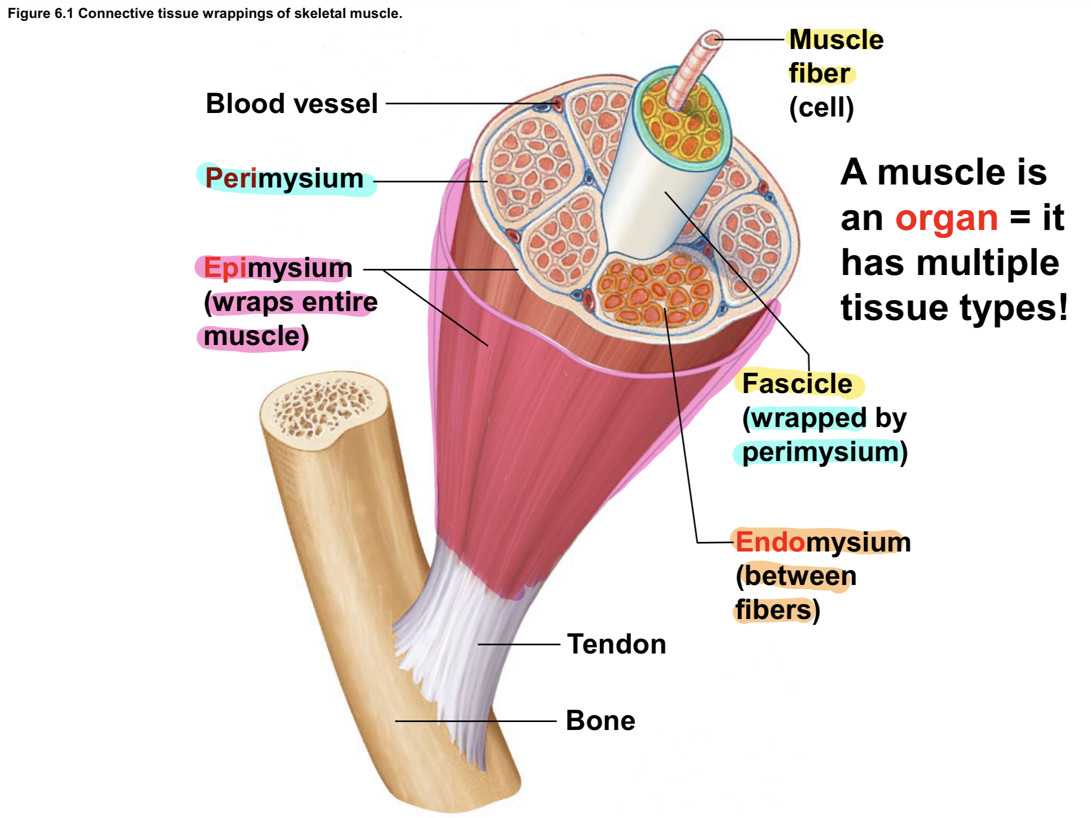
CT in Skeletal Muscles EXTERIOR to INTERIOR (Muscular System)
Fascia (Hypodermis) EXTERIOR
EPImysium
PERImysium
ENDOmysium INTERIOR
![<p>Fascia (Hypodermis) <span style="color: yellow;"><strong>LOCATION</strong></span><span style="color: red;"> </span><span style="color: rgb(255, 251, 251);">[</span><span style="color: red;"><span>CT</span></span> in <span style="color: red;"><span>Skeletal Muscles</span></span> <span style="color: rgb(116, 254, 154);"><span>EXTERIOR to INTERIOR</span></span><span style="color: rgb(254, 254, 254);"><span>] (Muscular System) </span></span></p>](https://assets.knowt.com/user-attachments/140bd574-8fea-4f77-874d-b985d467f2a9.png)
Fascia (Hypodermis) LOCATION [CT in Skeletal Muscles EXTERIOR to INTERIOR] (Muscular System)
BINDS muscle & skin
![<p><span style="color: rgb(8, 236, 251);"><span>EPI</span></span>mysium <span style="color: yellow;"><strong><span>LOCATION</span></strong></span> <span style="color: rgb(255, 251, 251);"><span>[</span></span><span style="color: red;"><span>CT</span></span> in <span style="color: red;"><span>Skeletal Muscles</span></span> <span style="color: rgb(116, 254, 154);"><span>EXTERIOR to INTERIOR</span></span><span style="color: rgb(254, 254, 254);"><span>] (Muscular System)</span></span></p>](https://assets.knowt.com/user-attachments/574634ca-dbf1-42ff-9c3a-5f4c28fee449.png)
EPImysium LOCATION [CT in Skeletal Muscles EXTERIOR to INTERIOR] (Muscular System)
covering around whole skeletal muscle
![<p><span style="color: rgb(8, 236, 251);"><span>PERI</span></span>mysium <span style="color: yellow;"><strong><span>LOCATION</span></strong></span> <span style="color: rgb(255, 251, 251);"><span>[</span></span><span style="color: red;"><span>CT</span></span> in <span style="color: red;"><span>Skeletal Muscles</span></span> <span style="color: rgb(116, 254, 154);"><span>EXTERIOR to INTERIOR</span></span><span style="color: rgb(254, 254, 254);"><span>] (Muscular System) </span></span></p>](https://assets.knowt.com/user-attachments/81d4d8e1-4bf4-4f7c-a380-dbc6dbe24de1.png)
PERImysium LOCATION [CT in Skeletal Muscles EXTERIOR to INTERIOR] (Muscular System)
covers fascicle (bundle) of muscle fibers
![<p><span style="color: rgb(8, 236, 251);"><span>ENDO</span></span>mysium <span style="color: yellow;"><strong><span>LOCATION</span></strong></span> <span style="color: rgb(255, 251, 251);"><span>[</span></span><span style="color: red;"><span>CT</span></span> in <span style="color: red;"><span>Skeletal Muscles</span></span> <span style="color: rgb(116, 254, 154);"><span>EXTERIOR to INTERIOR</span></span><span style="color: rgb(254, 254, 254);"><span>] (Muscular System) </span></span></p>](https://assets.knowt.com/user-attachments/aac68159-b787-40ee-8048-2df0a6ce250c.png)
ENDOmysium LOCATION [CT in Skeletal Muscles EXTERIOR to INTERIOR] (Muscular System)
covers individual muscle fibers
Fascia (Hypodermis) TISSUE [CT in Skeletal Muscles EXTERIOR to INTERIOR] (Muscular System)
adipose CT
EPImysium TISSUE [CT in Skeletal Muscles EXTERIOR to INTERIOR] (Muscular System)
fibrous CT