Haemoglobinopathies & Mutation
1/36
There's no tags or description
Looks like no tags are added yet.
Name | Mastery | Learn | Test | Matching | Spaced |
|---|
No study sessions yet.
37 Terms
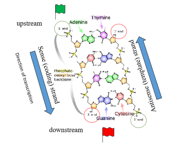
Explain the structure of DNA
Sense (coding) strand: Same sequence as the mRNA (except T → U). It runs 5’ → 3’ downstream.
Antisense (template) strand: The strand actually used by RNA polymerase to synthesize mRNA. It runs 3’ → 5’ upstream.
Transcription occurs in the 5′ → 3′ direction, moving downstream.
Mutation
A permanent heritable change in the nucleotide sequence of a gene or chromosome
Mutation may be described in terms of change in what 3 things
Mutation may be described in terms of change in:
• genomic DNA (prefix g.)
• complimentary (coding) DNA (prefix c.)
• protein ( prefix p.)
What does this mutation mean: G > A
G changes to A (Guanine is replaced by Adenine)
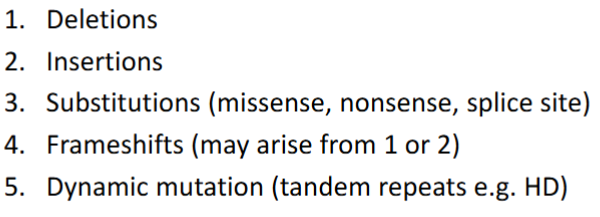
Classification of Mutations (5 groups)
Match each of these point mutations to their names:
A. Deletion
B. Insertion
C. Substitution
Normal: GAT CGT CAT GTG CAT
GAT CGT CAC GTG CAT
GAT CGT CAT CGT GCA T
GAT CGT CA_G TGC AT
1C
2B
3A
Name the mutations represented by each of these: CAN YOU LET THE DOG OUT
CAN YOU LET THE LOG OUT
CAN YOU LET XXX
CAN YOU _______ OUT
CAN ____ LET THE DOG OUT
CAN YUL ETT HED OGO UT
CAN YOU YOU YOU YOU YOU….. LET THE DOG OUT
Missense (altered but usually readable)
Nonsense (premature stop codon → loss of function)
Splice site (introns left in or exons spliced out)
In frame (most amino acids (words) the same)
Frameshift (protein (sentence) unreadable)
Trinucleotide repeat (sequence repeated excessively → gene silencing or abnormal protein)
What are 3 Functional consequences of mutations
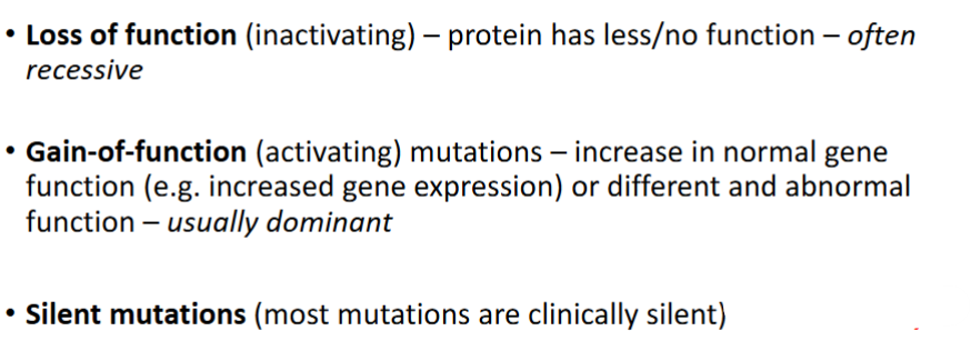
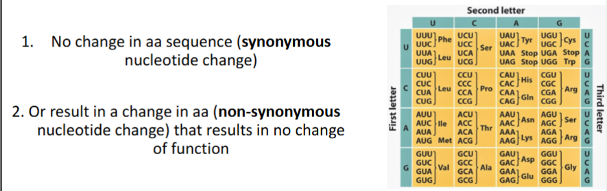
What is the result of Silent Mutations
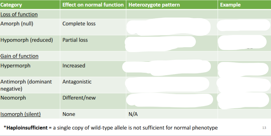
Fill in Muller Morph’s table
Antimorph is where you have an antagonistic mutation - interference of a mutant protein with the normal proteins
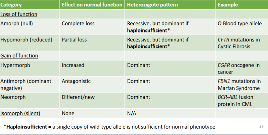
When proteins are spliced together, it usually leads to what type of mutation (using Muller’s Morphs)
Neomorph
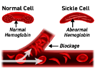
What is at risk of developing in patients with sickle cell anaemia
The Risk is Sickle Cell Crisis:
Acute Chest Syndrome (ACS)
Chronic pain
Organ Damage
Swelling in hands & feet
Bacterial infections
Autosplenectomy (repeated splenic infarctions from sickled RBCs eventually lead to fibrosis and shrinkage of the spleen)
What treatment is required for people with sickle cell disease and autosplenectomy
Require immunisation against common pathogens and
prophylactic ABs
How can a sickle cell prenatal diagnosis be made
Test a sample of the parents blood. If 1 or neither are carriers, the child won’t have sickle cell. If both are carriers, you can use Chorionic Villous Sampling (@ 9-10 weeks) or amniocentesis (test amniotic fluid @ 15-18 weeks)
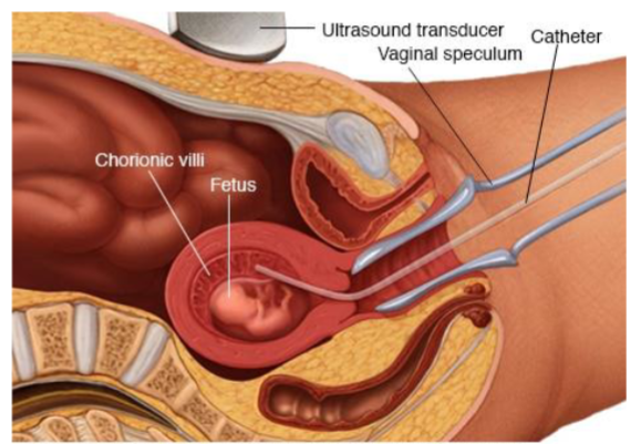
How does chorionic villous sampling work
Placental tissue containing fetal cells is collected
They are amplified using PCR to analyse the β-globin gene (which is mutated in sickle cell and β-thalassemia)
Oligonucleotide Probes (short, single-stranded DNA probes) are designed to specifically bind to either the normal or mutant sequence of the β-globin gene.
Hybridization / Sequencing confirms whether the fetus has the normal gene, is a carrier (heterozygote), or is affected (homozygote).
How does amniocentesis work
Foetal cells collected from amniotic fluid are grown & PCR is done to analyse them
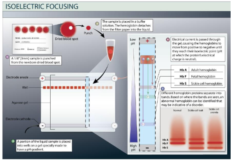
Name a method of newborn sickle cell screening
Blood spot test
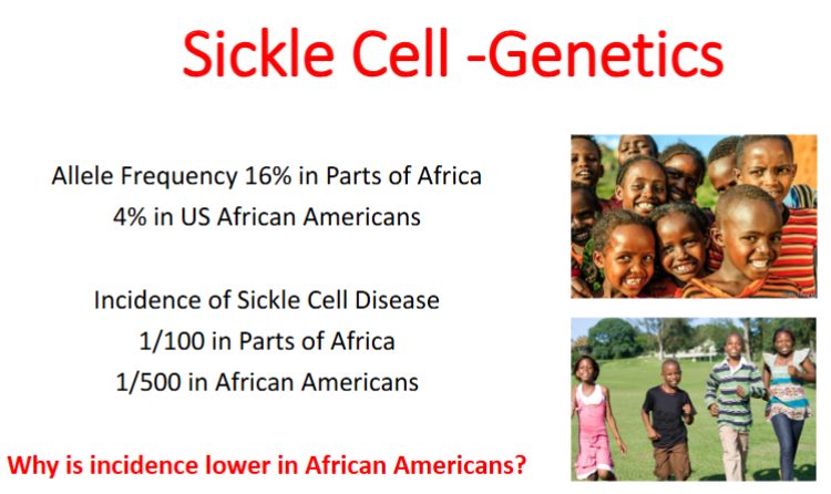
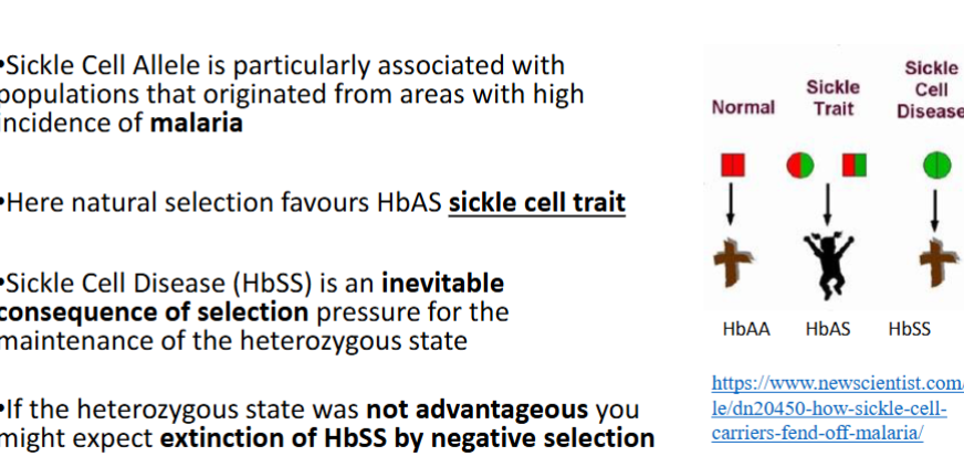
There is an advantage to having the gene for sickle cell in Africa as it protects against malaria - aids in survival of the fittest
Evolution
In population genetics, evolution is defined as the change in the frequency of an allele in a population over time
Adaptation
A heritable trait that aids the survival and reproduction of an organism in its current environment
Polymorphism
two or more discontinuous (i.e. clearly different) forms occur in a single population in the same place at the same time
Single (panmictic) population
Mean there is random (unrestricted) mating within the group
E.g. blood groups A, B, AB, & O
Balanced Genetic Polymorphism
The simultaneous occurrence in the same population of two or more “discontinuous” genetic forms in “such proportions” that the frequency of occurrence of the rarest of them cannot be explained just by recurrent mutation or immigration.
“Such proportions” = with a frequency of at least 1% of alleles.
The Implication is that something in the environment is acting to select for maintenance of the equilibrium (balance) between the different forms in the population
i.e. Natural selection
How do Sickle cell anaemia (HbSS) and Sickle cell trait (HbAS) differ
HbSS - Clinically Manifest Phenotype, Pattern of Inheritance is recessive, usually Parents Do Not Have sickle Cell Anaemia
HbAS - “Cryptic” Phenotype (present genetically but is very mild, hidden, or not easily recognized clinically), Pattern of inheritance is dominant, Parent Almost Always Has HbAS
Name an inherited Disorder of Balanced Haemoglobin Biosynthesis
Thalassaemia
How is Thalassaemia a good example of the complexity of genetic context
The same β-globin mutation can give very different outcomes depending on:
Co-inherited modifying factors
Level of fetal globin (HbF) expression
Level of α-globin expression
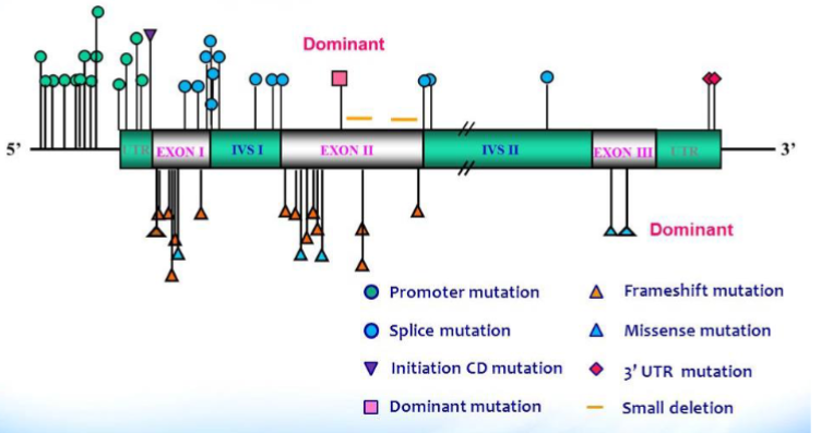
What are the different β mutations & their genotype & phenotype
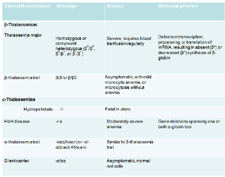
What are some β-Globin Gene Mutations Associated with β-Thalassaemia
There are two main types of thalassaemias: α-thalassaemia & β-thalassaemia.
Explain the genetics behind β-thalassaemias
HBB gene promoter (where transcription machinery binds) position -34 is normally A Mutation A-G = no binding and no transcription
What races are more prone to β-thalassaemias
Black & chinese people
How do Diseases Associated with the β-thalassaemias Point Mutation Differ in Different Ethnic Groups
Due to differences in Ability to Compensate by Synthesis of HbF in response to erythroid stress
There is a rare form of β-thalassaemias that is an LCR - explain
LCR = Locus Control Region
If a large deletion includes the LCR, the β-globin gene itself may be structurally intact and with a normal coding sequence, but cannot be expressed.
β-globin gene is structurally normal, DNA sequence is normal for 500 bp 5’ and 3’ but there is a large 5’ deletion
Far less β-globin produced (no enhancer)
Why wouldn’t the baby’s blood work for the prenatal diagnosis of β-thalassaemia?
Blood of baby won’t work – not expressing β-globin to sufficient levels for clear delineation of hetero/homozygous
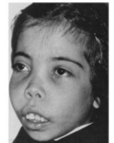
In β-thalassaemias why is the face an abnormal shape?
Many of the Features of the Syndrome related to physiological response that represents an effort to compensate for the physiological deficit associated with the inherited mutation Hypoxia > high EPO> bone marrow hyperplasia Increased haematopoiesis distorts bones
haemoglobinopathies vs thalassaemias
haemoglobinopathies - production of abnormal globin chains
thalassaemias – imbalance in production of structurally normal globin chains
Balanced polymorphism
Refers to the persistence of a number of alleles in a stable equilibrium in a population and is generally discussed in the context of one allele that is disadvantageous in homozygous state