Orbit Contents - Lab 19
1/23
There's no tags or description
Looks like no tags are added yet.
Name | Mastery | Learn | Test | Matching | Spaced | Call with Kai |
|---|
No analytics yet
Send a link to your students to track their progress
24 Terms
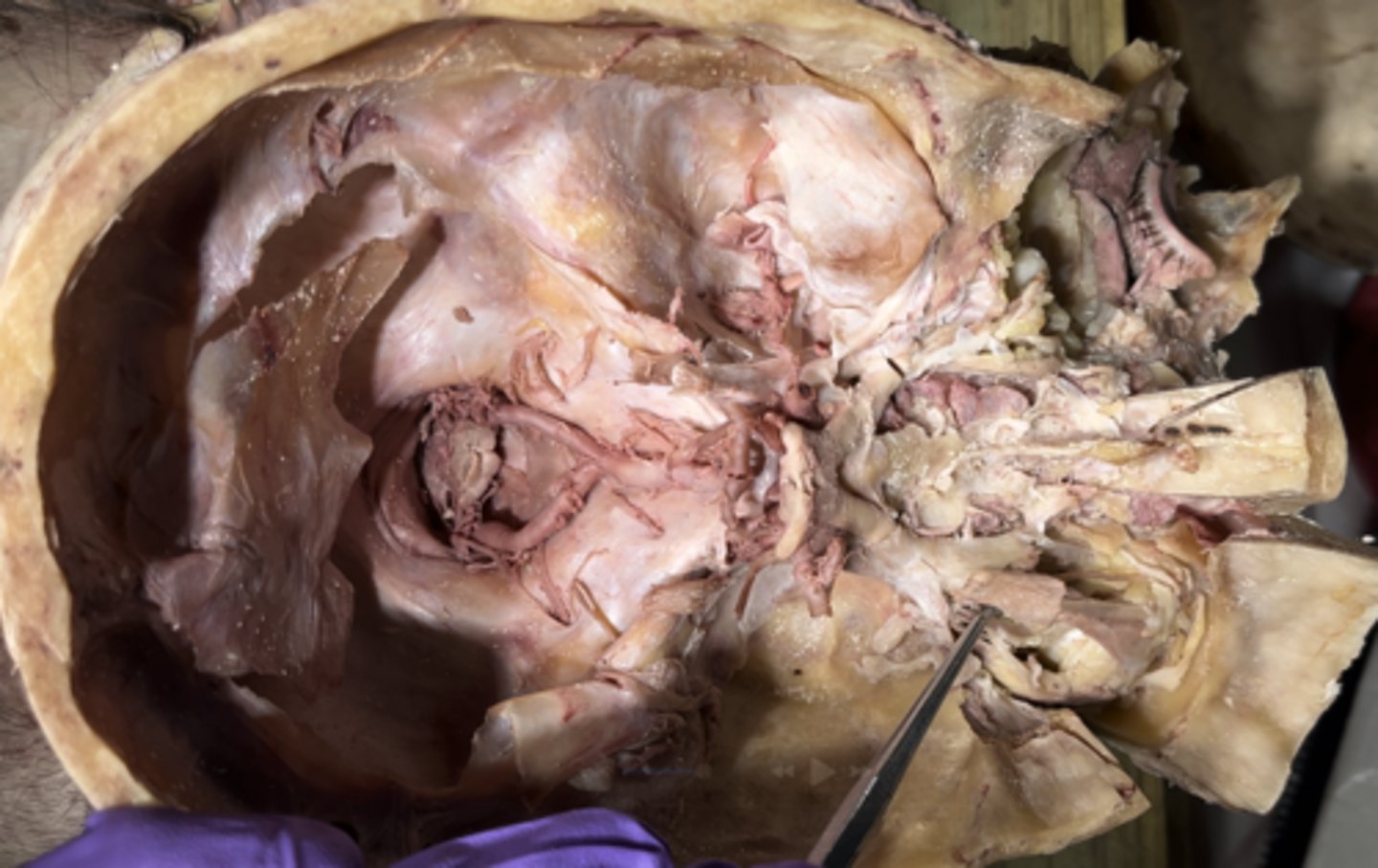
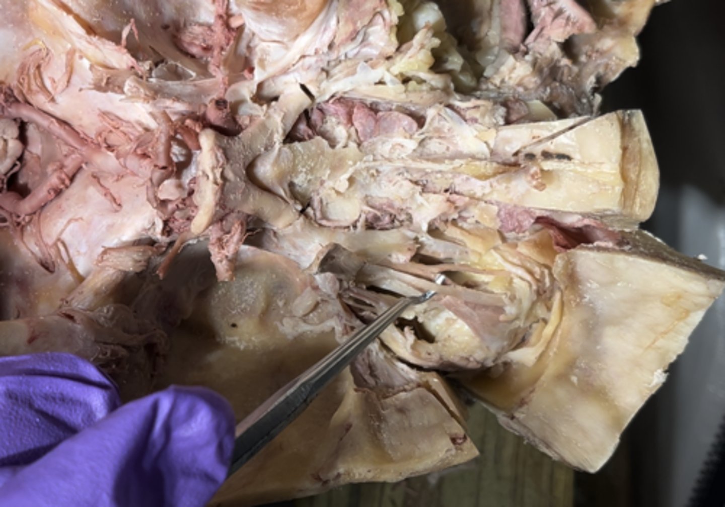
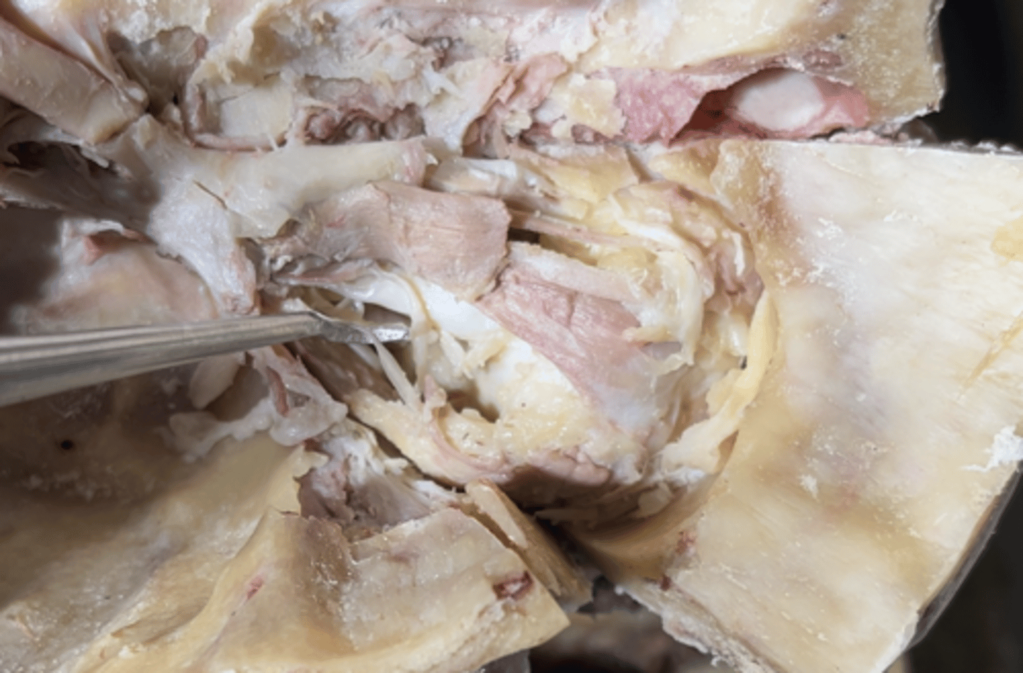
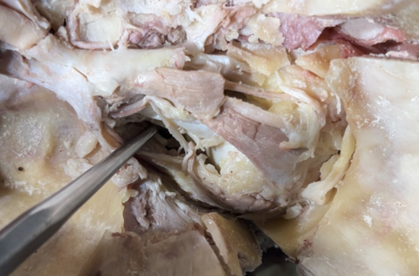
Periorbital fat and Fascia
Around the eye you will see
periorbita
The thin layer covering shown is the
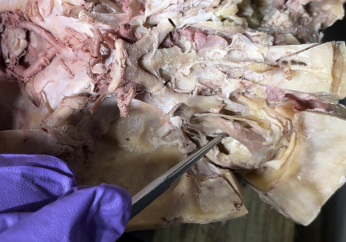
Levator palpebrae superioris muscle
In the top area of the eye the most superficial muscle is
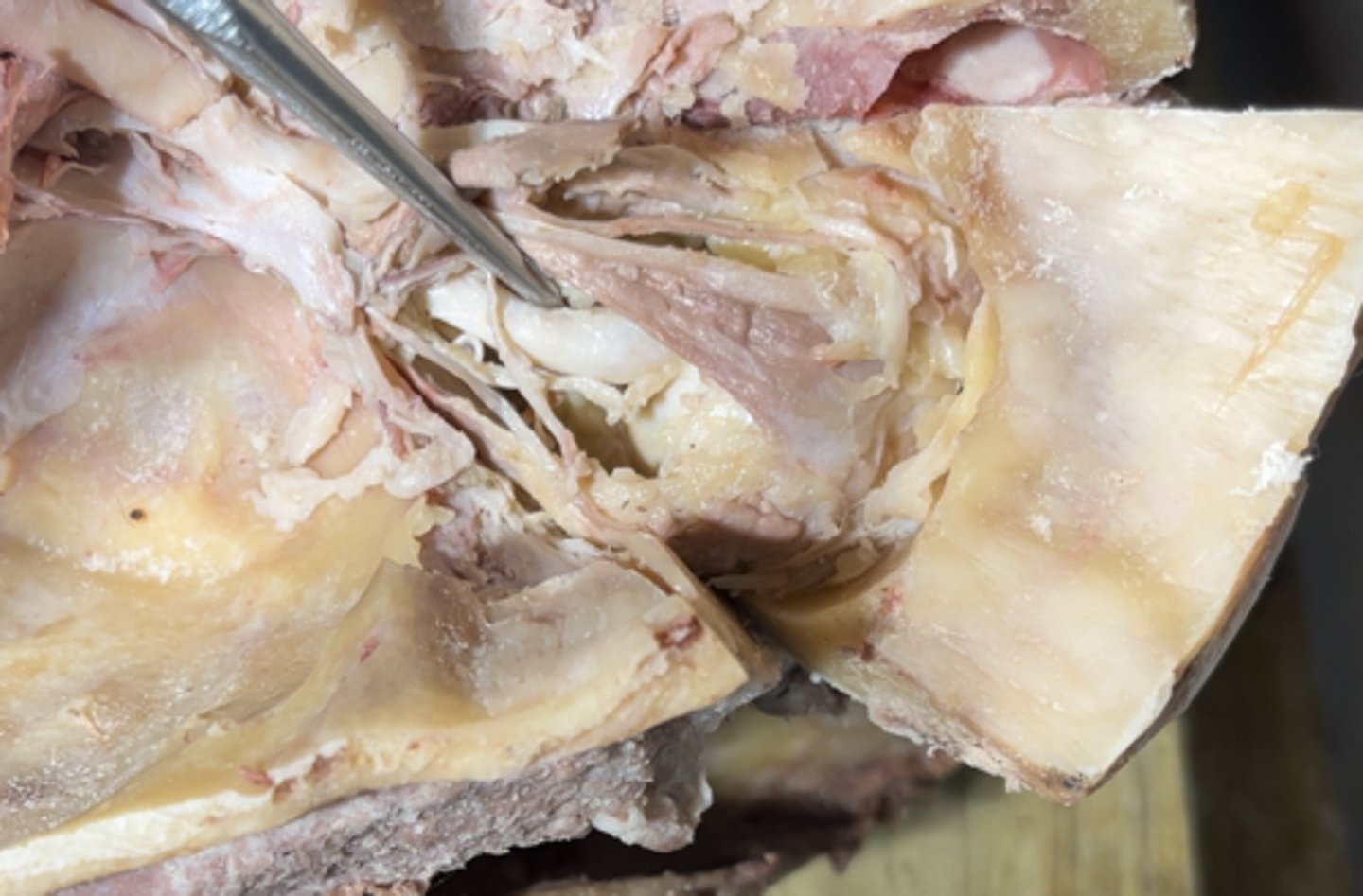
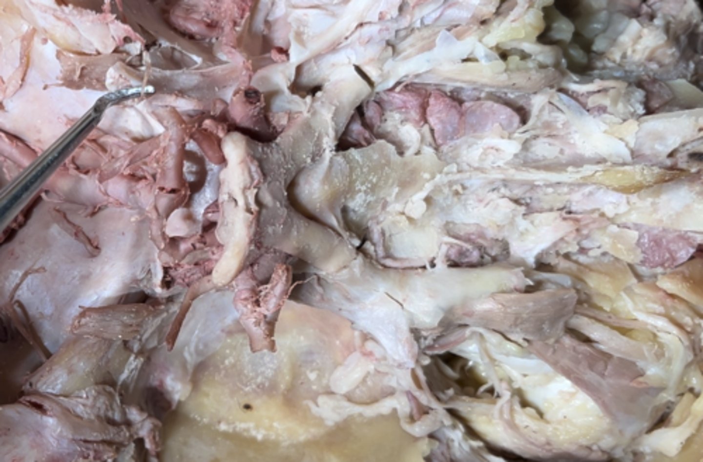
Frontal nerve
The nerve running in the same "plane" as the levator palpebrae superioris muscle is the
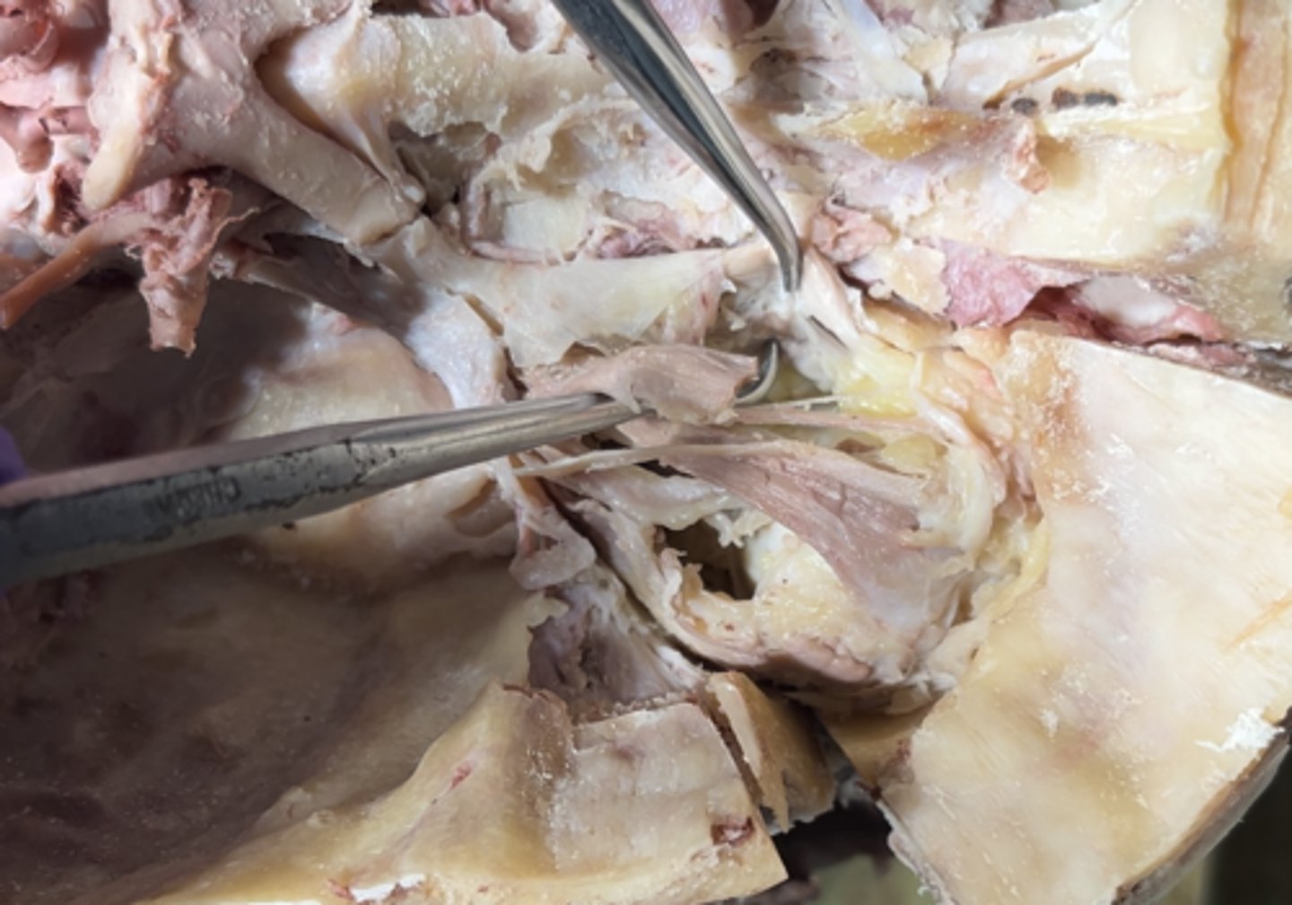
Superior rectus muscle
The muscle running on the top of the eye that actually CONNECTS to the eye is the
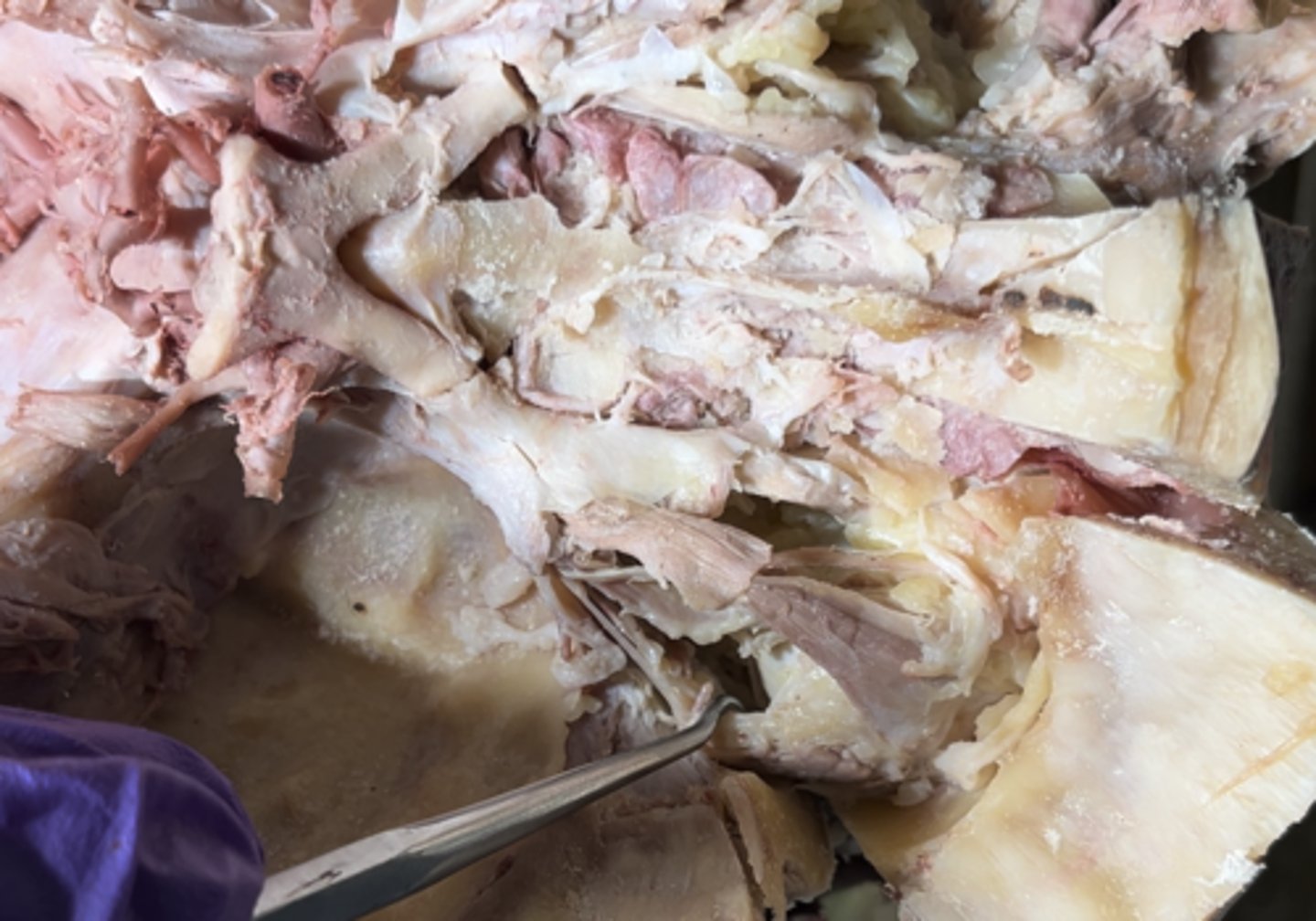
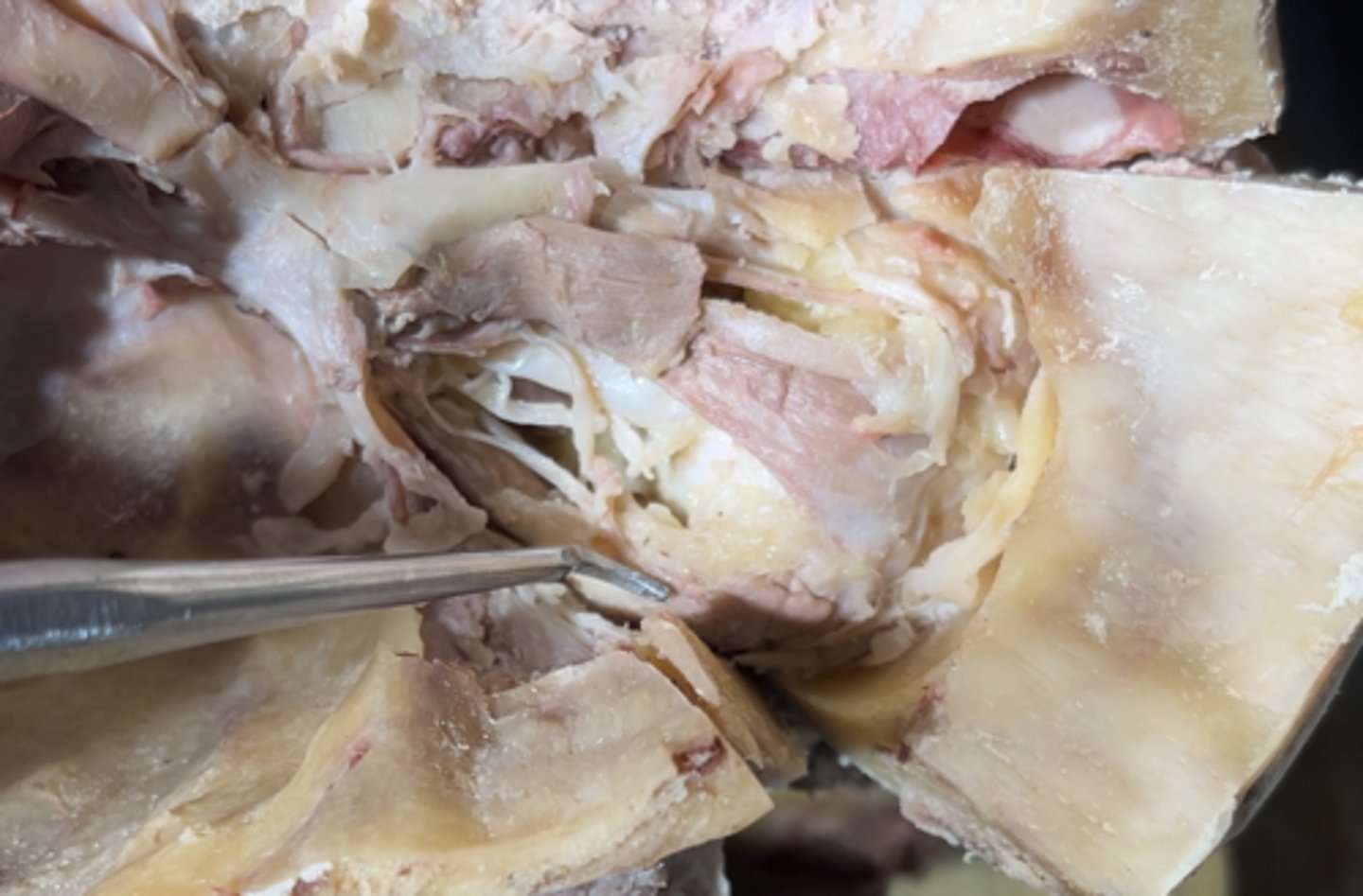
Superior oblique muscle
On the medial side of the eye the top muscle is the

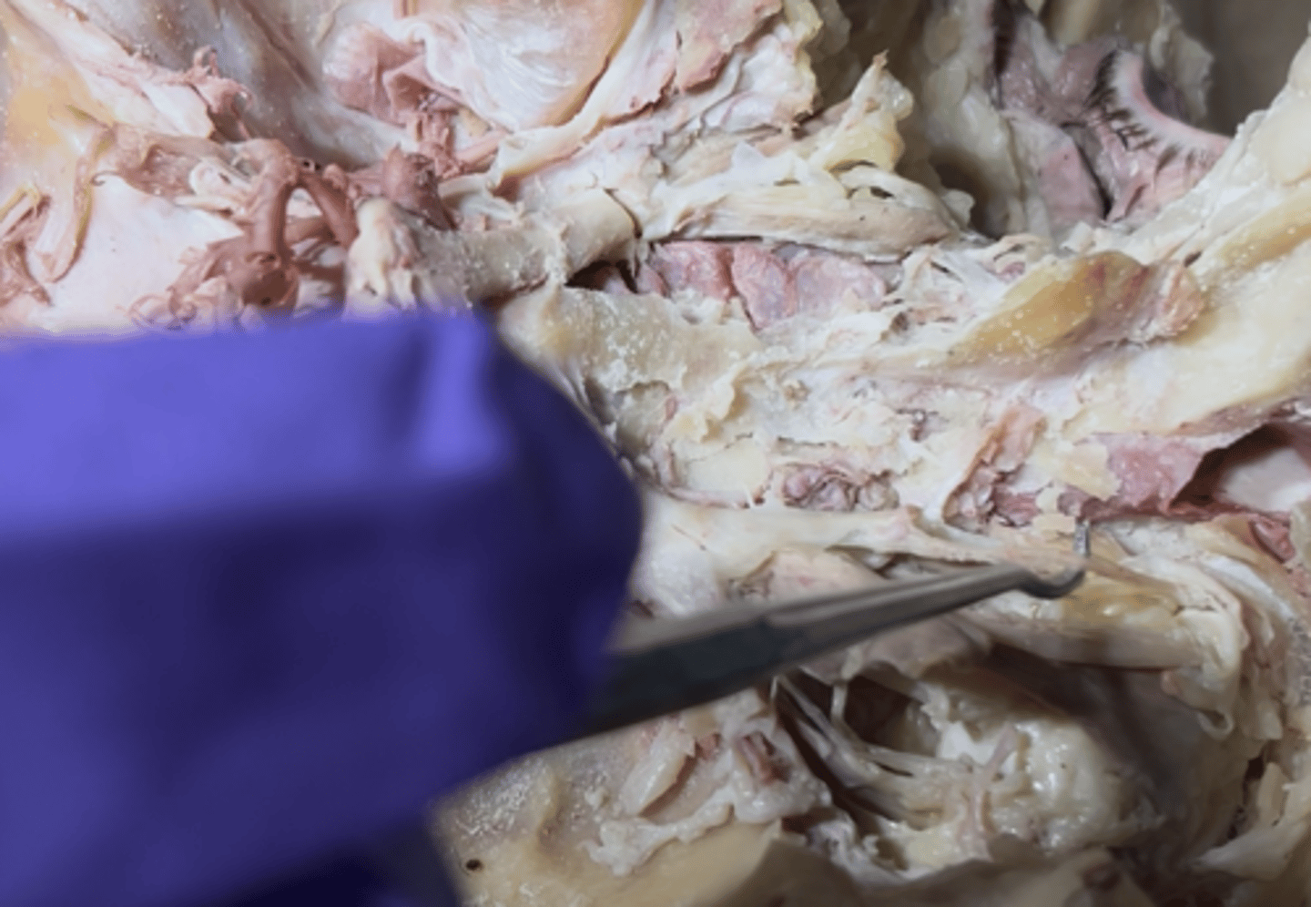
medial rectus muscle
Underneath the superior oblique muscle on the medial of the eye is going to be the
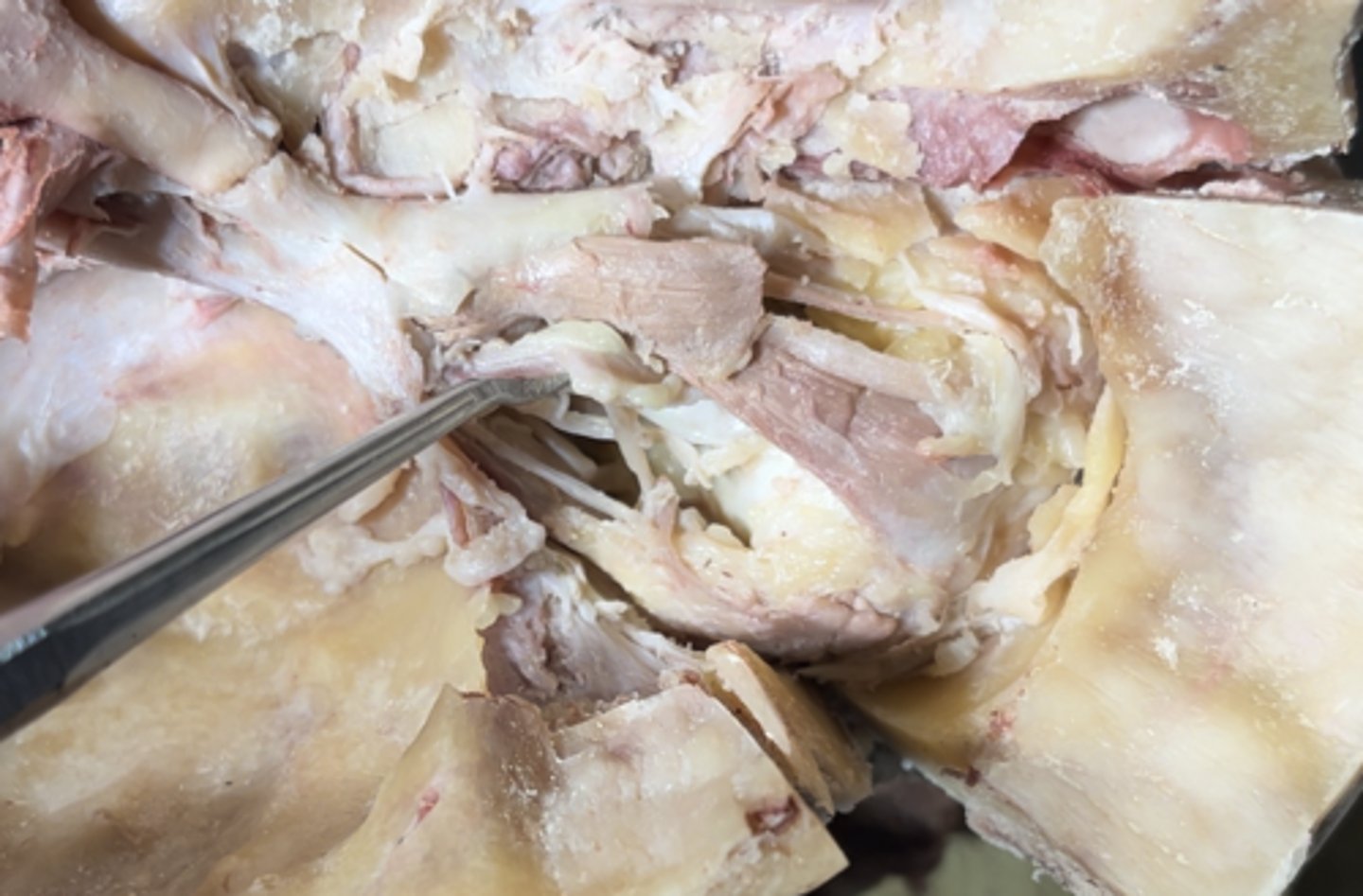
Lateral rectus muscle
On the lateral side of the eye the muscle is the
Lacrimal nerve
The nerve running lateral to the eye is the
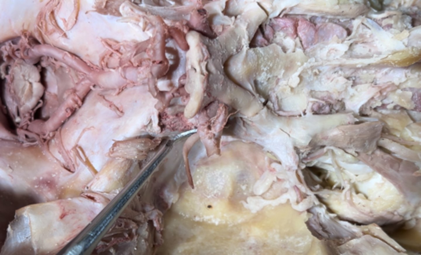
optic nerve
(this was mahdiyars idea btw)
Fat a** white piece of MEAT THICK AF running to the eye is the
Lacrimal gland
Lacrimal nerve leads into the
Opthalmic artery
The artery running ontop of the optic nerve is the
inferior rectus muscle
The muscle running under the eye is the
trochlear nerve
can be seen near the eye too will show later
The nerve that is super duper tiny eetsy weensy point at is the
Trochlear nerve
The nerve leading into the superior oblique muscle is the
Oculomotor nerve
The nerve pointed at is the
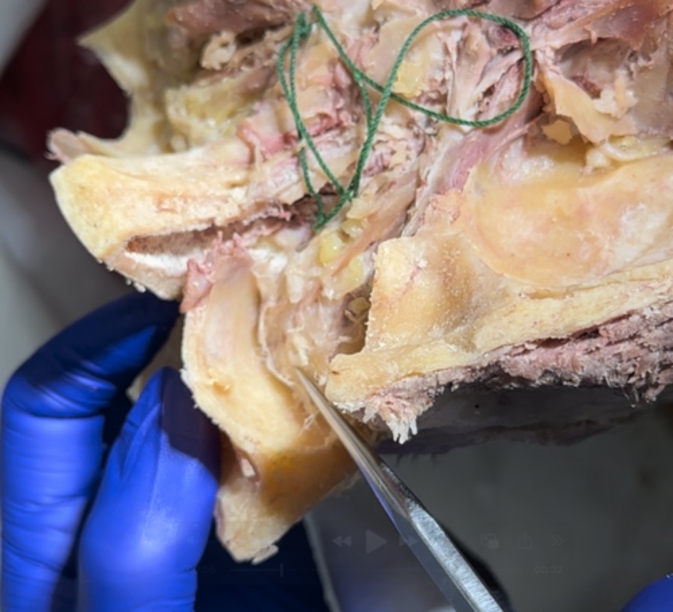
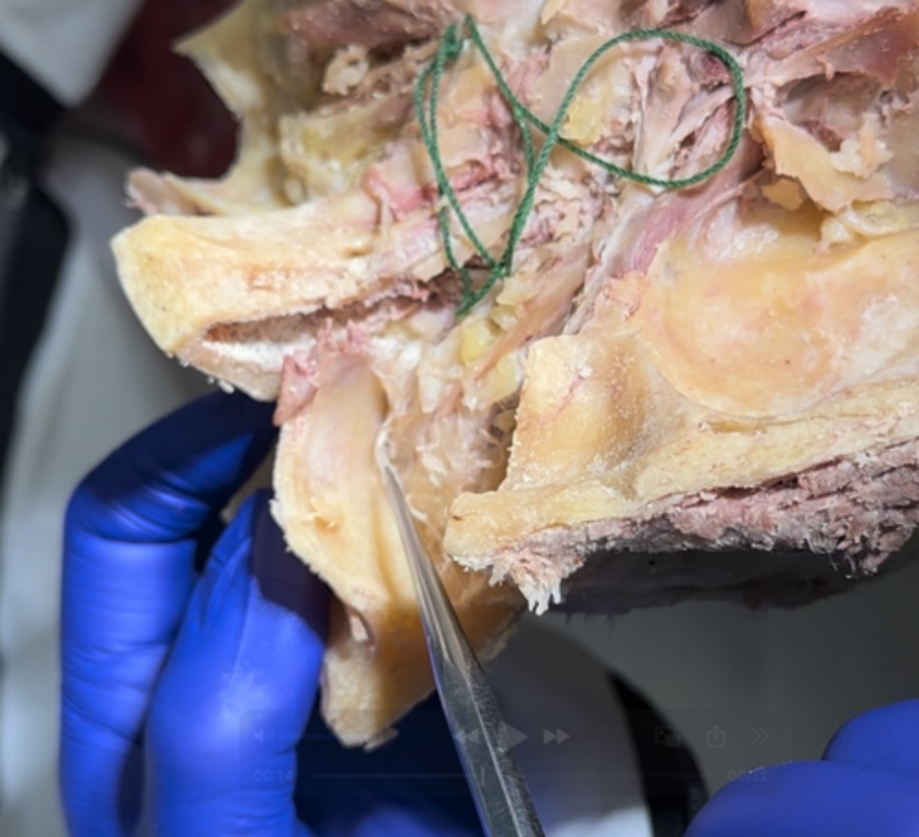
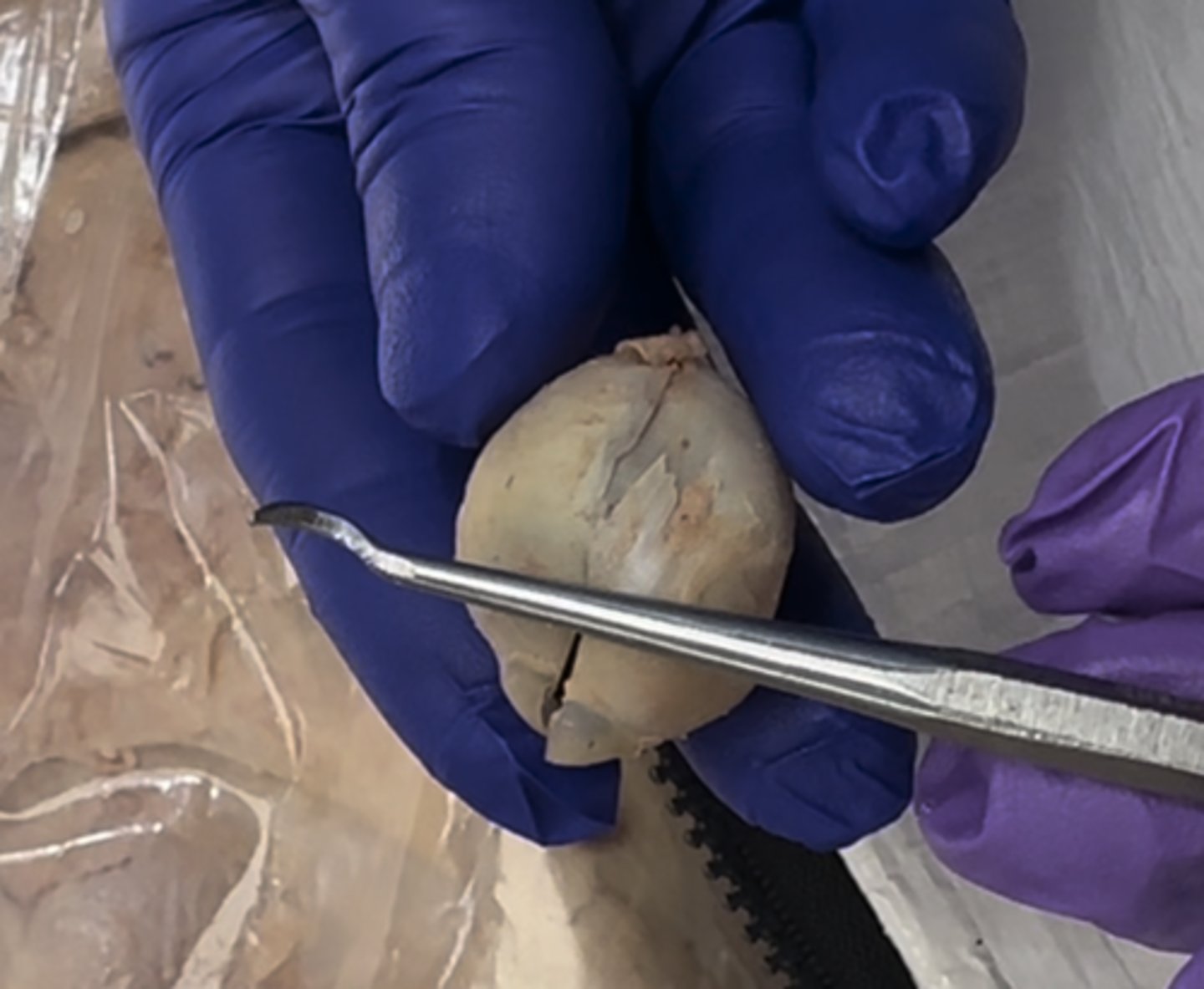
Sclera
The outer white part of the eye is the
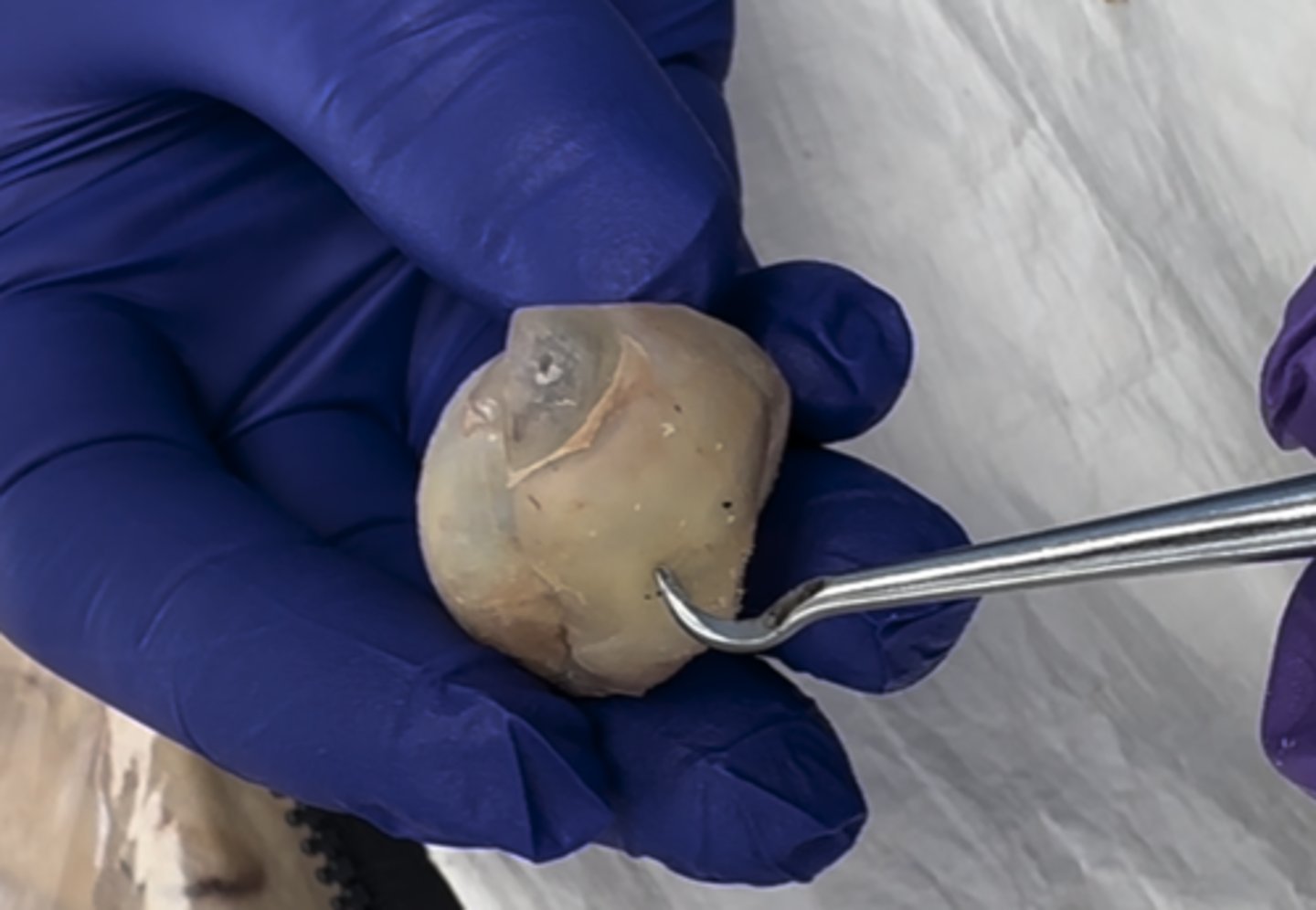
Cornea
The outer bulge part in the front of the eye is the
iris
The black part behind the cornea is the
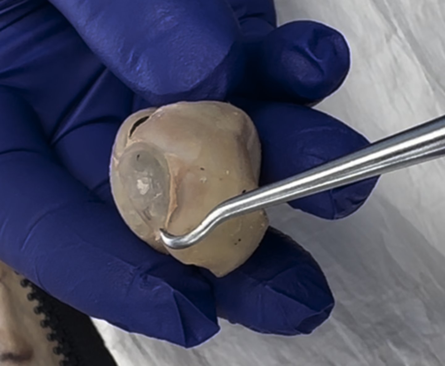
Anterior chamber
The area between the cornea and the iris (black) is the
Ciliary bodies
The "white ring" area around the iris is the
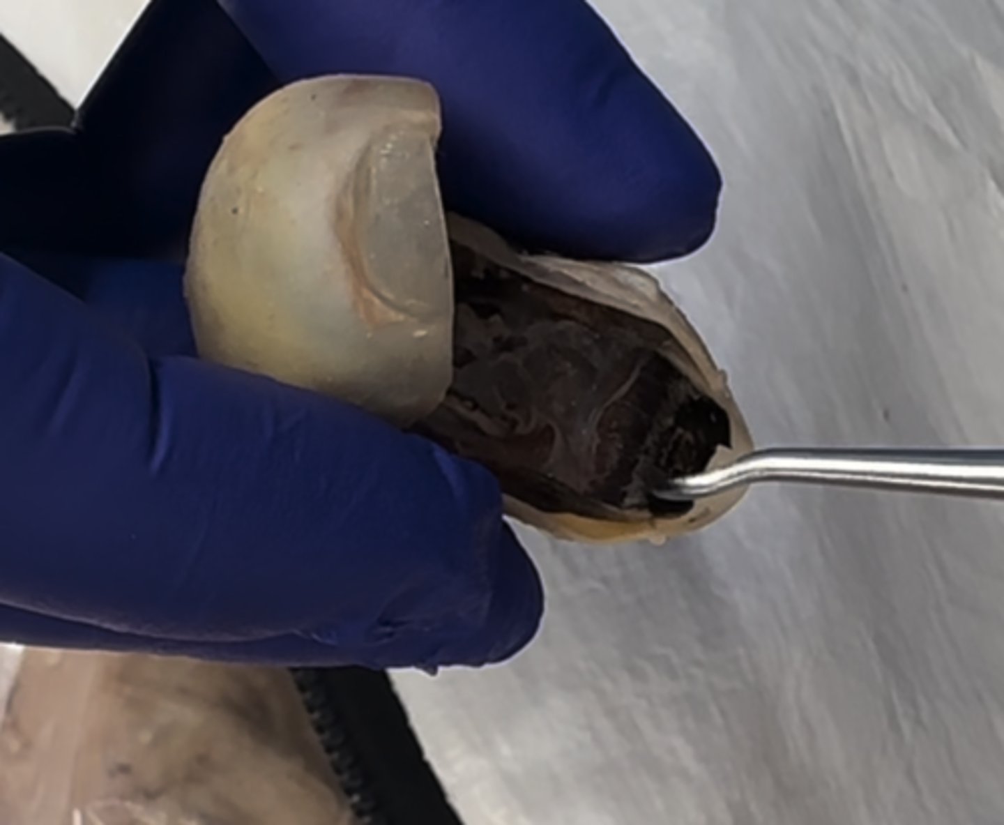
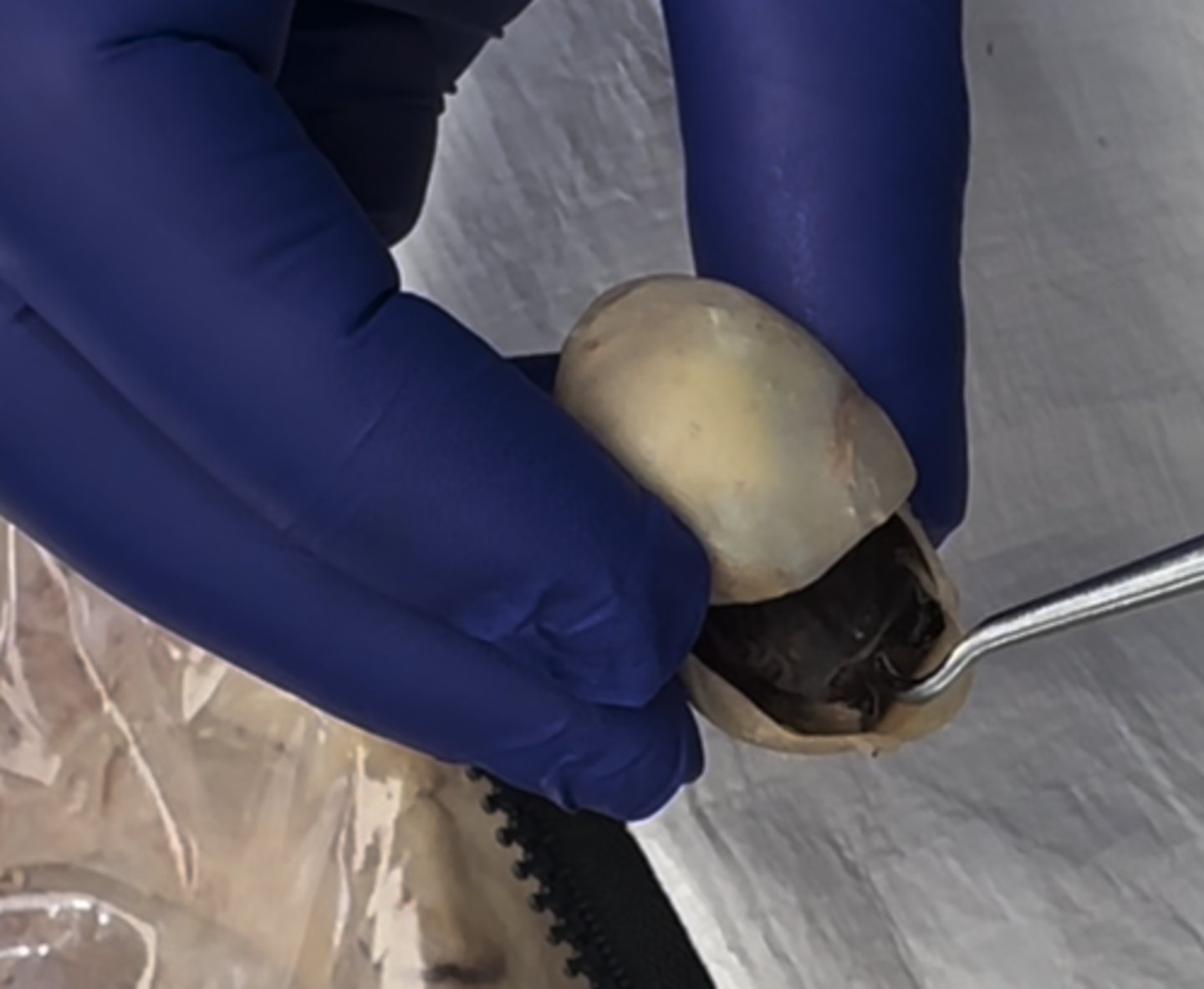
Retina
What is the black part surrounding the inner body of the eye
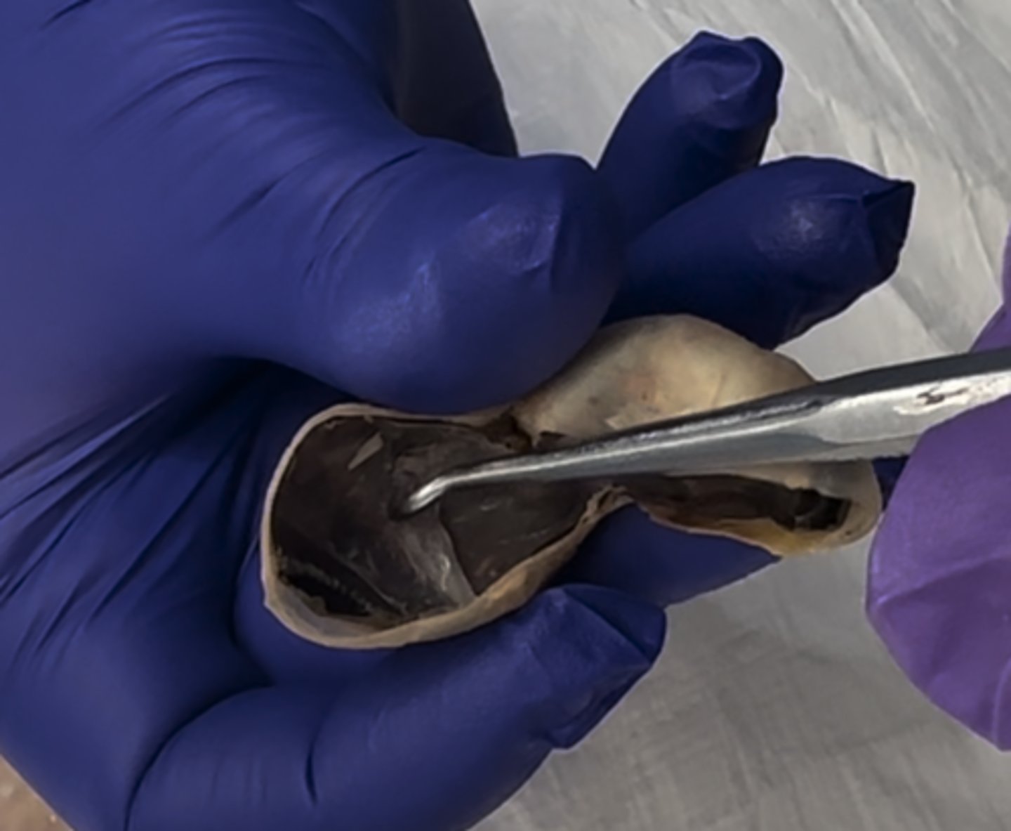
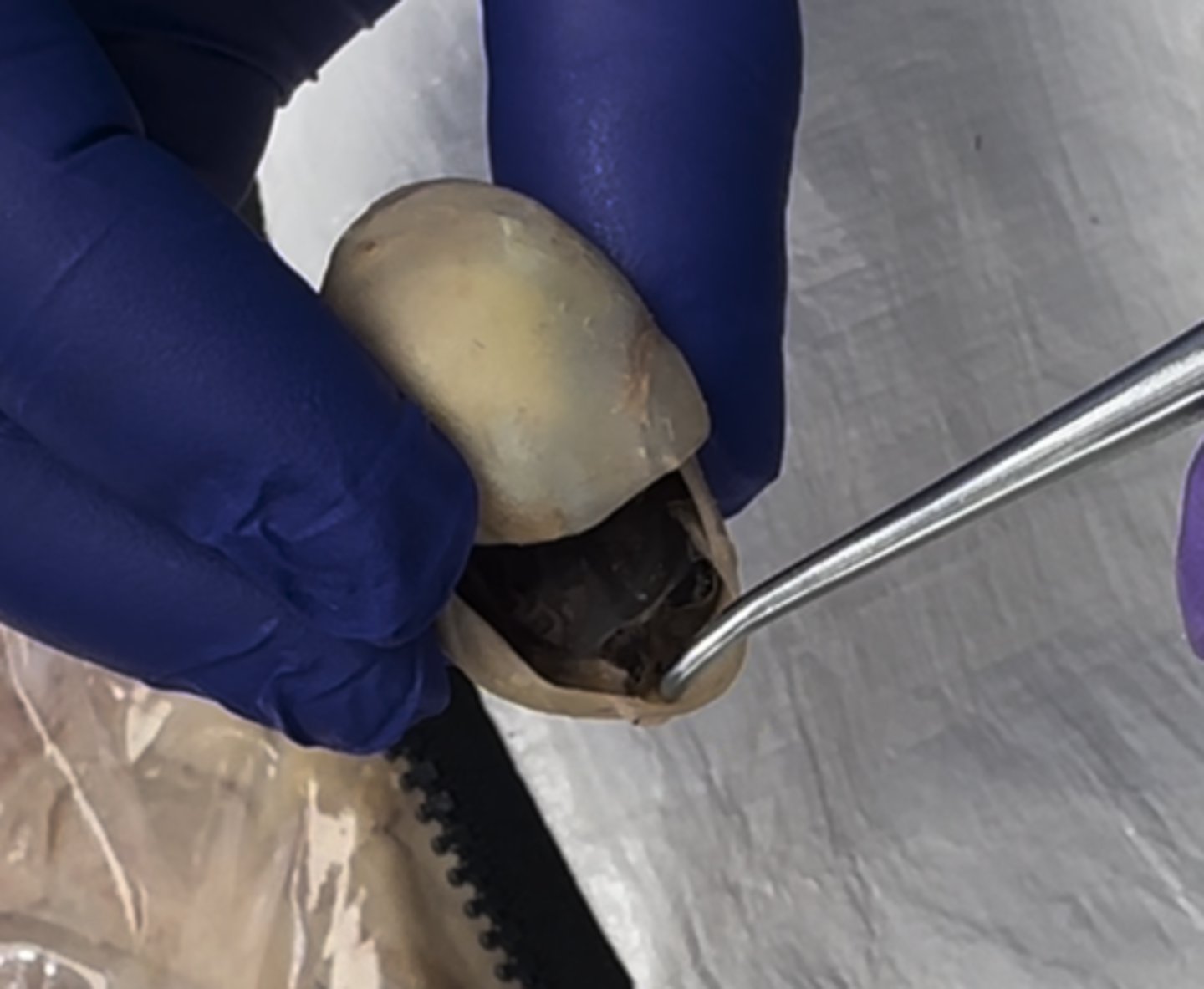
Vitreous chamber
Taking the eye and splitting it into thirds, the lower 2/3 will form the
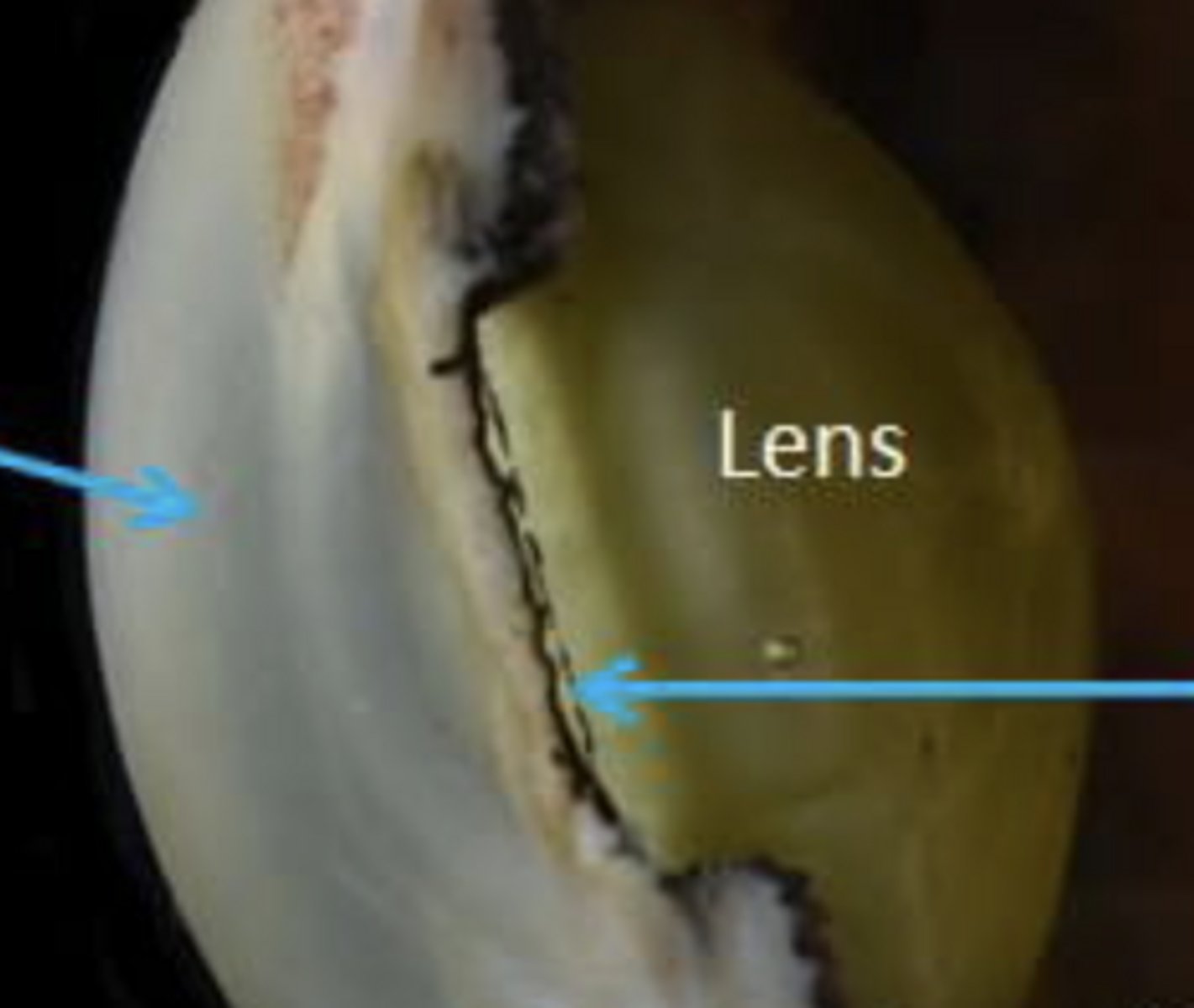
Lens
Added lens just because it's not on the actually eyes in lab.
Bulby looking area on the inside of the eye