Bis2D M1 EVQs/LGs
1/86
There's no tags or description
Looks like no tags are added yet.
Name | Mastery | Learn | Test | Matching | Spaced |
|---|
No study sessions yet.
87 Terms
(T/F) According to the cell theory all cells are surrounded by a membrane
F
(T/F) Cells are made of the following macromolecules: lipids, polysaccharides, proteins and nucleotides
F
(T/F) Precursors that make macromolecules in the protocells may have been functionally and structurally different from the ones observed in cells nowadays
T
Defining characteristics of a protocell
presence of a barrier that creates a compartment
the barrier is a membrane of lipidic nature since it’s made of fatty acids
membrane allows the transport of solutes and water across it, as it is permeable
chemical reactions can be facilitated inside the compartment. for example, protonucleotides combine into protonucleic acids
compartment can grow in size
compartment can be divided into smaller and independent compartments by the action of external forces
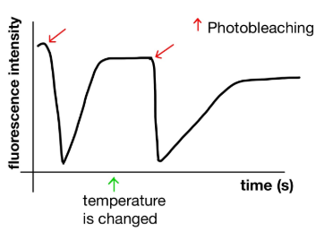
This graph represents a FRAP experiment in which the recovery of fluorescence is measured before and after changing the temperature at which the experiment is done. Has the temperature been reduced or increased? Justify your answer.
The temperature has been reduced because the fluorescence is recovered more slowly after the temperature is changed (the slope of the graph is smaller or less steep after the second photobleaching event that happens once the temperature has been changed).
Lower temperature implies less fluidity and as a consequence it takes more time for membrane components to move to recover fluorescence.
(T/F) In a given eukaryotic cell, the membrane composition of the different organelles is the same.
F
(T/F) Peripheral membrane proteins are partially embedded in the lipidic membrane
F
(T/F) The higher the temperature the more fluid a biological membrane is
T
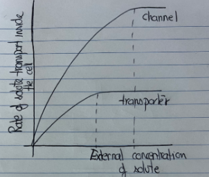
In a graph similar to the one below we have represented simple and facilitated diffusion together in class. We also started to draw the differences if the facilitated diffusion was mediated by a channel or a transporter. How would you finish the representation? MAKE SURE to: (1) include the representation of both facilitated diffusion mediated by a channel and by a transporter (2) explain briefly the reasoning behind the curves you have drawn
Facilitated Diffusion via Channel:
I would draw a steep, almost linear curve at first, which then plateaus at high solute concentrations.
The initial slope is steeper because channels allow many solute molecules to pass rapidly when open.
The plateau is higher and occurs later (i.e., at higher solute concentrations) because channels can sustain a higher rate of transport than transporters. This is due to their mechanism: channels act like gates that open and allow continuous flow of solutes, without needing to change shape for each molecule.
Facilitated Diffusion via Transporter:
I would draw a less steep, hyperbolic curve that plateaus earlier and at a lower maximum rate than the channel curve.
This is because transporters undergo conformational changes for each transport cycle, and can usually move only one or a few molecules at a time.
Therefore, the rate of diffusion increases more slowly, and saturates earlier since there is a limit to how many solutes can be moved at once (due to finite numbers of transporter proteins and time needed for their conformational cycling).
The difference in slopes: The channel has a steeper initial slope because it allows fast, bulk transport when open.
The difference in plateaus: The channel’s curve plateaus later and at a higher level because it can handle a greater maximum transport rate than the transporter.
The mechanism: Channels work by simply opening, allowing many solutes to pass at once, while transporters are slower, requiring a step-by-step conformational change per solute molecule.
(T/F) Channels can participate in active transport
F
(T/F) ATP is required for the conformational change of carriers
F
(T/F) The rate of diffusion of a solute varies according to the concentration gradient
T
You are studying two different peroxisomal transmembrane proteins (prot A and prot B). Prot A participates in the synthesis of cholesterol and prot B is an essential part of the peroxisome translocation machinery. Mutations which cause the lack of each of these proteins have a very different phenotype. Protein A mutant has some problems in cholesterol synthesis but the other functions of the peroxisome are totally fine. Protein B mutant renders a completely dysfunctional peroxisome. How can you explain this difference if in both cases only one protein is absent from the cell? Explain your reasoning.
The main difference comes down to the importance and function of the proteins. Protein A is probably more of an accessory protein; it helps with the efficiency of cholesterol synthesis, but it’s not essential to the peroxisome as a whole. Protein B is critical for the basic function and structure of the peroxisome because it’s likely involved in importing the enzymes that the peroxisome needs to work. A mutation in Protein B would be seriously damaging, maybe even lethal, basically shutting down all peroxisomal functions. In contrast, a mutation in Protein A would still affect the cell, specifically with its cholesterol production, but the peroxisome itself would still be intact and functional, just less efficient in cholesterol synthesis.
(T/F) In George Palade’s experiments secreted proteins were first observed in the Golgi
F
(T/F) Mitochondrial proteins encoded in the nuclear DNA are modified with the addition of a sorting signal once they are translated
F
(T/F) Vesicles mediate the traffic between compartments of the secretory pathway
T
Identify the steps of the scientific method
observation and research
hypothesis
prediction
experimental design
analysis of results
Hypothesis definition
an informed, logical, and plausible explanation for observations of the natural world. it must be constructed in such a way that it is potentially falsifiable or refutable
Model organism + main characteristics
It needs to share (with the organism we are studying) some features of the biological process we are interested in understanding
We should have abundant data about it: full genome, collection of mutants... How to find a good model organism?
It should be easy to maintain and suitable for experimental manipulation
Discuss how information obtained using different organisms can be transferable among them as far the phenomenon under study is conserved among those organisms
Principles of cell theory
All living organisms are composed of one or more cells.
The cell is the basic unit of structure and organization in organisms.
Cells come from preexisting cells.
Energy flow (metabolism) occurs within cells
Heredity information is passed on from cell to cell (DNA)
All cells have the same basic chemical composition
Main components of the cell at an atomic level
carbon
hydrogen
oxygen
phosphorus
sulfur
nitrogen
Building blocks/precursors to macromolecules
monosaccharides
fatty acids
amino acids
nucleotides
What are the advantages of building macromolecules from precursors?
Energy efficient- one precursor for many different macromolecules → more efficient regulation
Macromolecules can also be degraded into precursors to be used for something else
dynamic process that can be tightly regulated
Protein primary structure
sequence/list of amino acids (3 letter code)
key structure of amino acids
amine group and carboxylic acid with a carbon backbone and R group in between
Protein secondary structure
alpha helices, beta strands
R groups of the individual amino acids interact with each other based on how they’re ordered in the primary structure and twist and turn into either the alpha or beta structure
Protein tertiary structure
fold helices and strands into domains
Protein quaternary structure
biological units: functional assemblies of chains (subunits)
Primordial Soup
primitive ocean
contained clay (providing surfaces for molecules to concentrate and interact) and other non-organic surfaces that could serve as catalyzers at high temperatures for spontaneous formation of amino acids.
thought to be where life originated.
high UV radiation
different atmospheric composition
combination of reduced gases in the atmosphere: methane, ammonia, hydrogen
low/no oxygen atmosphere
strong electrical activity - lightning
hot temperature
Amphipathic nature of fatty acids/lipids
polar carboxyl group → hydrophilic
in phospholipids, includes the phosphate group and part of the glycerol backbone
nonpolar carbon chain → hydrophobic
fatty acid tails
self-organization leads to the formation of the phospholipid bilayer where:
hydrophilic heads face outwards towards the environment
hydrophobic tails face inward, away from water
How does the amphipathic property of fatty acids lead to the protocell?
early protocell membranes were likely made of simple fatty acids
in the primordial soup, fatty acids with these properties could spontaneously form:
micelles (single-layer spheres)
liposomes (bilayer vesicles)
this allows the protocell “membrane” to form and:
create a protected internal environment
allow for a concentration of molecules
provide a boundary between self and environment
Micelle
Singular layer circular made of simple fatty acids. They can come together to create liposomes and make up the protocell
Liposome
Linear bilayer molecule (made up of micelles) that can close up and become a proto-membrane + compartment.
How could the first macromolecules have formed in the protocell?
The protocell provided crucial advantages for macromolecular formation:
Protected Environment: The protocell membrane created a protected space where reactions could occur with less interference from the external environment.
Concentration Effect: By containing molecules within a small space, protocells increased the likelihood of molecular interactions necessary for forming macromolecules.
Dynamic Membrane: The protocell membrane was "firm but dynamic," allowing smaller molecules to enter while retaining larger macromolecules inside.
The replication of macromolecules within protocells likely involved:
Growth and Division: Protocells could grow through the addition of micelles to their membranes, and eventually divide through mechanical forces
Nucleic Acid Role: The lectures mention the potential role of nucleic acids in primitive replication processes within these protocells.
Self-Assembly: Some macromolecules may have had self-assembly properties that facilitated their replication.
Osmosis
simple diffusion for water - a type of passive transport
Protocell division (shearing forces)
mechanical forces in the ocean environment could induce division-like processes in these early protocells
when a protocell grows too large from eater intake and micelle incorporation, it develops a branched structure and becomes vulnerable to the ocean’s physical forces (current and shearing)
division relied on physical properties of the membrane and help of the environment for replication
What are shearing forces
a type of mechanical stress that causes layers of a material to slide past one another in opposite but parallel directions. Unlike tensile forces (which pull apart) or compressive forces (which push together), shearing forces act parallel to the cross-section of a material.
Fluid mosaic model
Describes the structure of biological membranes as a mosaic of components—including phospholipids, proteins, cholesterol, and carbohydrates—that are able to move fluidly within the membrane
Structural components:
The membrane consists of a phospholipid bilayer, with the hydrophilic (water-loving) heads facing outward toward the aqueous environment and the hydrophobic (water-fearing) tails facing inward
The membrane contains various proteins, including transmembrane proteins that span the entire membrane and peripheral proteins that associate with the membrane surface
Cholesterol (sterols) is present in the membrane and affects its fluidity
Oligosaccharides may be attached to proteins or lipids on the cell surface
Fluidity properties:
The membrane is fluid, allowing lateral movement of its components (BIS 2D Lecture).
Higher temperatures increase fluidity, while lower temperatures decrease it
Longer fatty acids can establish more interactions with surrounding molecules, affecting membrane fluidity
Cholesterol content affects the rigidity and fluidity of the membrane
Mosaic: it is composed of different elements all mixed with each other
Fluid: the lipid bilayer can re-seal, is dynamic, fluid and semipermeable
Functions of the biological membranes
self-sealing
fluid and deformable
adaptable
dynamic
semipermeable
Main lipid components of the biological membranes
Phosphoglycerides(phospholipids): They have a hydrophilic head and hydrophobic tails, which allows them to form bilayers in aqueous environments. This amphipathic nature is essential for membrane formation, as the hydrophilic heads face the water while the hydrophobic tails face inward. Phosphoglycerides contribute significantly to membrane fluidity and can move laterally within the membrane.
Sphingolipids: Based on a sphingosine backbone rather than glycerol. They also have hydrophobic and hydrophilic regions, allowing them to integrate into the membrane bilayer. Sphingolipids are particularly abundant in certain specialized membranes and can form membrane domains with distinct compositions
two regions of hydrophobicness and a polar head
Sterols: Rigid lipid molecules that play a crucial role in regulating membrane fluidity. Sterols like cholesterol are found in the membrane and affect its properties. Sterols can make membranes less fluid at higher temperatures and more fluid at lower temperatures, essentially acting as membrane stabilizers. Different organisms may use different sterols - for example, ergosterol is found in some bacterial membranes
tiny polar head represented by an OH group
the hydrophobic section is very rigid
How can proteins interact with membranes
Transmembrane proteins: proteins that go fully across the membrane
Peripheral proteins: proteins are associated with another membrane protein or lipid but they do not penetrate the membrane
Anchored proteins: proteins are anchored but not fully embedded in the membrane
FRAP Principle
Fluorescent Labeling: First, membrane components (lipids or proteins) are labeled with fluorescent markers, making them visible under a fluorescence microscope.
Initial Observation: The entire membrane appears fluorescent as the labeled molecules are distributed throughout.
Photobleaching: A high-intensity laser is used to bleach (destroy the fluorescence of) molecules in a specific small area of the membrane.
Recovery Monitoring: The return of fluorescence to the bleached area is monitored over time (typically at intervals like 10 seconds, 30 seconds, 1 minute).
Analysis: The rate and extent of fluorescence recovery provide quantitative measures of membrane fluidity.
FRAP Logic
Non-Destructive Measurement of Molecular Movement
A critical aspect of FRAP is that photobleaching only affects the fluorescent properties of the molecules, not their physical or chemical properties.
The bleached molecules continue to behave normally within the membrane, allowing for an accurate assessment of natural membrane dynamics.
Direct Visualization of Lateral Diffusion
The recovery of fluorescence in the bleached area can only occur through lateral movement of unbleached fluorescent molecules from surrounding areas into the bleached region. This directly visualizes the lateral mobility that defines membrane fluidity. Your lecture explained that membranes exhibit several types of movement:
Lateral movement (molecules moving sideways within the membrane)
Rotation (molecules spinning in place)
Flexion (bending of fatty acid chains)
FRAP specifically measures lateral movement, which is the primary indicator of membrane fluidity.
Quantifiable Parameters
FRAP provides quantitative measurements that can be used to compare membrane fluidity under different conditions:
Recovery Half-time (t½): The time it takes for the fluorescence to recover to half of its final intensity
Mobile Fraction: The percentage of molecules that are mobile within the membrane
Diffusion Coefficient: A measure of how quickly molecules move within the membrane
These parameters allow researchers to objectively compare membrane fluidity across different:
Cell types
Membrane compositions
Temperature conditions
Chemical treatments
Ability to Study Specific Membrane Components
As discussed in your lecture, FRAP can be used to study different membrane components
This versatility allows researchers to compare the mobility of different membrane components (e.g., lipids vs. proteins) within the same membrane, providing insights into how membrane composition affects fluidity.
Real-Time Observation in Living Cells
FRAP can be performed on living cells, allowing for the study of membrane dynamics under physiological conditions. This is crucial for understanding how membrane fluidity relates to cellular function.
Passive transport
movement of molecules without consuming energy.
Active transport
movement up or against an electrochemical gradient:
facilitated diffusion
Ions and other solutes
Source of energy: ATP or electrochemical gradient
osmosis
simple diffusion for water. A type of passive transport
simple diffusion
transport of gases and small lipid solutes (ethanol, urea, fatty acids..) across the lipid membrane without help. Passive transport
Negated diffusion
how certain molecules are prevented from moving freely across biological membranes
Facilitated diffusion
assistance of diffusion
Carrier mediated diffusion
transports large charged or uncharged molecules (poorly lipid soluble solutes) with an available transporter- glucose/amino acids
Channel diffusion
ion transportation through specific channels
ATP as a source of energy for active transport is used by?
Pumps
A previously established electrochemical gradient that can be utilized as a source of energy for active transport is used by?
Transporters/Symporters
Prokaryotes
Organisms | Bacteria and Archaea |
Nucleus or membrane surrounded / enclosed compartments / organelles | X |
Size | Smaller |
DNA | Circular (no histones) |
Cytoskeleton | X |
Division | Binary Fission |
Ribosomes | Smaller |
Transcription Regulation | Operon |
Eukaryotes
Organisms | Fungi, Protozoans, Plants and Animals |
Nucleus or membrane surrounded / enclosed compartments / organelles | Y |
Size | Larger |
DNA | Linear (yes histones → packed DNA) |
Cytoskeleton | Y |
Division | Mitosis or Meiosis (germ cells) |
Ribosomes | Bigger |
Transcription Regulation | Yes, but not through Operons |
Order of events of the appearance of eukaryotic cells
Invaginations of the membrane, that eventually separated, and are the origin of the subcellular compartments
Prokaryotic cells were engulfed by primitive eukaryotic cells (endosymbiosis theory)
Engulfed prokaryotic cells transfer part of their genetic information to the nuclear DNA
Identifying characteristics of post-engulfed organelles
Double Membrane Structure: These organelles possess a double membrane, with the inner membrane derived from the engulfed organism and the outer membrane from the host cell
Their Own DNA: They contain their own circular DNA, separate from the nuclear DNA, which is similar to bacterial DNA in structure
Independent Replication: These organelles can replicate somewhat independently of the cell division cycle.
Ribosomes: They contain their own ribosomes that are more similar to prokaryotic ribosomes than to the eukaryotic ribosomes found in the cytosol.
Protein Synthesis Machinery: They have their own protein synthesis machinery, though they also rely on proteins encoded by nuclear genes
Size and Structure: Their size and internal structure resemble those of free-living prokaryotes.
Evolutionary Relationship: Genetic analysis shows clear evolutionary relationships between these organelles and existing free-living prokaryotes.
Advantages of Subcellular compartments
Specialized Functions: Each compartment contains specific proteins that enable specialized functions
Concentration of Molecules: Compartmentalization allows for higher concentrations of specific molecules within a confined space, which increases the efficiency of biochemical reactions.
Separation of Incompatible Processes: Different cellular processes that might interfere with each other can occur simultaneously in separate compartments. For example, protein degradation can occur in lysosomes while protein synthesis happens in the endoplasmic reticulum.
Creation of Different Environments: Compartments can maintain different pH levels, ion concentrations, or redox states that are optimal for specific biochemical reactions.
Protection of Cellular Components: Harmful substances or reactions can be isolated in specific compartments to protect the rest of the cell. For example, peroxisomes contain enzymes that detoxify harmful compounds.
Disadvantages of Subcellular compartments
Protein Sorting and Trafficking: Cells need complex mechanisms to ensure proteins reach their correct destinations. This requires sorting signals, sorting receptors, and translocation machinery.
Membrane Maintenance: Each compartment requires its own membrane, which must be maintained and repaired. This increases the cell's energy requirements and complexity.
Communication Between Compartments: Compartments need to communicate with each other to coordinate cellular activities, requiring sophisticated signaling mechanisms.
Transport Across Membranes: Cells need specialized transport proteins to move molecules across membranes. This includes both passive and active transport mechanisms.
Osmotic Balance: Maintaining proper osmotic balance between compartments is crucial.
Organelles in Eukaryotic cells
Translocation
process in which a protein moves from outside the compartment to inside (passing through the membrane)
Post-translational translocation
Protein is completely synthesized in the cytosol
Sorting signal is exposed on the completed protein
Sorting receptors in the cytosol recognize the signal
Protein is delivered to the translocation machinery
Protein unfolds to pass through the translocation channel
Co-translational translocation
Translation begins in the cytosol
Signal sequence emerges from the ribosome and is recognized by Signal Recognition Particle (SRP)
SRP directs the ribosome-mRNA-nascent protein complex to the target membrane
Translation continues as the protein is threaded through the translocation channel
Signal sequence may be cleaved during translocation
Sorting signal
any signal present in a protein sequence that gives information about that protein’s final subcellular destination
Translocation machinery function
Creating membrane passages: "...the translocation machinery will make it happen across this particular case."
Maintaining membrane integrity: Allows proteins to cross membranes without compromising the permeability barrier
Ensuring directional transport: Proteins move in only one direction (from cytosol to organelle)
Facilitating protein unfolding/refolding: Many proteins must partially unfold to pass through the channel
Coordinating with sorting receptors: "...this certain signal receptor is going to find that certain signal and then make possible the following this complex goes to the [membrane] of the compartment that we may be discussing. And then here we are going to have the translocation machinery"
Translocation machinery importance
It maintains the unique protein composition of each organelle
It enables compartmentalization of cellular functions
It allows proteins to reach their functional destinations
Without it, proteins would be unable to cross membrane barriers
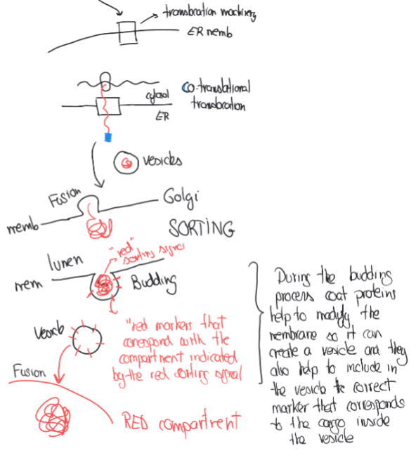
Translocation Machineries in Post-translational translocation
After the specific sorting signal receptor binds to the sorting signal on the completely synthesiszed protein, it gets brought over to the translocation machinery at the organelle membrane, where it facilitates the movement of the protein across the membrane where the protein can eventually be released into the lumen or membrane
Translocation Machineries in co-translational translocation
When the signal sequence emerges during the translation of a protein belonging to the secretory pathway, it gets recognized by the SRP (signal recognition particle) which pauses translation and directs the ribosome to the ER. The ribosome is on top of the translocation machinery, of which opens with the mechanism, allowing the growing protein to actually enter the ER lumen as it’s being synthesized.
George Palade’s Experiments
Used pulse & chase in pancreatic acinar cells
His question at that time, as well as ours now is: How are the cells taking the secreted proteins outside the cell?
The point of radioactively labelling the cells was so he could watch and follow them through their journey
He radioactively labelled the Met amino acid (methionine)
The labels only lasted 2-5 minutes
Then washed out the excess of radioactive Met
At first, they were in the ER
After 7 minutes, they were in the golgi
After 37 minutes, they were in the vesicles
After 117 minutes, they were outside the cell
This is the Secretory Pathway/Biosynthetic Pathway
Requires vesicles!!
Series of steps for transportation:
ER → Golgi →
Lysosome
Plasma membrane
Vacuole
Extracellular media
Peroxisome membrane
Nuclear membrane
Vesicle → outside cell
Secretory pathway
Requires vesicles to get proteins to their final destination. It's the primary route for proteins destined for various cellular compartments or for secretion outside the cell.
Secretory/Biosynthetic Pathway organelles
ER
Golgi
Lysossome
Plasma membrane
Vacole (plants and yeast)
Extracellular media
Peroxisome (membrane)
Nucleus (membrane)
Organelles NOT a part of the Secretory Pathway
Peroxisome (lumen)
Mitochondria
Chloroplast and plastids
Nucleus (nucleoplasm)
Signal sequence
defines the protein as one that will follow the secretory pathway to get to its final destination, focusing on how to get the protein there specifically
Secretory Pathway Protein Sequence
Initial Entry to ER:
- Begins with co-translational translocation - Signal sequence on the nascent protein is recognized by Signal Recognition Particle (SRP) - SRP directs the ribosome-mRNA-nascent protein complex to the ER - Translation continues as the protein is simultaneously translocated into the ER lumen through the translocation machinery (BIS 2D Lecture)
ER to Golgi Transport:
- Proteins are packaged into vesicles - These vesicles bud from the ER membrane - Vesicles travel to and fuse with the Golgi apparatus
Processing in Golgi:
- Proteins move through the Golgi cisternae (from cis to trans) - Undergo modifications like glycosylation - Sorting occurs based on sorting signals in the protein structure
Golgi to Final Destination:
- Proteins are packaged into specific vesicles based on their sorting signals - Coated vesicles help define the destination - Vesicles travel along cytoskeletal elements to reach their targets - Fusion with target membrane delivers proteins to final location
Coated Vesicles
vesicles covered with proteins on their cytosolic surfaces (when budding from the donor compartment). These proteins covering them help:
To read the sorting signals in the cargoes and ensure they go to their final destination
To deform the membrane so it can indeed create the shape of avesicle and eventually bud
(not a part of the list): the molecular mechanism behind the specificity of the proess and the capacity to bud and fuse with a biological membrane.
How do coated vesicles ensure specificity?
Recognition of Sorting Signals: Coated vesicles have proteins on their surface that specifically recognize and read the sorting signals on cargo proteins. This recognition is the first step in ensuring specificity.
Selective Packaging: The coat proteins selectively package only those proteins with the appropriate sorting signals into vesicles destined for specific compartments.
Directional Transport: From your lecture: "The proteins that cover the vesicles are going to pack on the surface proteins that correlate directly with the sorting signal of the protein inside, so they know that these vesicles need to go the place where this protein is indicating" (BIS 2D Lecture).
Vesicle Formation: The coat proteins also help physically form the vesicle by inducing membrane curvature and "chopping a piece of a membrane" to create the vesicle.
Targeted Fusion: Coated vesicles ensure that vesicles only fuse with the correct target membrane, preventing inappropriate mixing of contents between different cellular compartments.
Vesicular traffic - why must it be specific?
Compartmentalization: Eukaryotic cells contain numerous distinct compartments (organelles), each with specialized functions that depend on having the right proteins and lipids in the right place
Functional Integrity: Proteins must reach their correct destinations to perform their intended functions. Mislocalized proteins can disrupt cellular processes or even cause disease.
Cellular Organization: The proper organization of the cell depends on the correct distribution of proteins and lipids to different compartments.
Secretory Pathway Efficiency: The secretory pathway must efficiently move thousands of different proteins to their correct destinations without mixing them inappropriately.
Cytoskeleton
a three-dimensional filamentous protein network that helps cells maintain their shape and internal organization, and it also provides mechanical support that enables cells to carry out essential functions like division and movement
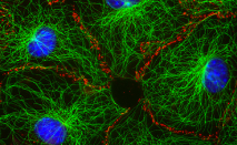
Microtubules (MTs)
In nondividing cells, microtubule networks provide the basic organization of the cytoplasm, including the positioning of organelles
Core of cilia and flagella
In dividing cells, they make the spindle that ensures the proper distribution of the chromosomes
Assembly of microtubules is such an unfavorable reaction that in at least animal cells onlyhappens in the presence of microtubule-organizing centers (MTOCs)
The centrosome is the main MTOC in animal cells. During interphase, it is located near the nucleus and MTs irradiate from it
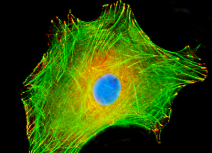
Microfilaments (MFs)
networks of actin filaments are part of the cell cortex, which is the meshwork of membrane associated proteins that supports and strengthens the plasma membrane
Such networks allows cells to hold and move specialized shapes such as the brush border of microbilli
Actin filaments are also involved in cytokinesis and cell movement
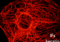
Intermediate filaments (IFs)
their functions are primarily mechanical and as a class, intermediat filaments are less dynamic than actin filaments or microtubules.
Commonly work in tandem with microtubules, providing strength and support for the fragile tubulin structures.
MTOCs (Microtubule Organization Centers) Structure
Centrosomes: In animal cells, the centrosome is the primary MTOC, consisting of centrioles surrounded by pericentriolar material
Centrioles: These are cylindrical structures made up of multiple microtubule rods arranged in a specific pattern.
Microtubules: These are hollow rods composed of alpha-beta tubulin dimers that assemble into the microtubule structure.
MTOCs (Microtubule Organization Centers) Functions
Microtubule Nucleation: MTOCs serve as the primary sites where microtubule assembly begins.
Cell Division: During mitosis, the centrosomes split and move to opposite poles of the cell, forming the spindle apparatus that separates chromosomes:
Spindle Fiber Formation: MTOCs organize the spindle fibers that attach to chromosomes during cell division:
Cell Structure Maintenance: In animal cells, microtubules radiate from the MTOC near the nucleus to the cell periphery, helping maintain cell shape and organization:
Intracellular Transport: Microtubules serve as tracks for motor proteins to transport vesicles and organelles throughout the cell.
Centrosome subcellular localization
Perinuclear Position: Centrosomes are typically located very close to the nucleus in the cytoplasm of interphase cells
Central Position: They are positioned centrally in the cell, serving as the main microtubule organizing center from which microtubules radiate outward toward the cell periphery.
Dynamic Repositioning: During cell division, centrosomes duplicate and migrate to opposite poles of the cell to form the mitotic spindle.
Centrosome Structure
Centrioles: A pair of cylindrical structures arranged perpendicular to each other, each composed of nine triplets of microtubules arranged in a ring.
Pericentriolar Material (PCM): A protein-rich matrix surrounding the centrioles that contains proteins necessary for microtubule nucleation, including γ-tubulin.
Centrosome Importance in animal cells
Chromosome Segregation Errors: Leading to aneuploidy and genomic instability.
Cell Division Abnormalities: Resulting in multinucleated cells or cell division failure.
Developmental Disorders: When centrosome dysfunction occurs during embryonic development.
Cancer: Centrosome amplification is a common feature of many cancers and may contribute to tumor progression.