Week 1: Introduction to Body systems and cells
1/61
There's no tags or description
Looks like no tags are added yet.
Name | Mastery | Learn | Test | Matching | Spaced | Call with Kai |
|---|
No analytics yet
Send a link to your students to track their progress
62 Terms
List the body systems
Skeletal system
Muscular system
Nervous system
Cardiovascular system
Respiratory system
Endocrine system
Urinary system
Reproductive system
Lymphatic and immune system
Digestive system
Integumentary system
Describe Physiology
is the study of the normal FUNCTION of the human body
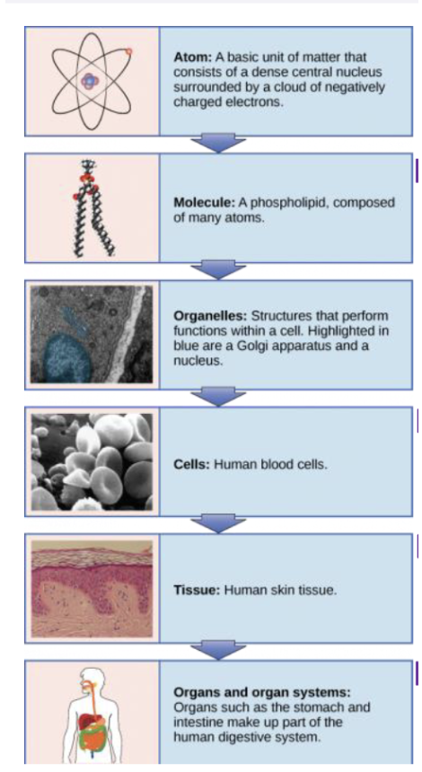
Explain organelles
are specialised structures within a cell that perform distinct functions, such as energy production, protein synthesis, and waste processing e.g mitochondria
What are tissues
groups of cells with the same or similar jobs
What are organs
a collection of tissues that structurally form a functional unit specialized to perform a particular function
What is an organism?
The complete, living individual, such as a human
what is the organisation of the body:
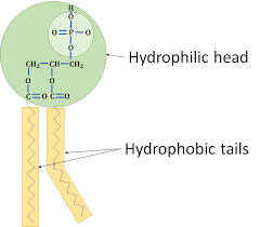
Explain the function of
cell membrane
Explain the function of the nucleus
acts as the control center of a eukaryotic cell, storing genetic information (DNA) and regulating essential activities like growth, metabolism, protein synthesis, and cell division
Explain the function of
mitochondria
generate most of the cell's supply of (ATP), used as a source of chemical energy
Explain the function of
Endoplasmic reticulum
(smooth versus rough)
many functions include;
-Protein synthesis(RIBOSOMES), Calcium storage, Steroid synthesis, Lipid metabolism
-two types: Rough ER(covered in ribosome) and smooth ER (no ribosome)
Explain a ribosome
primary site for protein synthesis translation
Explain function of Golgi body (Golgi apparatus)
Assist with protein synthesis. Packaging of protein molecules to export from cell. Materials from ER go to Golgi bodies in from of vesicles
Explain function of lysosome
"garbage disposal" and recycling center
Contain enzymes that degrade and recycle cellular waste–autophagy.
-Help eliminate bacteria, virus, and foreign bodies that get into cells
Explain function of Vesicles
storing, transporting, and digesting cellular products, waste, and materials
they move substances across the cell
Explain function of Vacuoles
maintaining structural integrity (turgor pressure) in plant cells
What is cytosol?
jelly-like, aqueous fluid contained within the cell membrane that surrounds the organelles
What is the cytoplasm?
allows transport, maintain cell shape and structure, protection, storage and acts as the host to metabolic processes
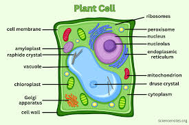
plant cell structure
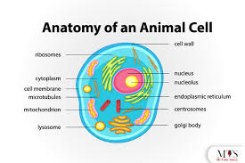
animal cell structure
What does hydrophilic mean?
water loving
What does hydrophobic mean?
do not mix with water
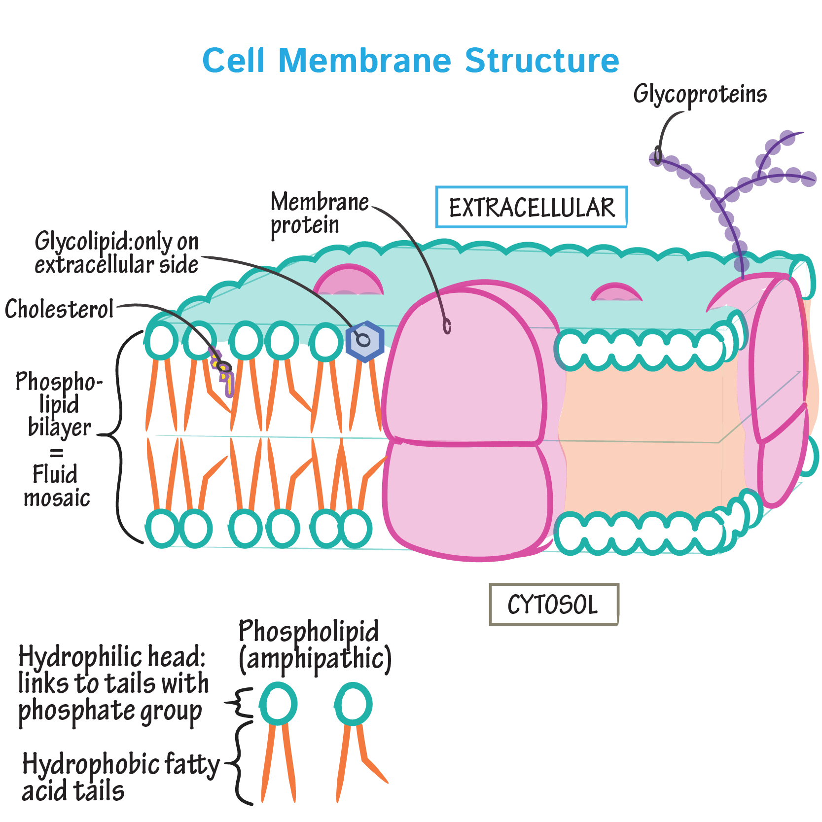
Why is cholesterol important in the cell membrane.
essential for the structure, elasticity and many different functions of cell membranes
Label the cell membrane and
its structures.
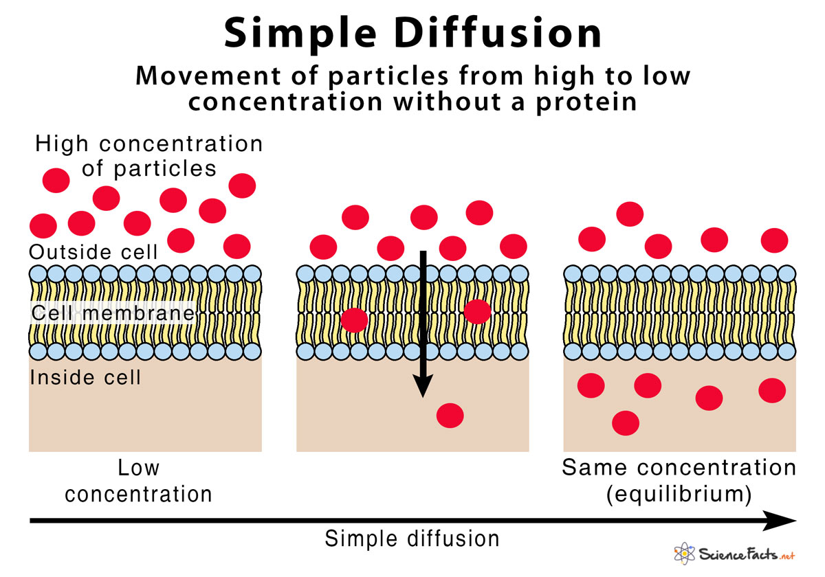
Explain simple diffusion
Direction: Down the concentration gradient (high to low).
Energy: No energy (ATP) required (passive).
Mechanism: Molecules pass directly through the membrane's lipid bilayer.
Substances: Ideal for gases (O₂, CO₂), small uncharged polar molecules (like urea), and hydrophobic substances.
Barrier: Cannot easily transport large polar molecules or ions because of the membrane's hydrophobic interior.
Factors Affecting Rate:
Concentration Gradient: Steeper gradient = faster rate.
Temperature: Higher temperature = faster rate (more kinetic energy).
Surface Area/Distance: Larger area/shorter distance = faster rate.
List the molecules that can diffuse across the membrane via simple diffusion
Gases: Oxygen Carbon dioxide, Nitrogen
Hydrophobic Molecules: Steroid hormones (testosterone, progesterone), lipids
Fat-Soluble Vitamins: Vitamins A, D, E, and K
Small Polar Uncharged Molecules: Ethanol, Urea, Water
Nonpolar Molecules: Benzene
Characteristics of Particles that Simple Diffuse:
Size: Very small molecules.
Charge: Uncharged (nonpolar) molecules.
Solubility: Lipid-soluble (lipophilic) molecules.
Explain facilitated diffusion
molecules move across a cell membrane from high to low concentration via specific transmembrane integral proteins
What is a concentration gradient?
gradual difference in the concentration of solutes in a solution between two regions, commonly separated by a membrane
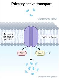
Explain primary active transport
moves ions or molecules across a membrane against their concentration gradient (from low to high concentration) using energy derived directly from ATP hydrolysis
Explain secondary active transport
uses energy indirectly
Antiporter: Na+ moves down the concentration gradient allowing H+ or Ca2+ to move against the concentration gradient. Doesn’t go through the same time and keeps shape
Symporter: carrier protein that simultaneously binds to sodium and another substance, and then changes it shape so that both substances cross the membrane at the same time
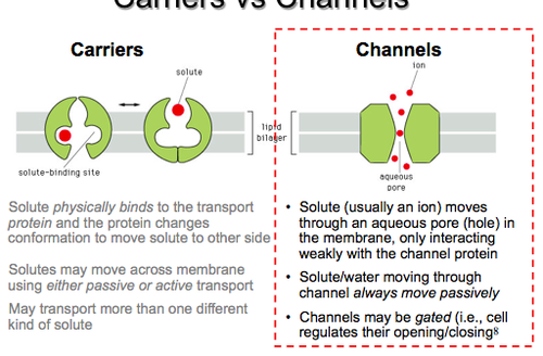
What is the difference between a channel mediated transporter and carrier mediated transporter.
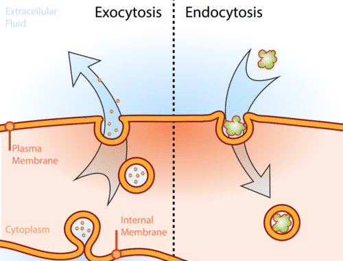
Compare endocytosis and exocytosis
Endocytosis: brings substances into the cell (invagination of membrane forming a vesicle)
Exocytosis: releases substances out of the cell (vesicle fusion with membrane)
Explain pinocytosis and phagocytosis
types of endocytosis (cellular uptake) used by cells to ingest external materials
Phagocytosis ("cell eating"): engulfs large, solid particles like bacteria using pseudopodia
Pinocytosis ("cell drinking"): involves the invagination of the cell membrane to ingest liquids, solutes, and small particles
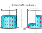
Explain osmosis
the natural movement of water molecules through a semipermeable membrane (like a cell wall) from an area with more water (less dissolved stuff/solute) to an area with less water (more dissolved stuff), aiming to balance the concentration on both sides
Define isotonic solution and the effect on cells
a fluid that has the same solute concentration and osmotic pressure as a cell's cytoplasm, resulting in no net movement of water across the membrane
Cells in an isotonic environment maintain their shape and volume, as water enters and exits at equal rates.
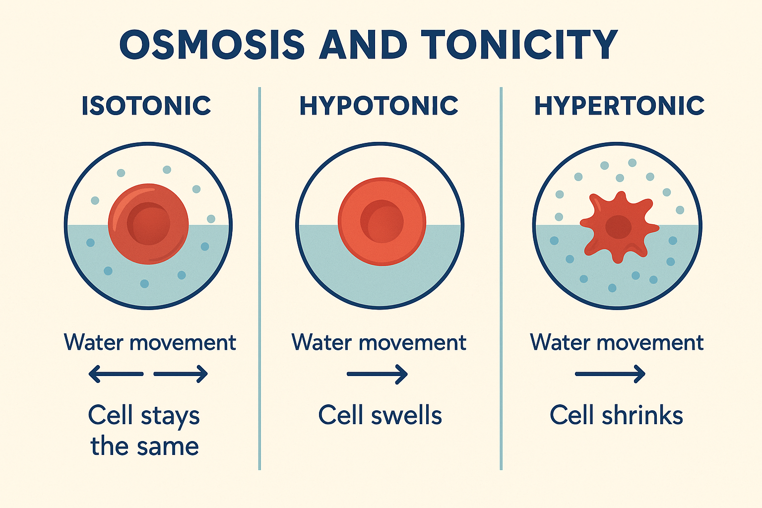
Define hypertonic solution and the effect on cells
has a higher concentration of solutes (like salt or sugar) outside the cell compared to inside, causing water to move out of the cell via osmosis.
This net loss of water results in cell shrinkage
Define hypotonic solution and the effect on cells
an external environment with a lower solute concentration compared to the fluid inside a cell, causing water to flow into the cell via osmosis. This net influx leads to cell swelling
Plant cells become turgid but are protected from bursting by their cell walls
What is the role of the sodium/potassium pump
uses ATP to actively transport 3 sodium ions (𝑁𝑎+) out of the cell and 2 potassium ions(𝐾+) into the cell
Explain symporters vs antiporters
Antiporter: Na+ moves down the concentration gradient allowing H+ or Ca2+ to move against the concentration gradient. Doesn’t go through the same time and keeps shape
Symporter: carrier protein that simultaneously binds to sodium and another substance, and then changes it shape so that both substances cross the membrane at the same time
List molecules that require facilitated diffusion
-vitamins
-amino acids
-proteins
-glucose
What type of transport requires ATP directly
Primary active transport
What type of transport requires ATP indirectly
Secondary active transport
List the 4 primary tissue types
epithelial, connective, muscle, and nervous tissues
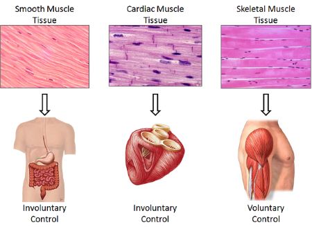
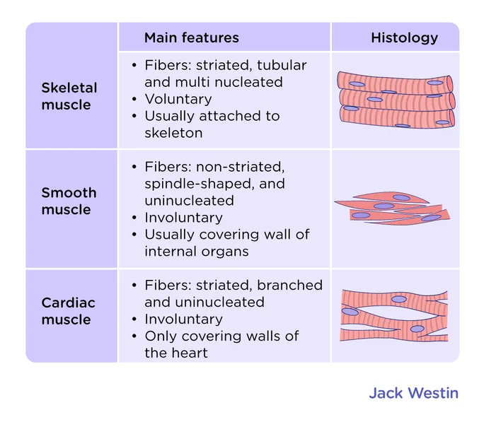
List the 3 types of Muscle tissue
skeletal, cardiac, and smooth
List the 2 division of Nervous tissue
Central Nervous System (CNS) and the Peripheral Nervous System (PNS)
List examples of connective tissue
tendons, ligaments, cartilage, bone, fat (adipose), and blood
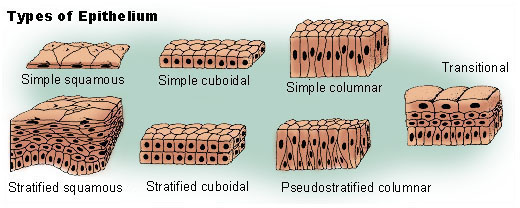
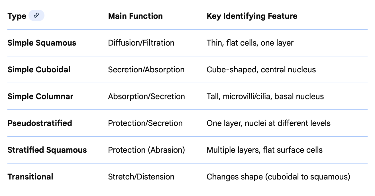
List the different types of epithelial tissue
List epithelial functions
Protection: Shields underlying tissues from abrasion, pathogens, chemicals, and dehydration.
Absorption: Absorbs nutrients and other substances, particularly in the lining of the intestines.
Secretion: Releases mucus, hormones, and enzymes from glandular epithelial cells.
Filtration/Diffusion: Controls permeability to allow selective, rapid passage of molecules and ions.
Excretion: Removes waste materials, such as sweat from the skin.
Sensory Reception: Provides sensory stimuli, such as in the olfactory epithelium.
Transportation: Moves particles or mucus across the surface using cilia (e.g., in the respiratory tract).
Location/ function of cardiac muscle, smooth muscle and
skeletal muscle
Skeletal muscles attach to bones
Cardiac muscle is exclusive to the heart
Smooth muscle lines hollow organs like intestines and blood vessels
Example of loose connective tissue
areolar tissue (found under the skin)
adipose tissue (fat for insulation/energy)
reticular tissue (supporting framework for organs)
Example of fluid connective tissue
blood and lymph
Examples of dense connective
tissue – regular versus irregular
versus elastic
Dense Regular Connective Tissue: Fibers are arranged in parallel bundles to resist high tensile strength in one direction.
Tendons: Connect muscle to bone.
Ligaments: Connect bone to bone.
Aponeuroses: Sheet-like tendons that attach muscles.
Dense Irregular Connective Tissue: Fibers are interwoven randomly, allowing resistance to stretching from multiple directions.
Dermis of the skin: Deep layer providing skin strength.
Organ capsules: Protective capsules around kidneys, liver, and spleen.
Submucosa of the digestive tract.
Heart valves and joint capsules.
Dense Elastic Connective Tissue: Contains a high amount of elastin fibers, allowing tissue to stretch and return to its original shape.
Arterial walls: Large arteries like the aorta.
Vocal cords/folds: In the larynx.
Ligamentum flavum: Specifically, the ligaments between vertebrae.
Examples of supportive connective tissue – compact bone versus cartilage
Compact bone: dense, mineralized tissue (e.g., shafts of long bones) utilizing osteons to provide rigid support. Cartilage: flexible, avascular tissue (e.g., hyaline, elastic, fibrocartilage) found in joints and ears that provides shock absorption
Examples of cartilage – hyaline, fibrocartilage, elastic
Hyaline Cartilage (Most Common)
Articular Cartilage: Covers the ends of long bones in synovial joints to reduce friction.
Respiratory Tract: Forms rings in the trachea and bronchi.
Costal Cartilage: Connects ribs to the sternum.
Nasal Cartilage: Supports the nose structure.
Embryonic Skeleton: Forms the precursor for bone development (growth plates).
Fibrocartilage (Strongest/Tough)
Intervertebral Discs: Pads between vertebrae.
Menisci: Shock-absorbing cartilage in the knee joint.
Pubic Symphysis: Joint between the left and right pubic bones.
Temporomandibular Joint (TMJ): Jaw joint cushioning.
Labra: Shoulder (glenoid) and hip (acetabular) socket cartilage.
Elastic Cartilage (Flexible/Resilient)
External Ear: Pinna and auditory tube.
Epiglottis: Flap in the throat that covers the larynx.
Laryngeal Cartilages: Specific structures like the corniculate and cuneiform cartilages.
Tip of the Nose: Provides shape and flexibility.
Distinguish between types of epithelia – know general role of each
Describe Homeostasis
body’s self-regulating process that maintains a stable, constant internal environment despite fluctuating external conditions
Define set point
a set point is the ideal, target physiological value (e.g.,
37∘C) for human body temperature) that the body works to maintain for a specific variable, such as blood pressure, nutrient levels, or temperature.
Define normal range
a set of values, typically derived from the central 95% of a healthy population, used by healthcare professionals to interpret laboratory test results
Explain negative feedback
To maintain homeostasis (constant internal conditions) and prevent instability, unlike positive feedback which amplifies changes.
Mechanism: When a system's output goes above or below a certain range, the system detects this, acts to reduce the output, and returns it to a target set point.
Explain positive
feedback
a self-amplifying, unstable cycle where the body’s response to a stimulus increases or accelerates that initial change, rather than reversing it
Provide examples of key mechanisms of homeostasis – positive feedback, negative feedback
Negative Feedback Mechanisms (Reversing changes, maintaining stability)
Body Temperature Regulation: When body temperature rises, the hypothalamus initiates sweating and vasodilation to cool the body. If it drops, shivering and vasoconstriction generate heat.
Blood Glucose Control: After eating, high blood sugar causes the pancreas to release insulin, allowing cells to take up glucose. If blood sugar falls, glucagon is released to signal the liver to release stored glucose.
Blood Pressure Regulation: Baroreceptors detect high blood pressure and signal the heart to slow down and vessels to dilate, reducing pressure.
Positive Feedback Mechanisms (Amplifying changes, driving processes to completion)
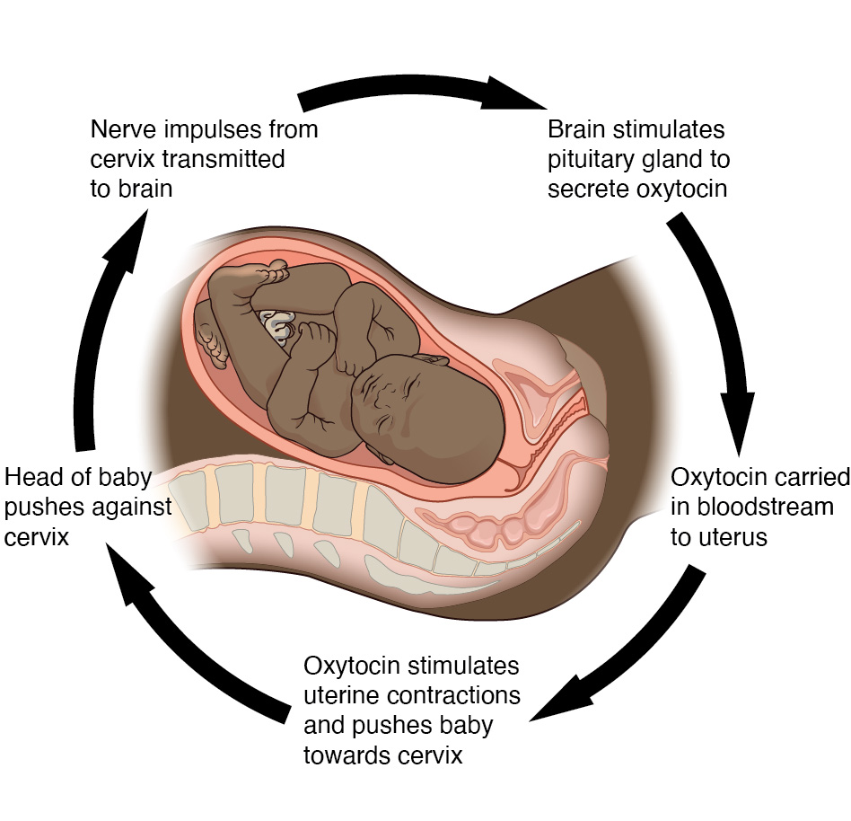
Childbirth (Labor): The hormone oxytocin triggers contractions, which push the baby against the cervix, causing more oxytocin release, further increasing the intensity and frequency of contractions until birth.
Blood Clotting (Hemostasis): When a vessel is damaged, platelets adhere to the site and release chemicals that attract more platelets, rapidly amplifying the process until the clot is formed.
Fruit Ripening: Ethylene gas is released by ripening fruit, which stimulates nearby fruit to ripen faster, leading to the rapid maturation of all fruit on a branch
Feedback loops: Be able to identify the stimulus, receptor,
control centre, effector, response – Body temperature, lactating, birth, blood pressure
Negative Feedback Examples (Return to Set Point)
Body Temperature (High):
Stimulus: Increased body temperature.
Receptor: Thermoreceptors in skin/brain.
Control Centre: Hypothalamus.
Effector: Sweat glands/blood vessels.
Response: Sweating and vasodilation (cooling).
Blood Pressure:
Stimulus: High/Low blood pressure.
Receptor: Baroreceptors in blood vessels.
Control Centre: Brain (Medulla oblongata).
Effectors: Heart and blood vessels.
Response: Reduced/increased heart rate and vessel dilation/constriction.
Positive Feedback Examples (Amplification)
Birth (Childbirth):
Stimulus: Baby's head pushes on cervix.
Receptor: Nerve cells in cervix.
Control Centre: Hypothalamus/Pituitary gland.
Effector: Uterus muscles.
Response: Uterine contractions (oxytocin released) until birth.
Lactation (Milk Production):
Stimulus: Baby sucking on nipple.
Receptor: Sensory receptors in nipple.
Control Centre: Hypothalamus/Pituitary gland.
Effector: Mammary glands (milk-producing cells).
Response: Milk letdown and continued production.