Central Nervous System Lab
1/94
There's no tags or description
Looks like no tags are added yet.
Name | Mastery | Learn | Test | Matching | Spaced | Call with Kai |
|---|
No analytics yet
Send a link to your students to track their progress
95 Terms
Central Nervous System (CNS)
Brain and spinal cord; integrates sensory input and coordinates motor output
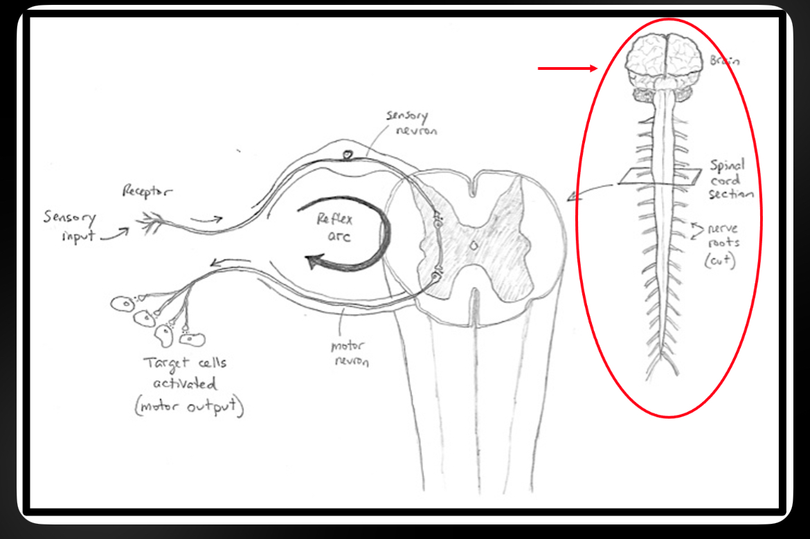
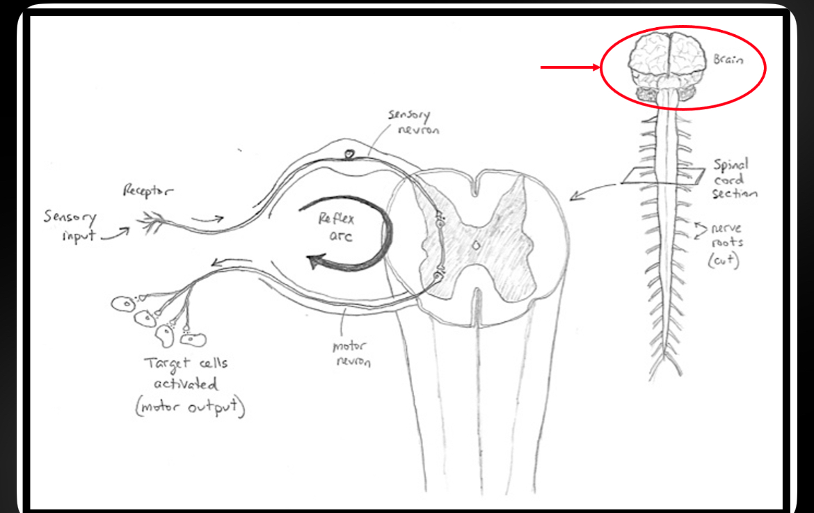
Brain
Control center of CNS; responsible for consciousness, memory, learning, sensation, and voluntary movement
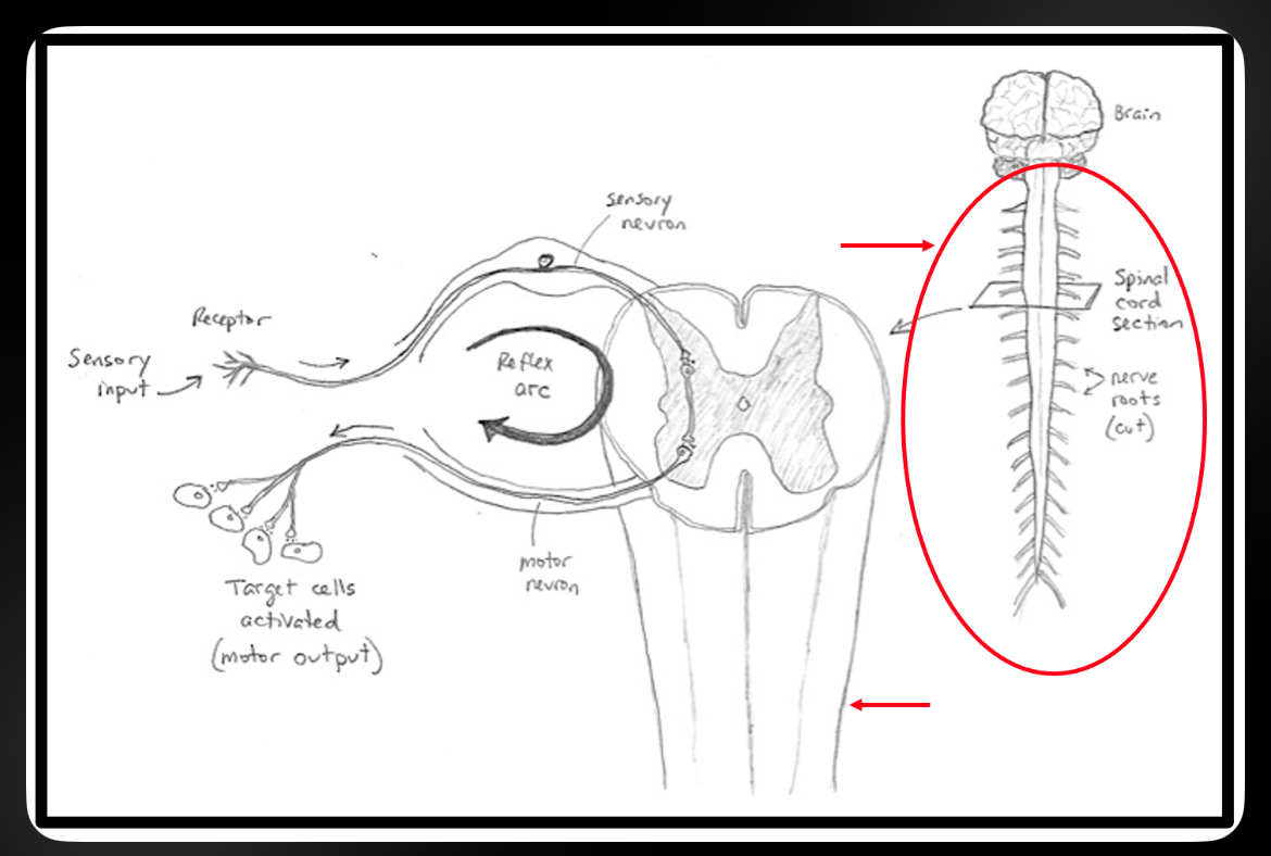
Spinal Cord
Conducts signals to and from brain; reflex center
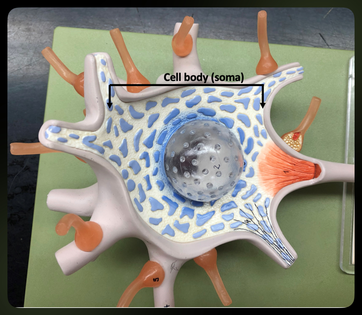
Cell Body (Soma)
Contains nucleus and organelles; metabolic center of neuron
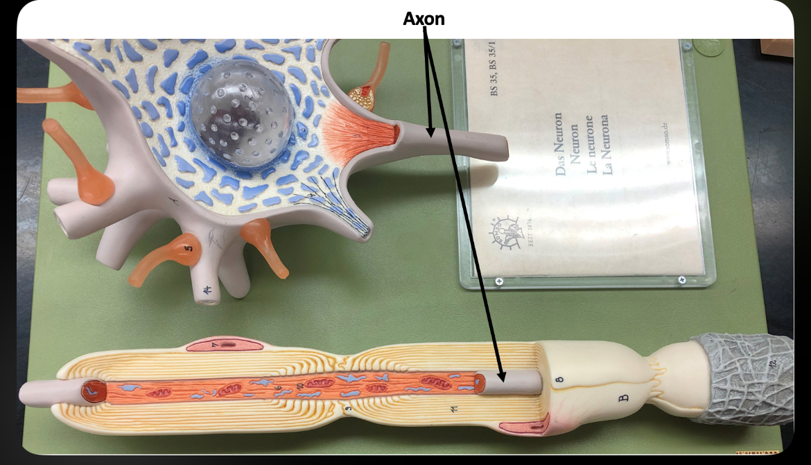
Axon
Conducts action potentials away from cell body
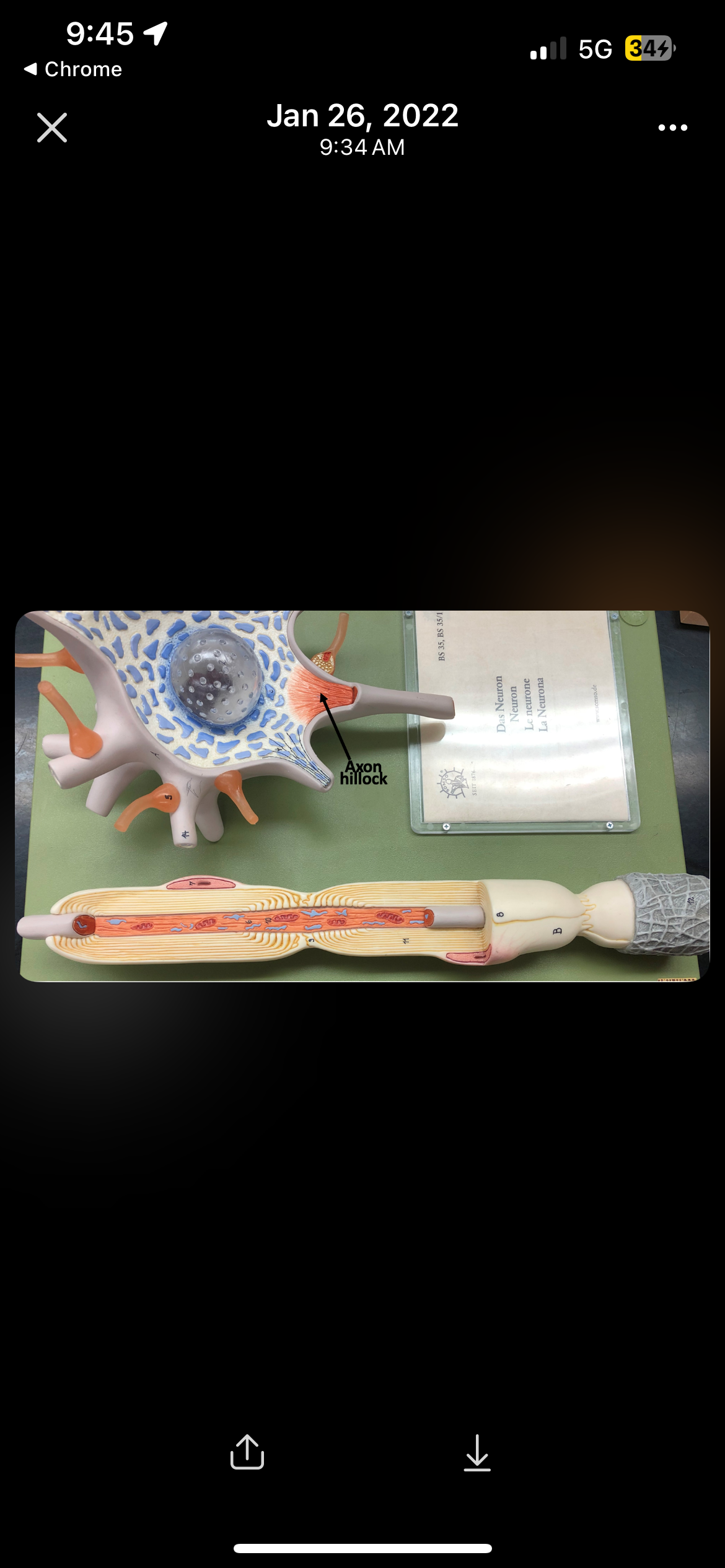
Axon Hillock
Site where action potentials are initiated
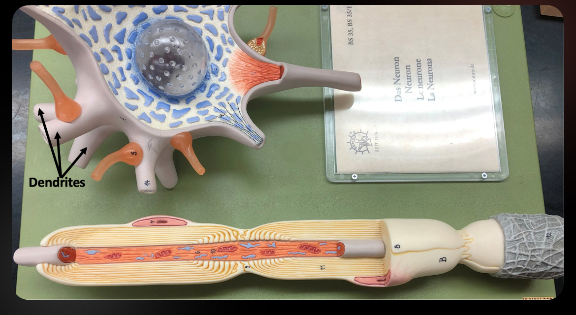
Dendrites
Receive signals and transmit them toward the soma
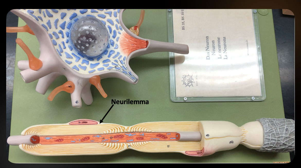
Neurilemma
Outer layer of Schwann cell in PNS; essential for axon regeneration
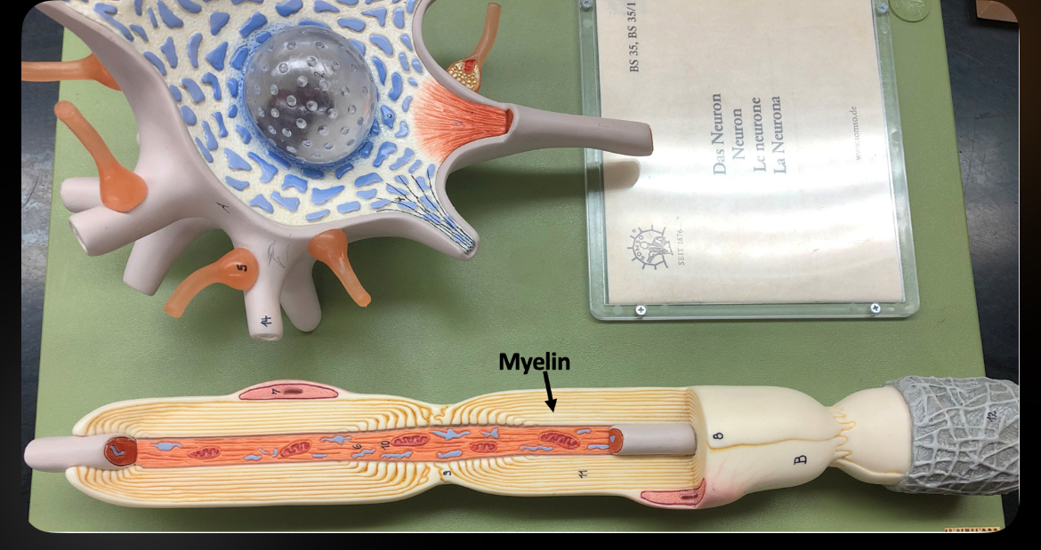
Myelin
Lipid covering around axon that increases conduction speed
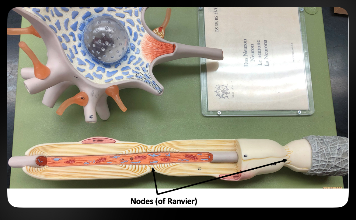
Nodes of Ranvier
Gaps in myelin sheath that allow saltatory conduction
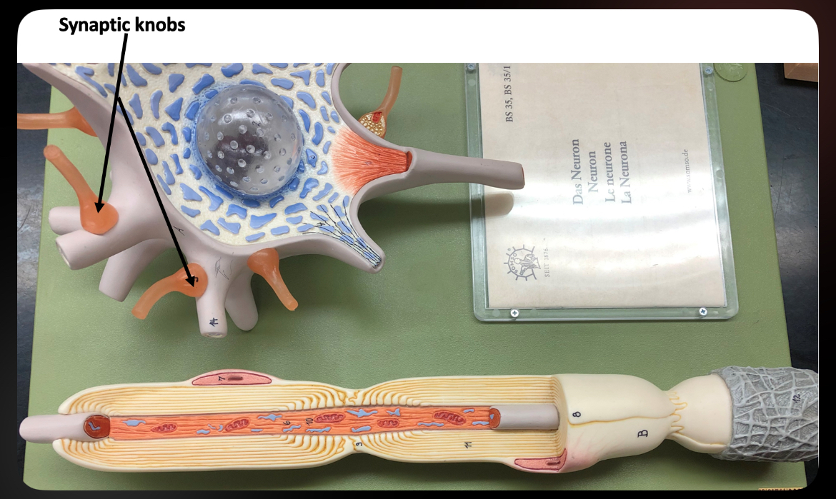
Synaptic Knobs
Swollen ends of axon terminals that release neurotransmitters
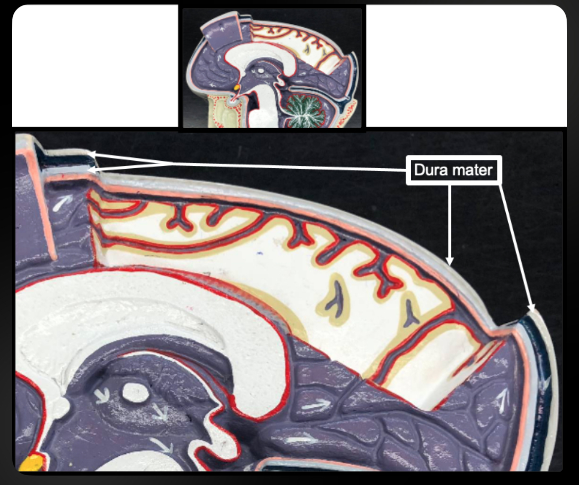
Dura Mater (Brain)
Tough outer meningeal layer protecting brain
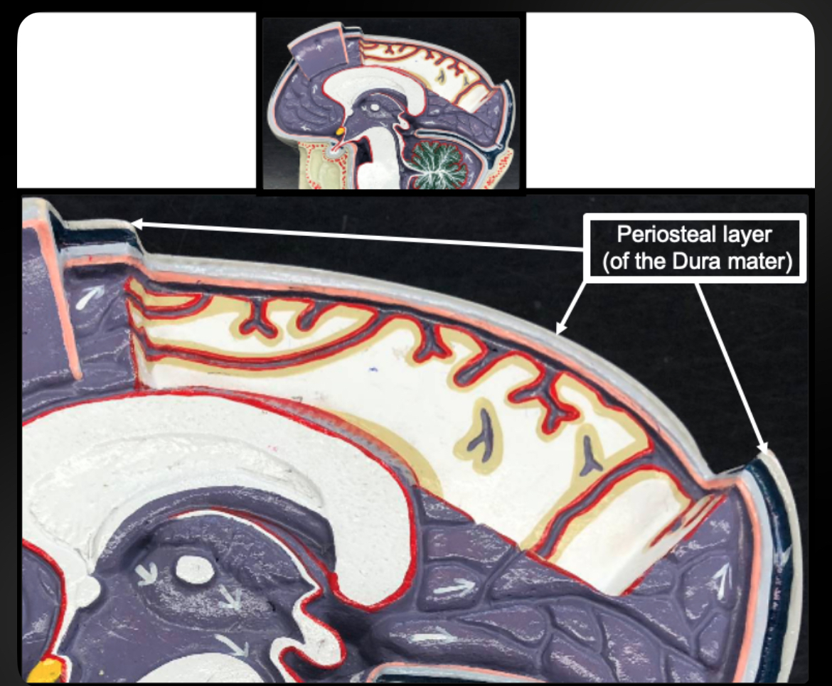
Periosteal Layer
Outer layer of cranial dura attached to skull
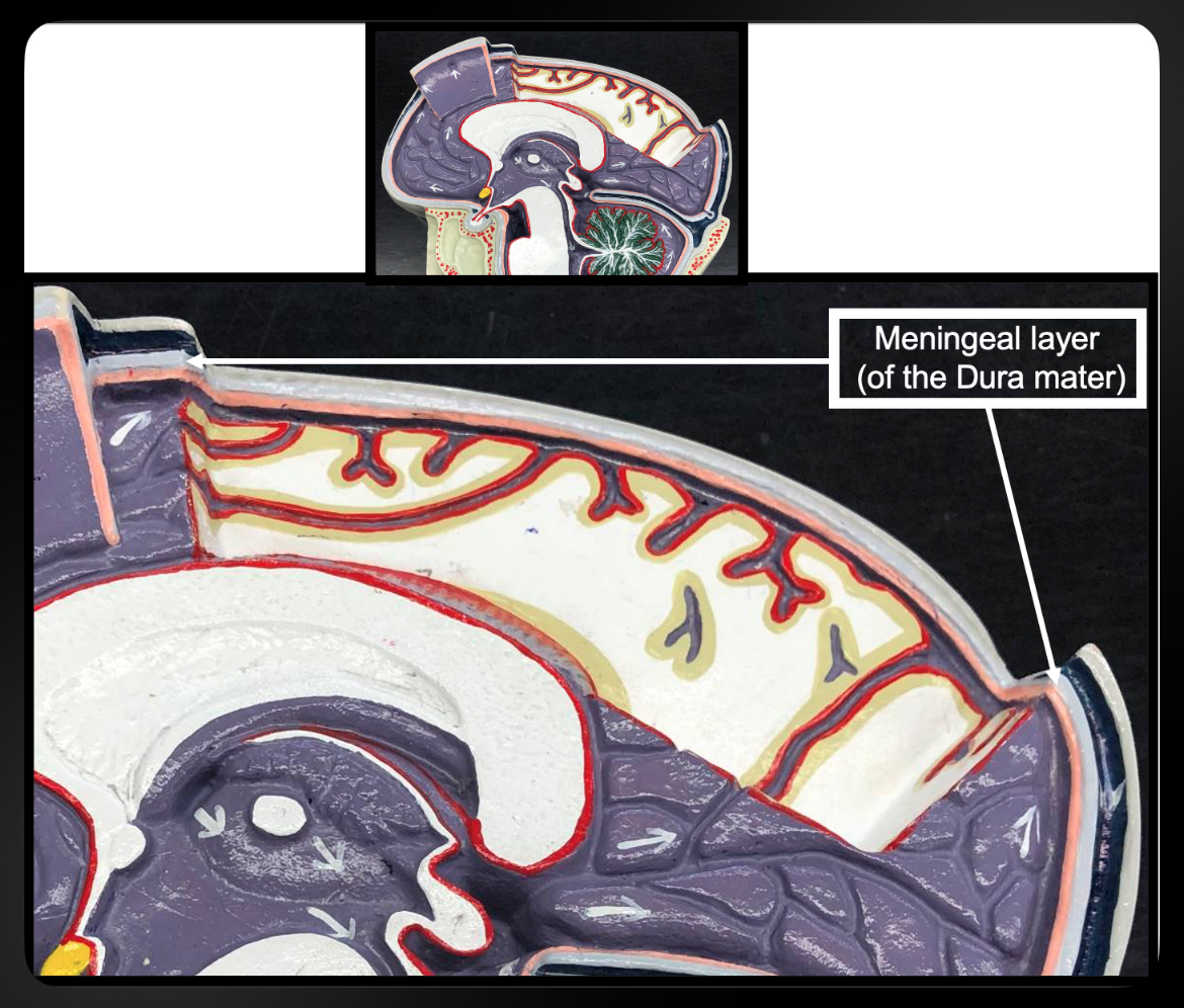
Meningeal Layer
Inner dura layer forming dural folds
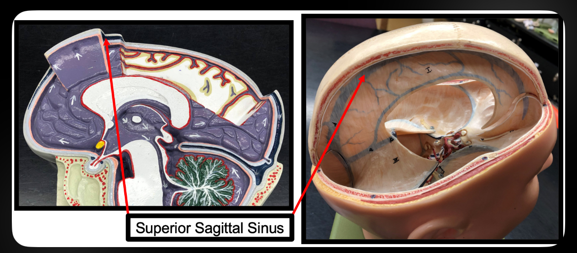
Superior Sagittal Sinus
Venous sinus that drains blood from brain
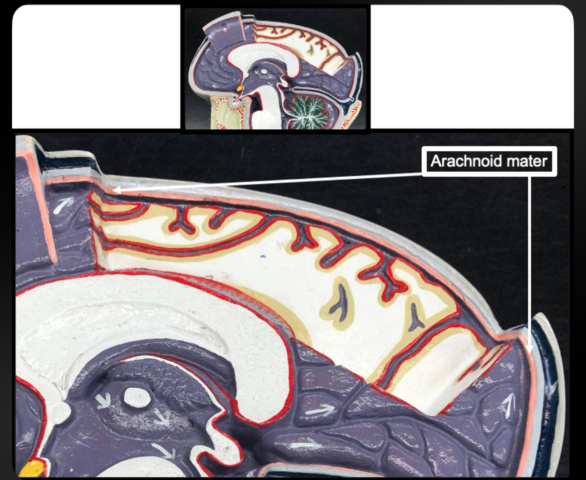
Arachnoid Mater
Middle meningeal layer; web-like

Subarachnoid Space
Space containing CSF and major blood vessels
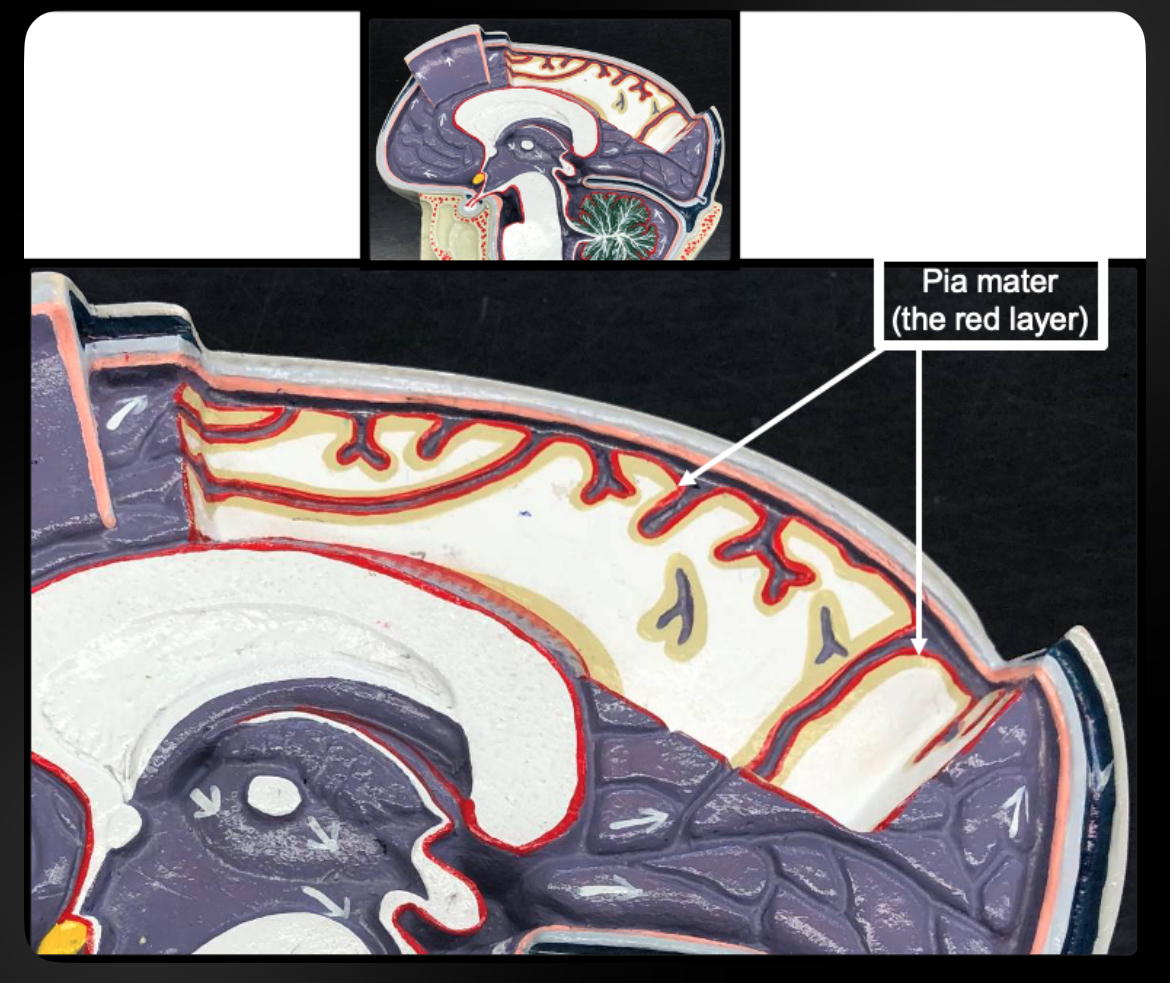
Pia Mater
Thin inner meningeal layer tightly adhering to brain
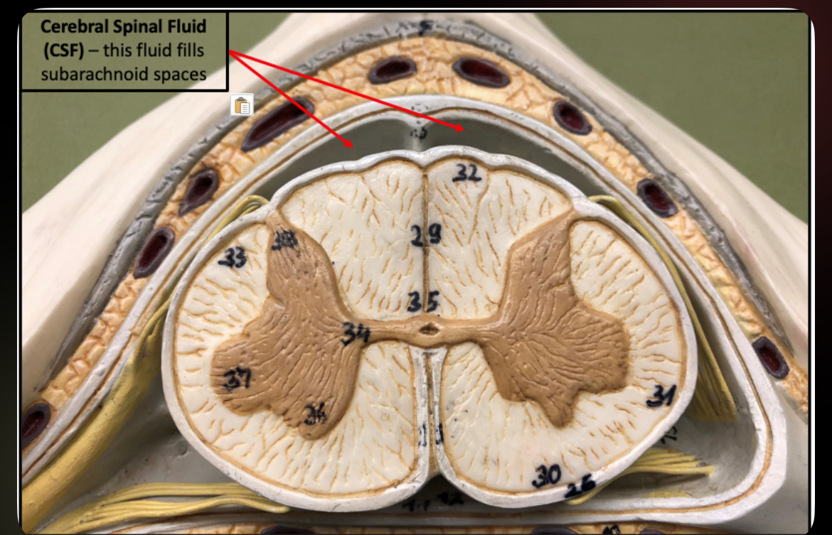
Cerebrospinal Fluid (CSF)
Cushions brain and spinal cord; provides nutrients and waste removal
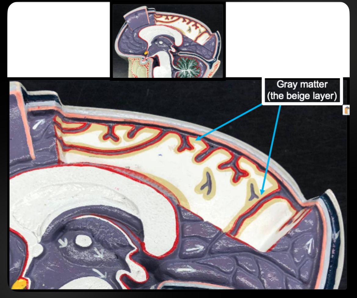
Gray Matter (Brain)
Contains neuron cell bodies; site of processing
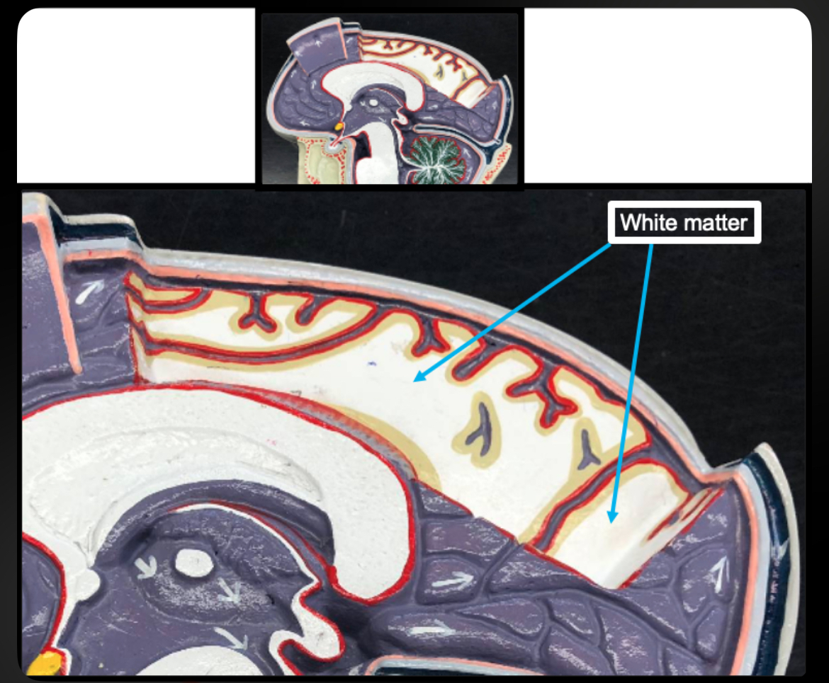
White Matter (Brain)
Contains myelinated axons; conducts impulses
Cerebral Hemispheres
Largest brain region; higher cognitive functions
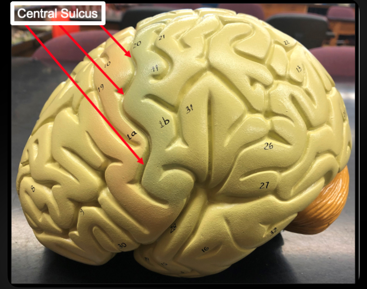
Central Sulcus
Separates frontal and parietal lobes
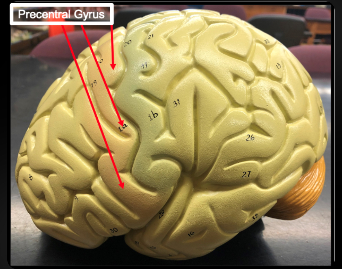
Precentral Gyrus
Primary motor cortex; voluntary skeletal muscle control
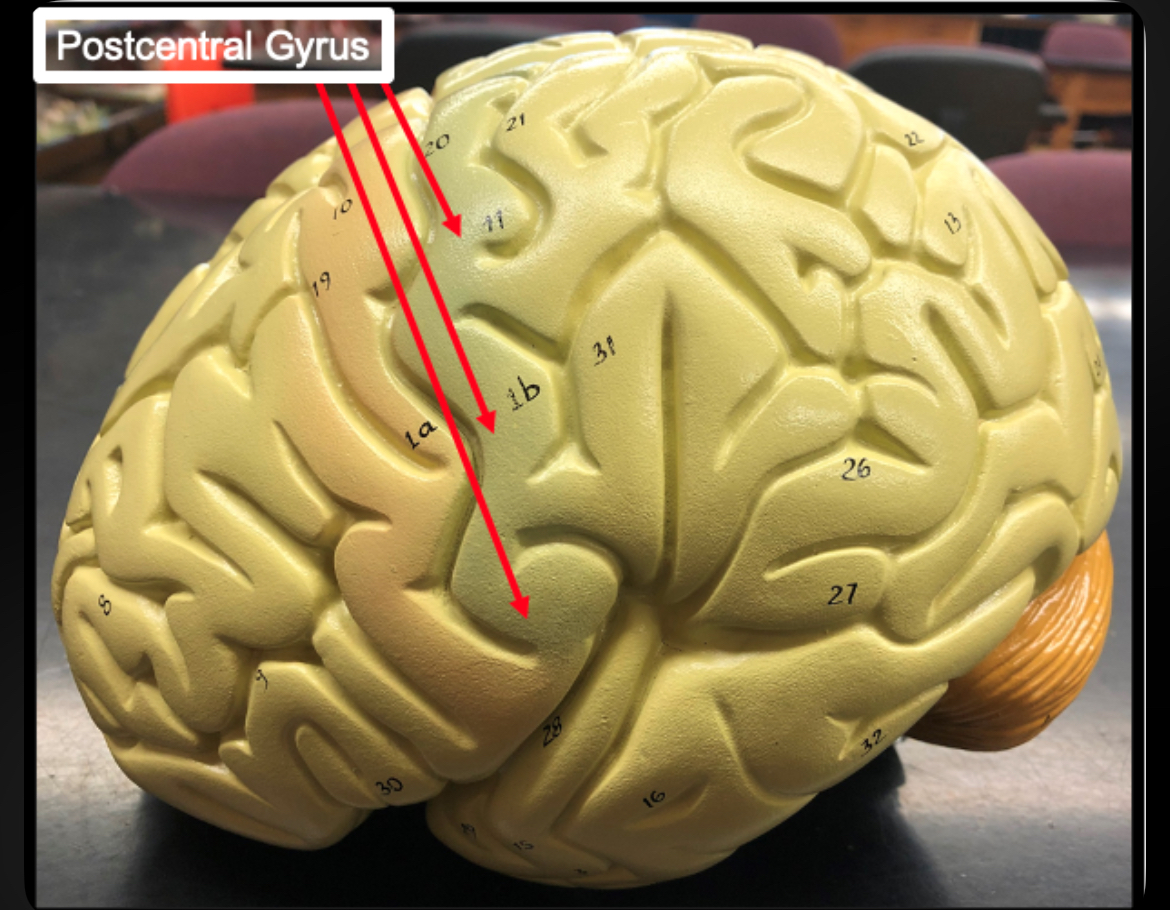
Postcentral Gyrus
Primary somatic sensory cortex; receives sensory input
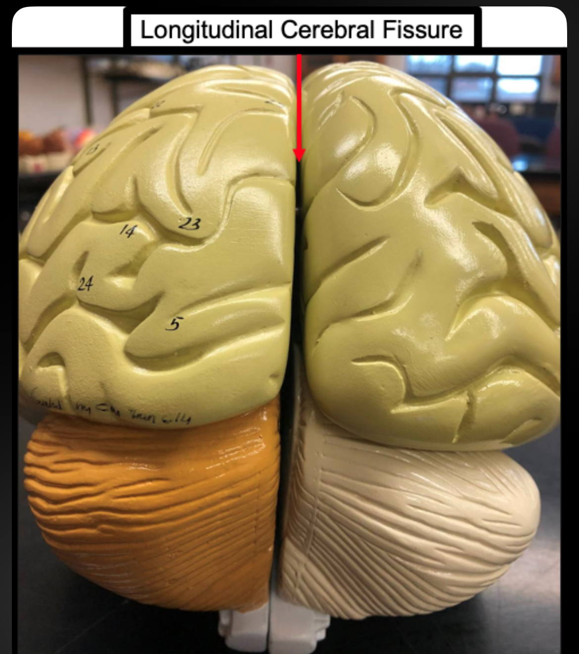
Longitudinal Fissure
Separates right and left hemispheres
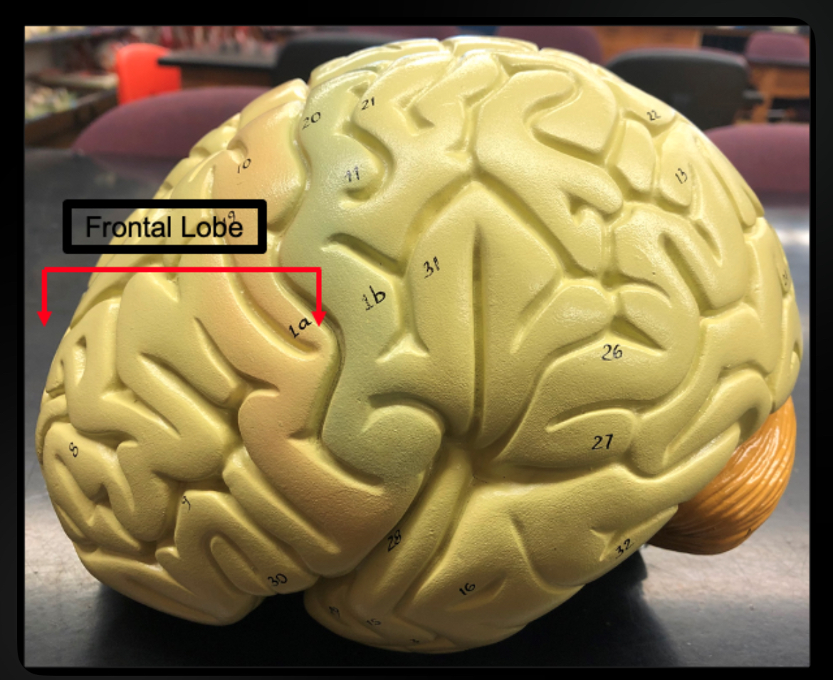
Frontal Lobe
Voluntary motor control, planning, personality
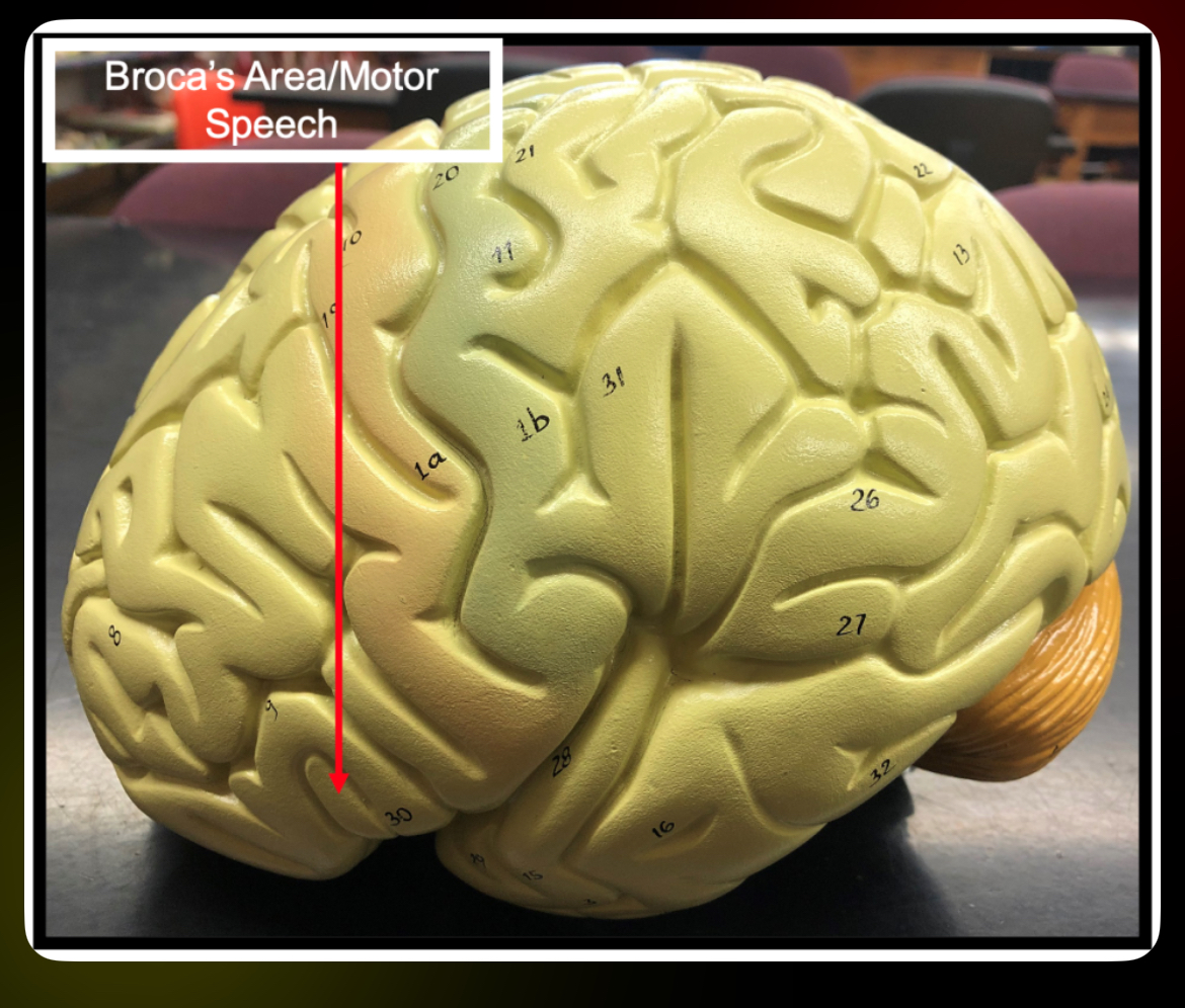
Broca’s Area of frontal
Motor speech area; speech production
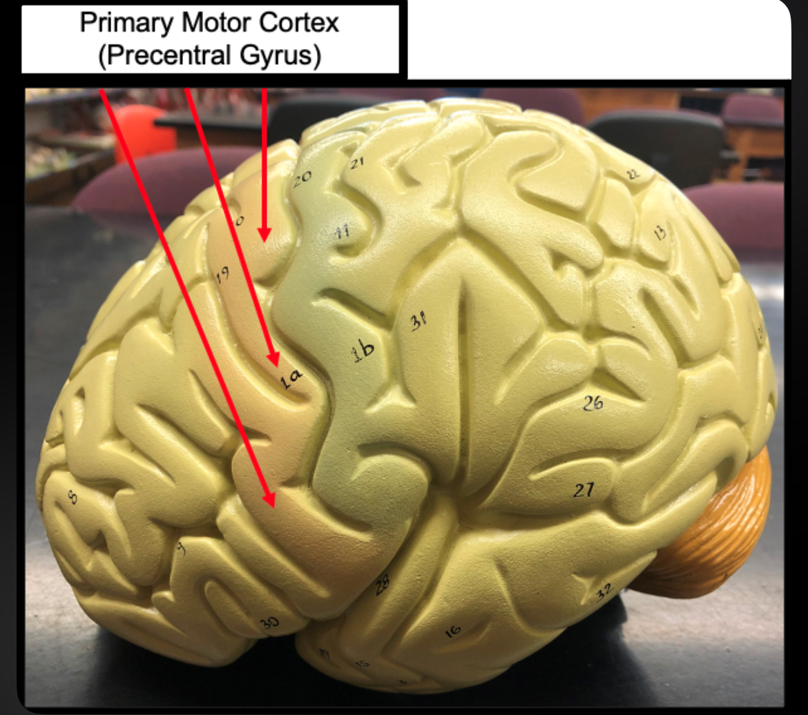
Primary Motor Cortex of frontal
Initiates voluntary movement
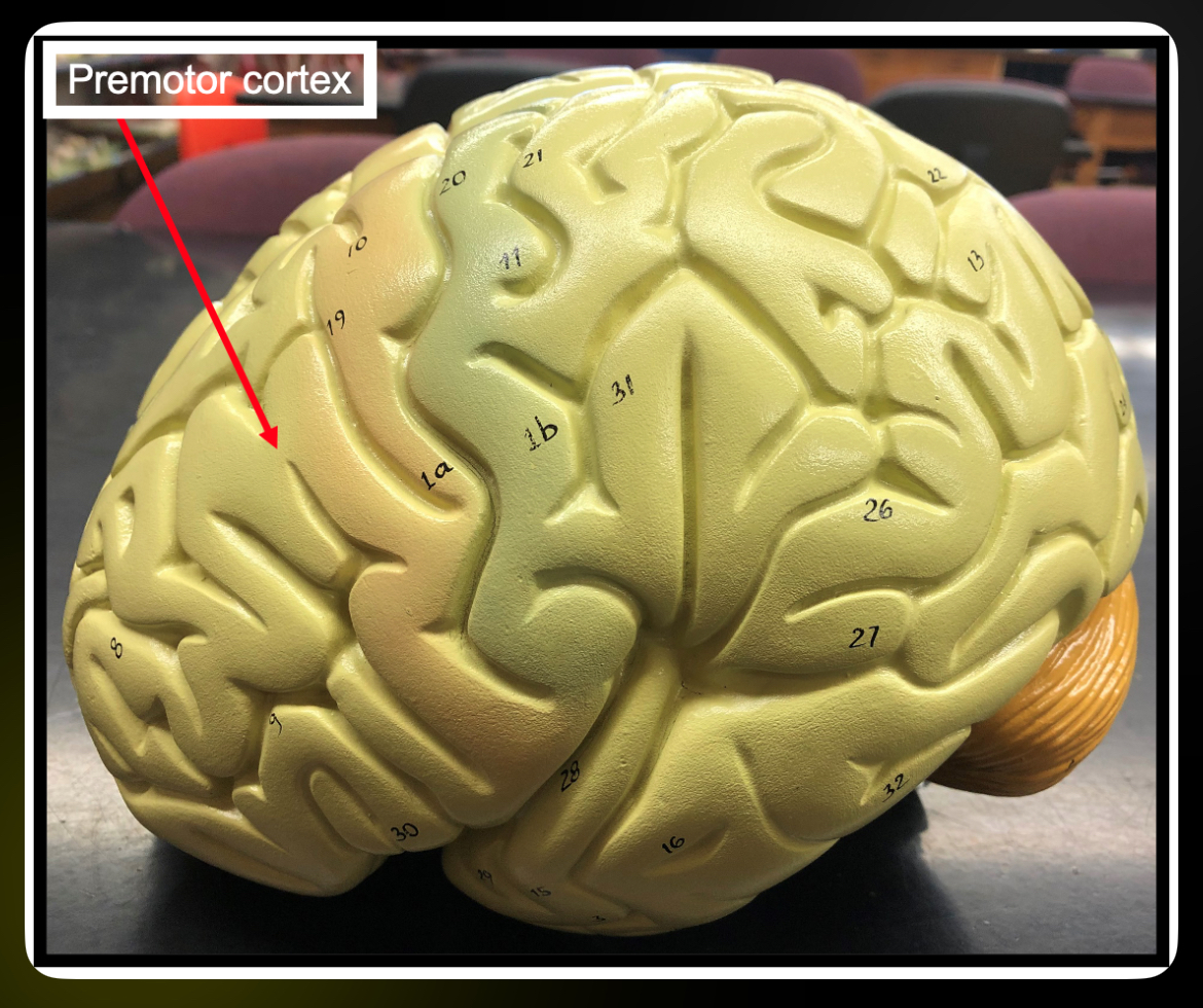
Premotor Cortex of frontal
Plans and coordinates learned movements
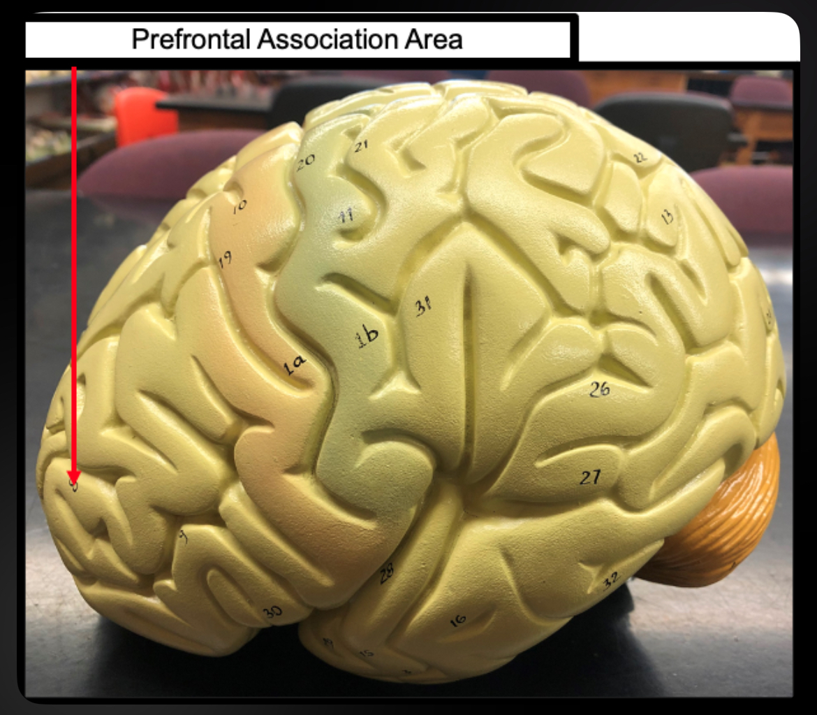
Prefrontal Association Area
Personality, judgment, decision making
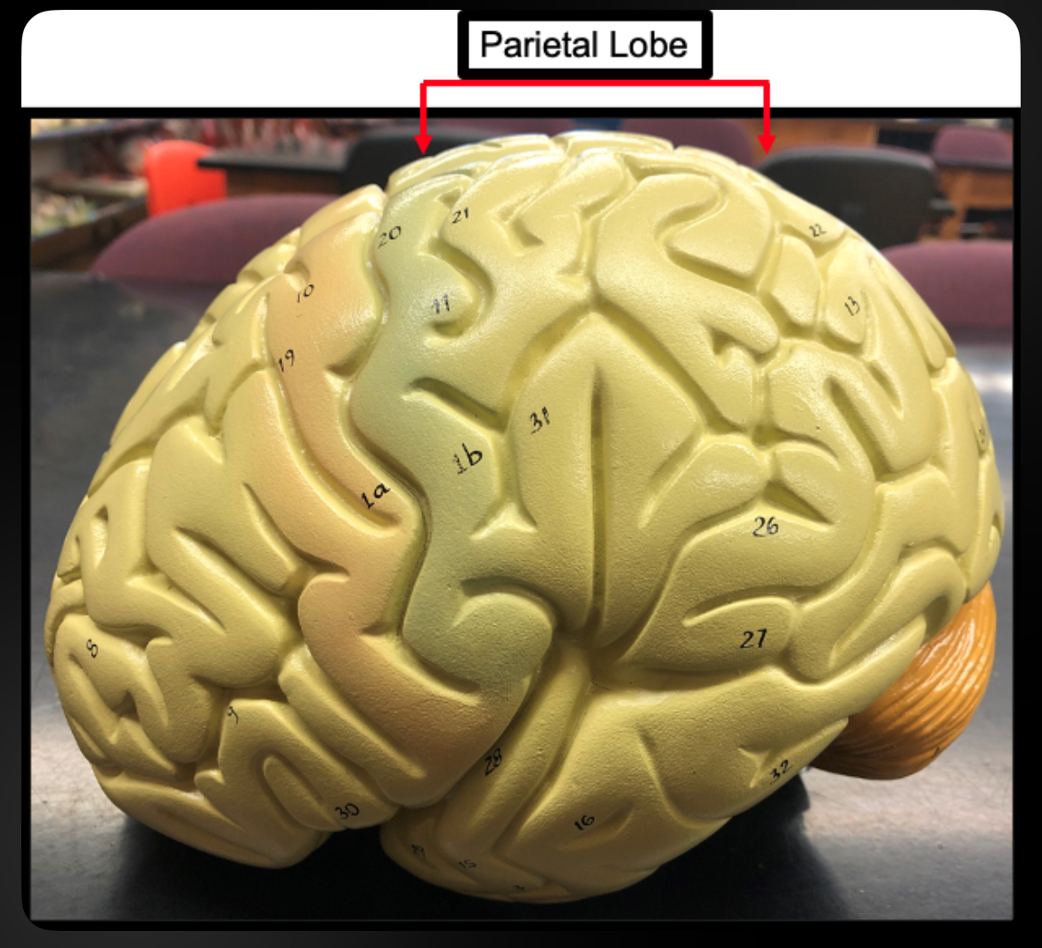
Parietal Lobe
Processes sensory information
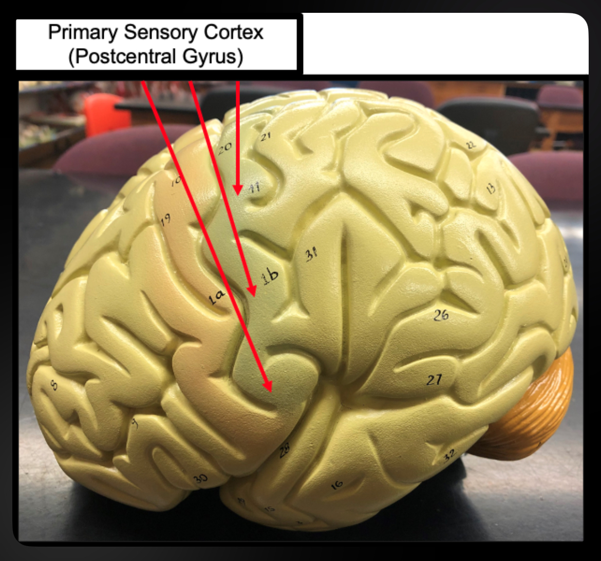
Primary Sensory Cortex of Parietal lobe
Receives touch, pain, temperature input
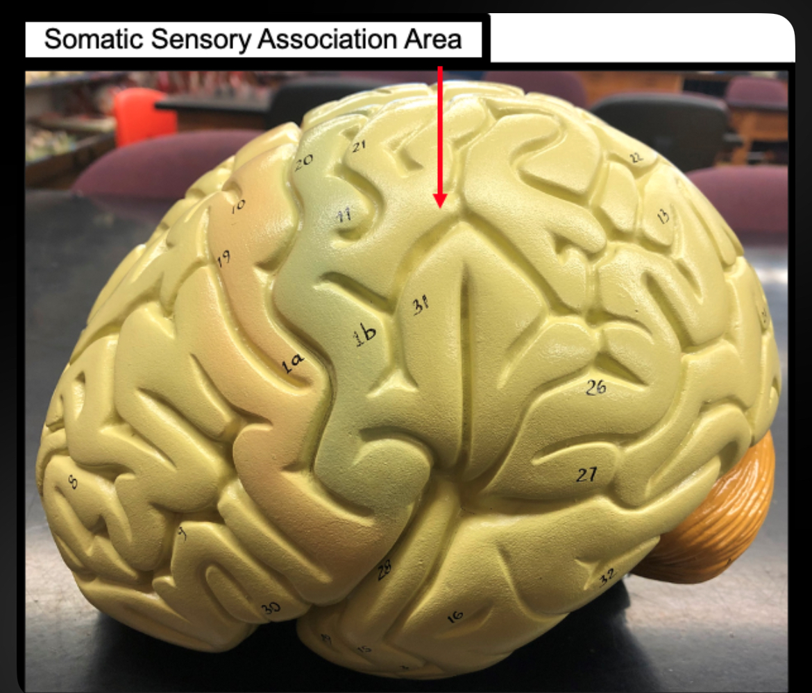
Somatic Sensory Association Area
Interprets sensory input
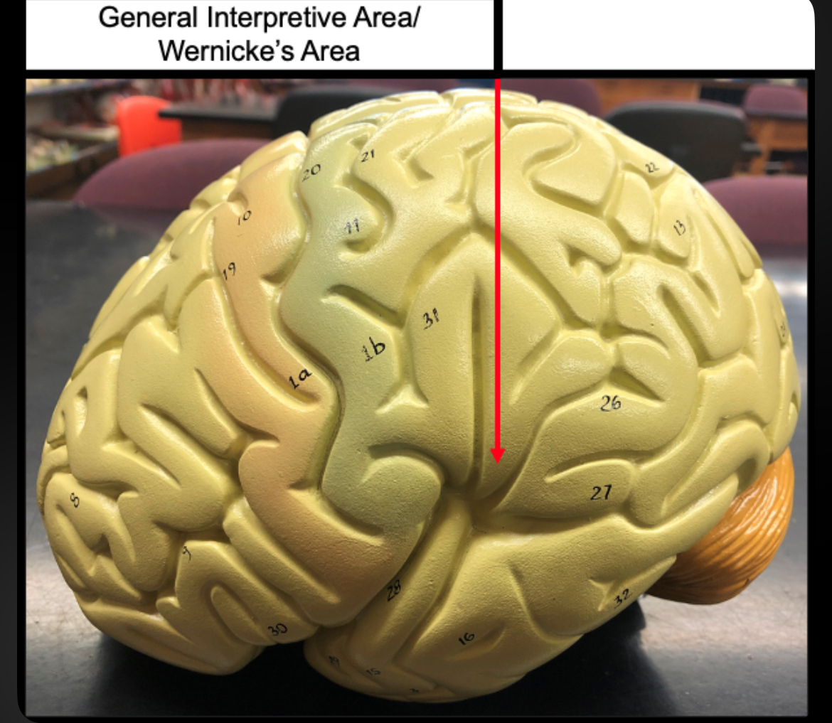
Wernicke’s Area
Language comprehension and interpretation
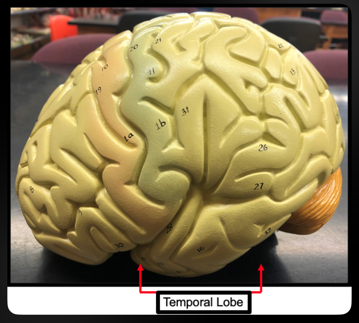
Temporal Lobe
Hearing, smell, memory
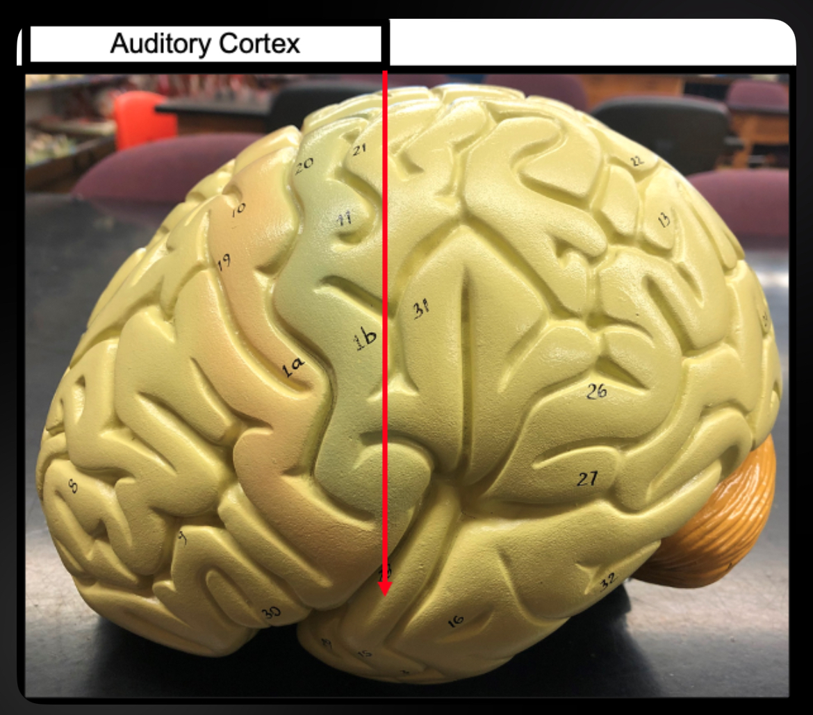
Auditory Cortex
Processes sound
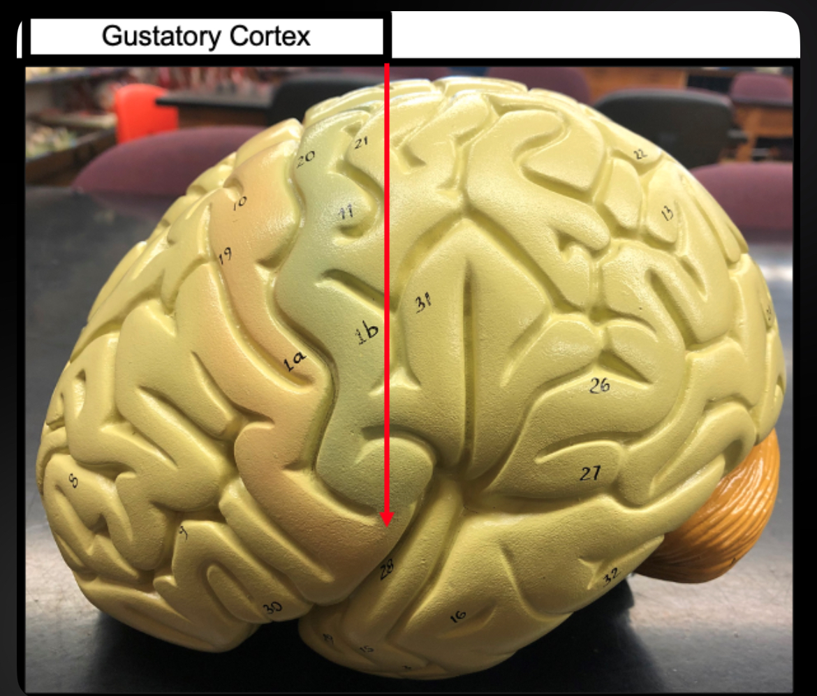
Gustatory Cortex
Processes taste
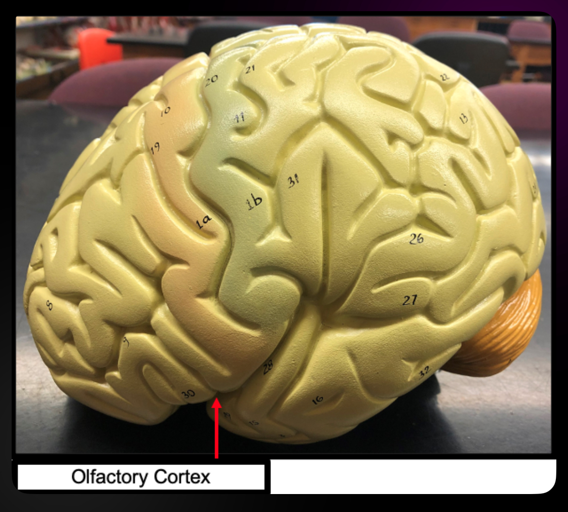
Olfactory Cortex
Processes smell
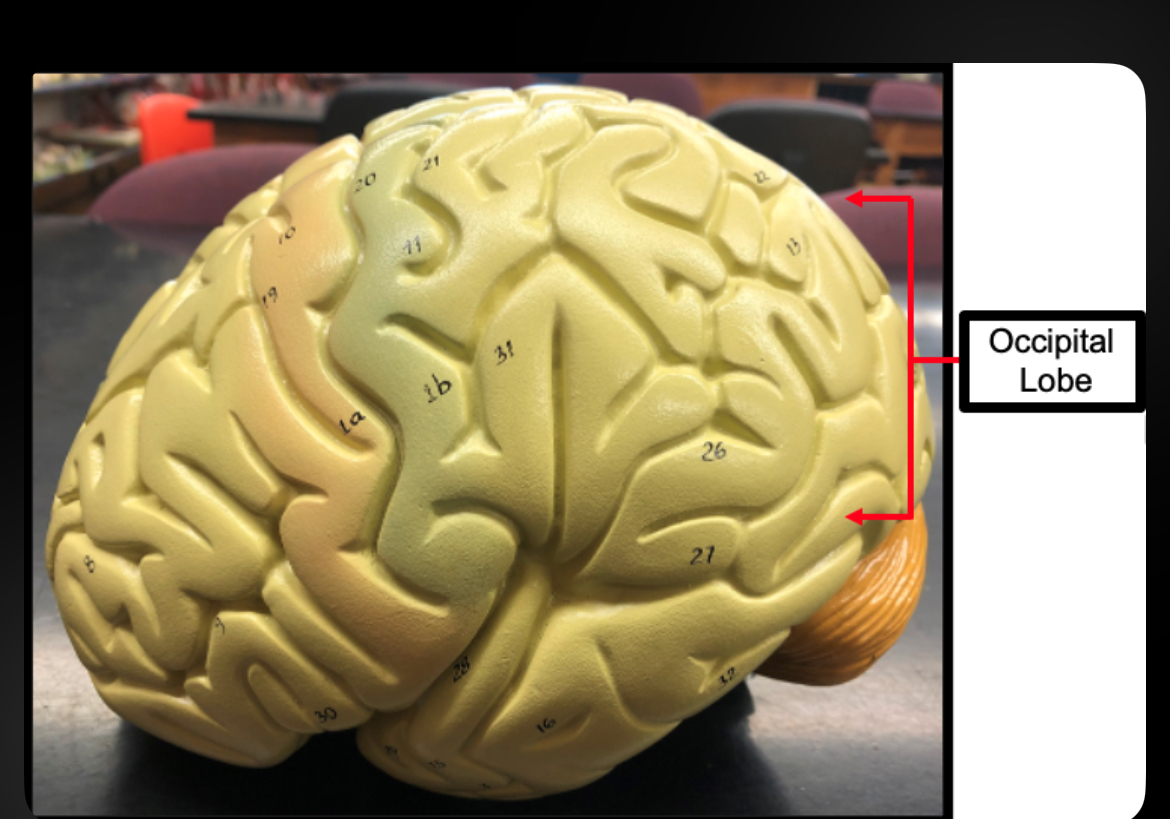
Occipital Lobe
Vision
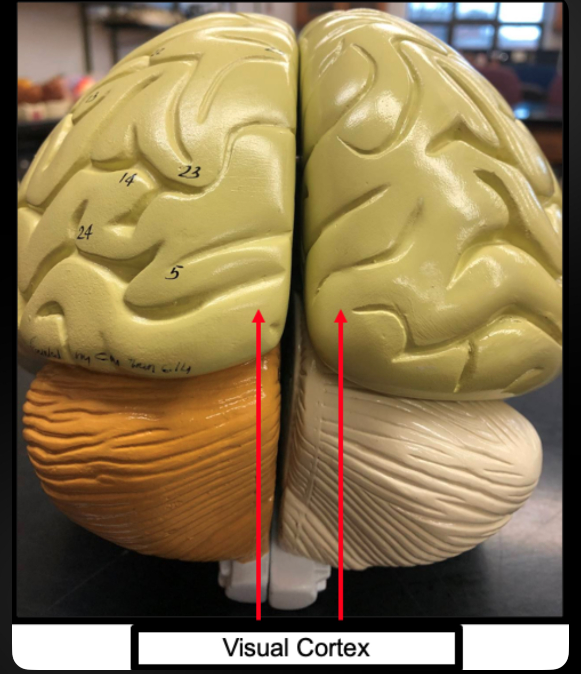
Visual Cortex
Processes visual information
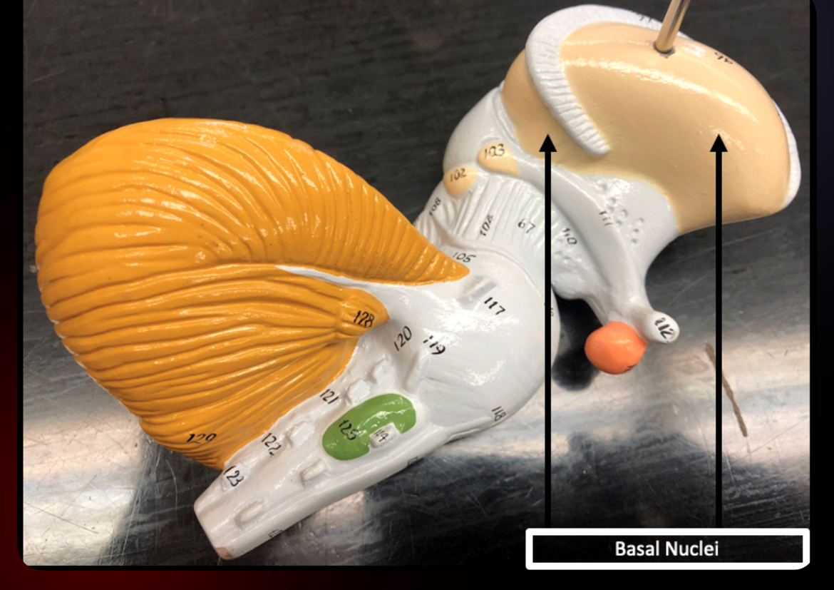
Basal Nuclei
Regulate motor control and muscle tone
Association Tracts
Connect areas within same hemisphere

Commissural Tracts
Connect two hemispheres (corpus callosum)
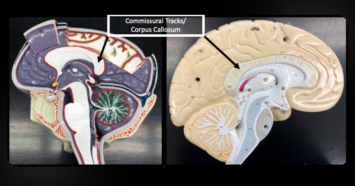
Corpus Callosum
Major commissural tract connecting hemispheres
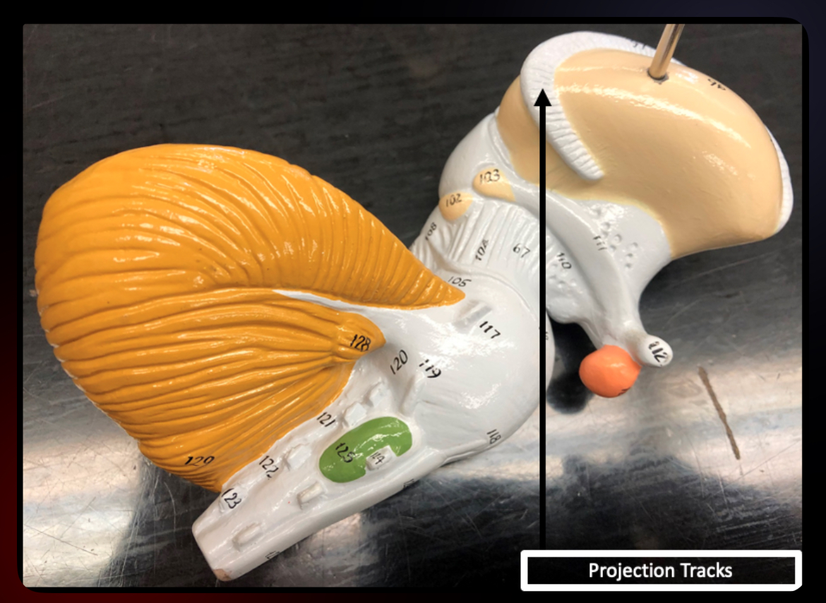
Projection Tracts
Connect cerebrum with lower CNS regions

Fornix
Connects hippocampus to other brain regions; memory processing
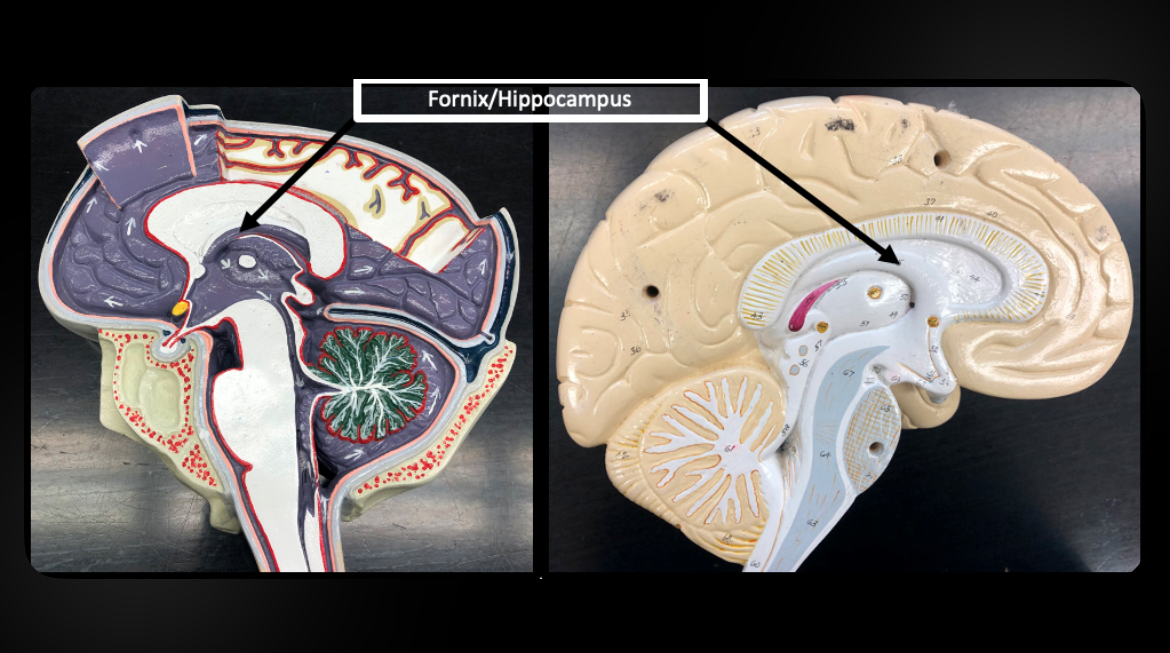
Hippocampus
Memory formation and learning
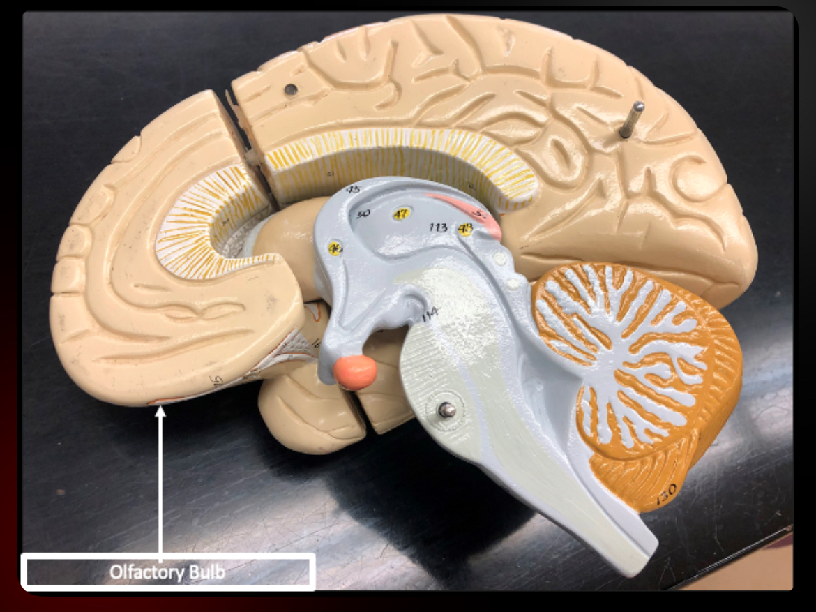
Olfactory Bulbs
Receive sensory input for smell
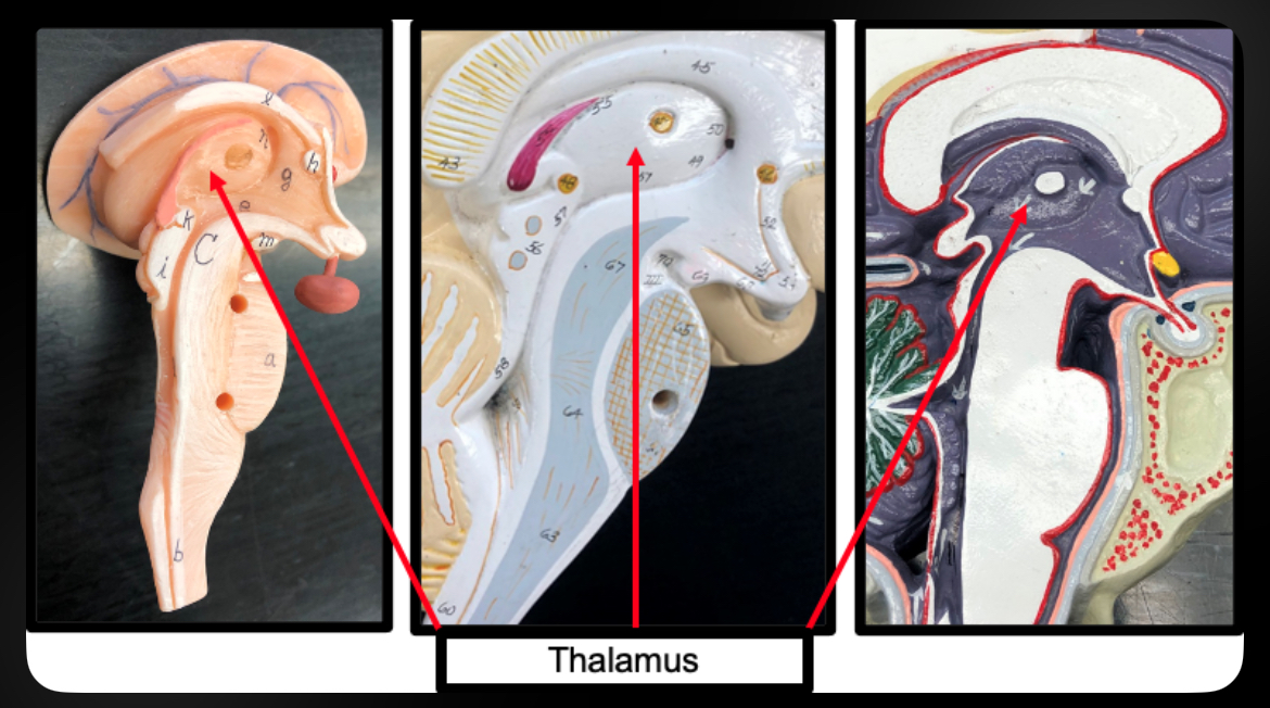
Thalamus
Sensory relay station to cerebral cortex
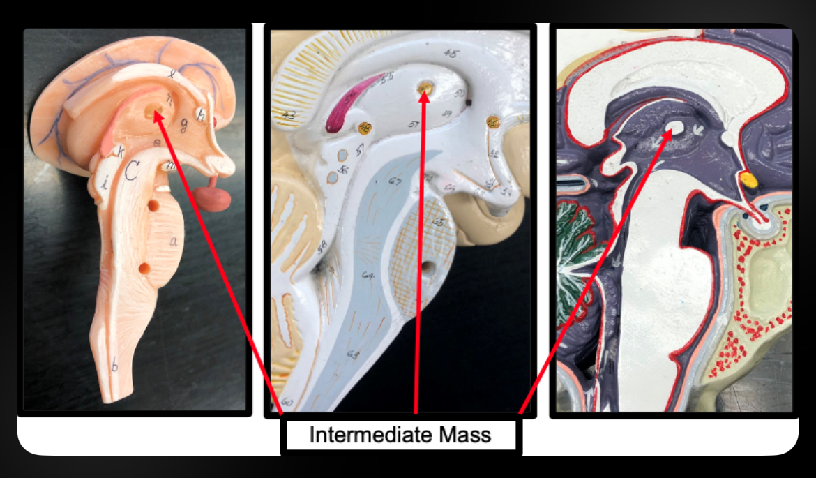
Intermediate Mass
Connection between right and left thalamus
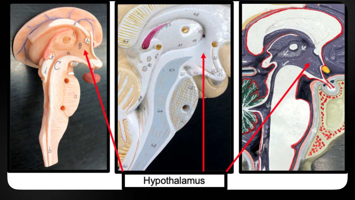
Hypothalamus
Maintains homeostasis; controls autonomic and endocrine systems
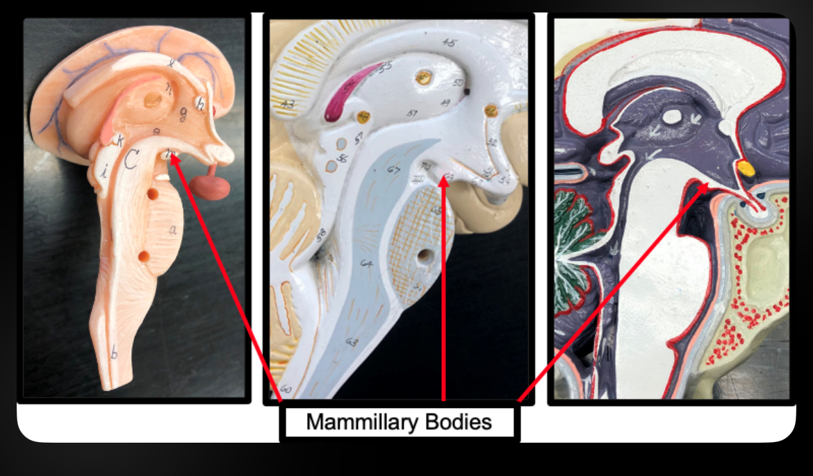
Mammillary Bodies
Relay stations for memory

Pituitary Gland
Master endocrine gland controlled by hypothalamus
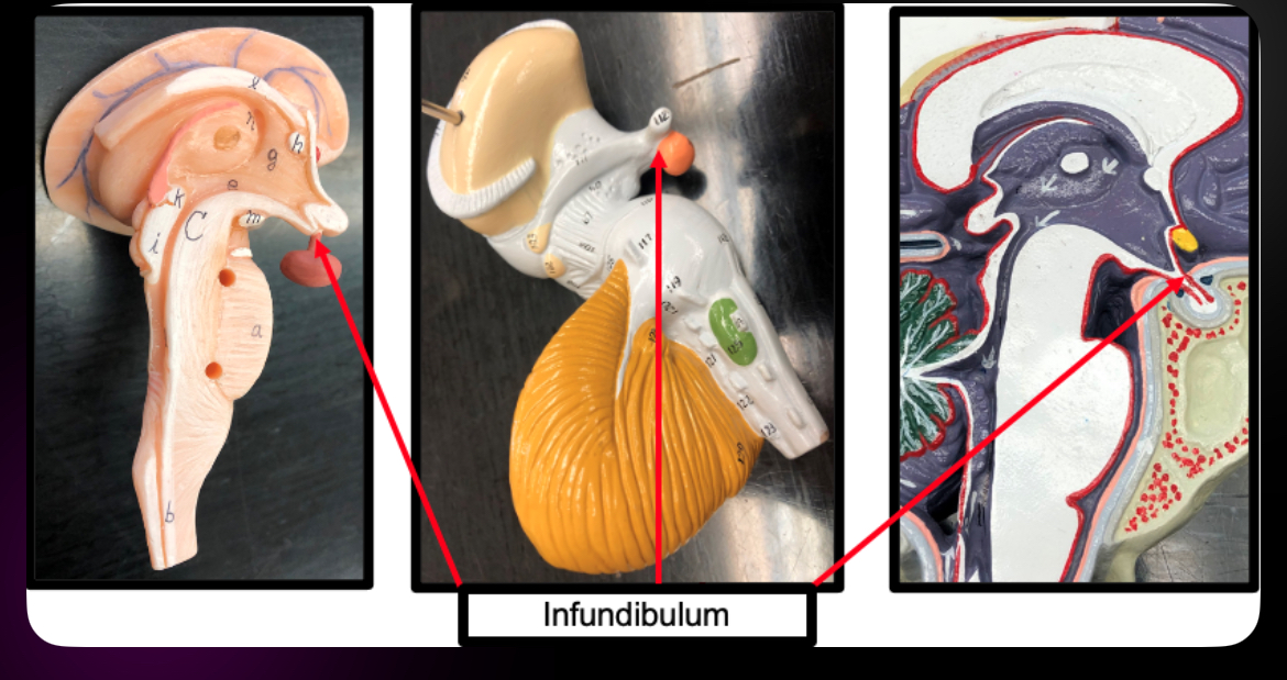
Infundibulum
Connects hypothalamus to pituitary gland
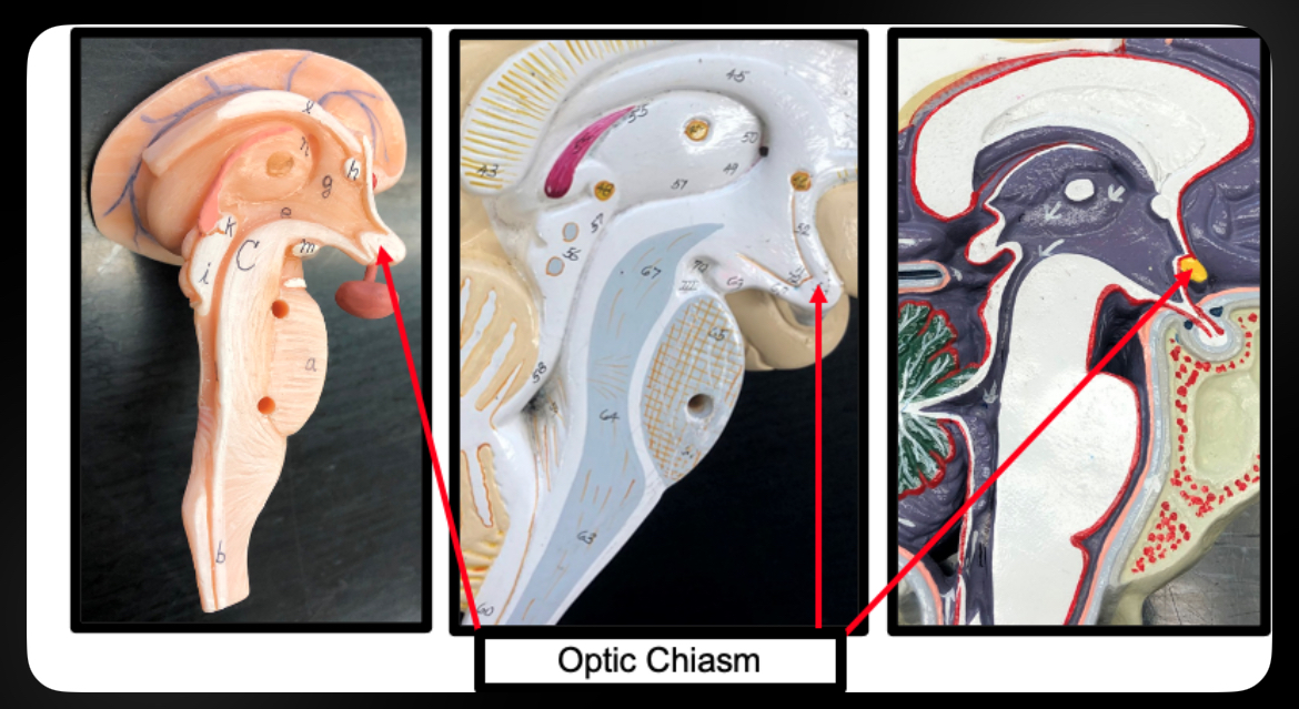
Optic Chiasm
Crossing of optic nerves
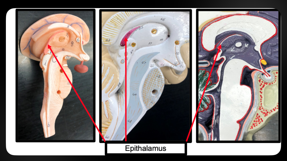
Epithalamus
Contains pineal gland; regulates circadian rhythms
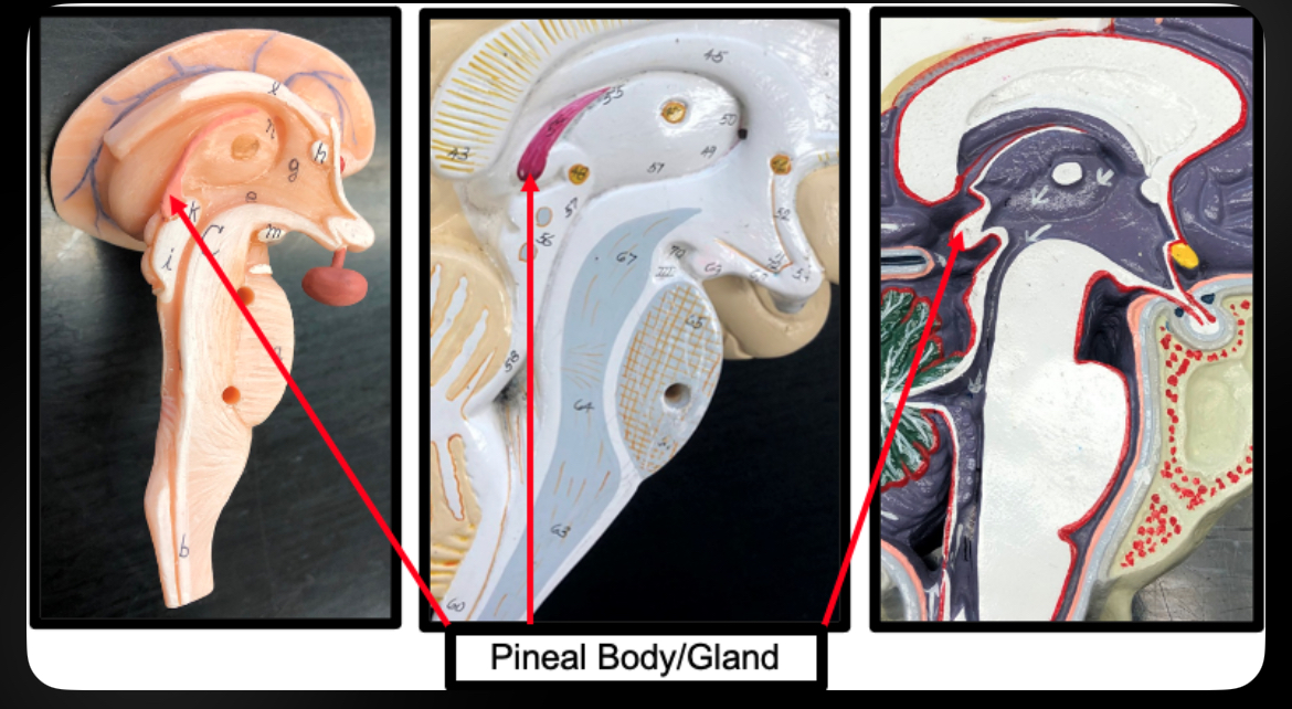
Pineal Body
Secretes melatonin
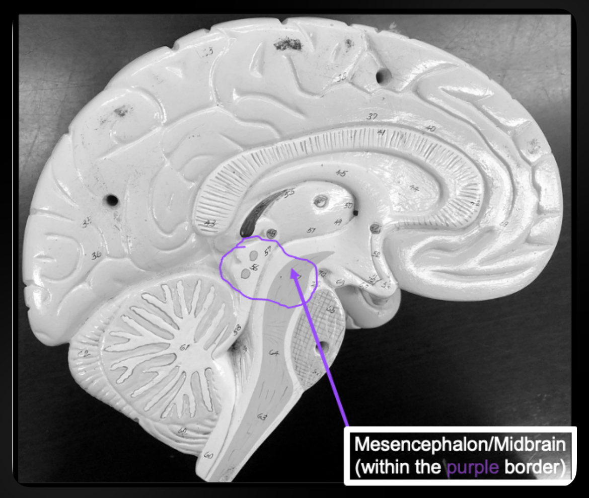
Midbrain
Visual and auditory reflex centers
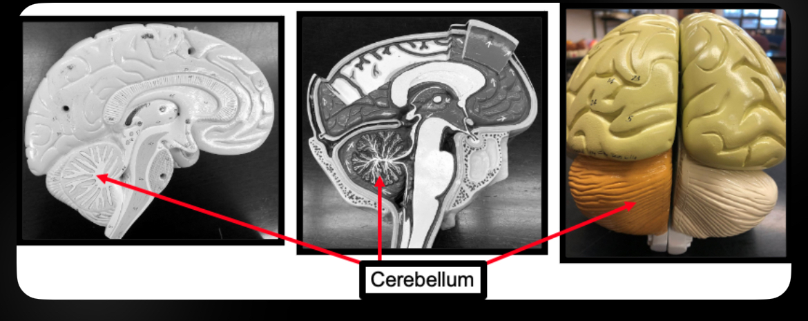
Cerebellum
Coordinates balance and fine motor movement
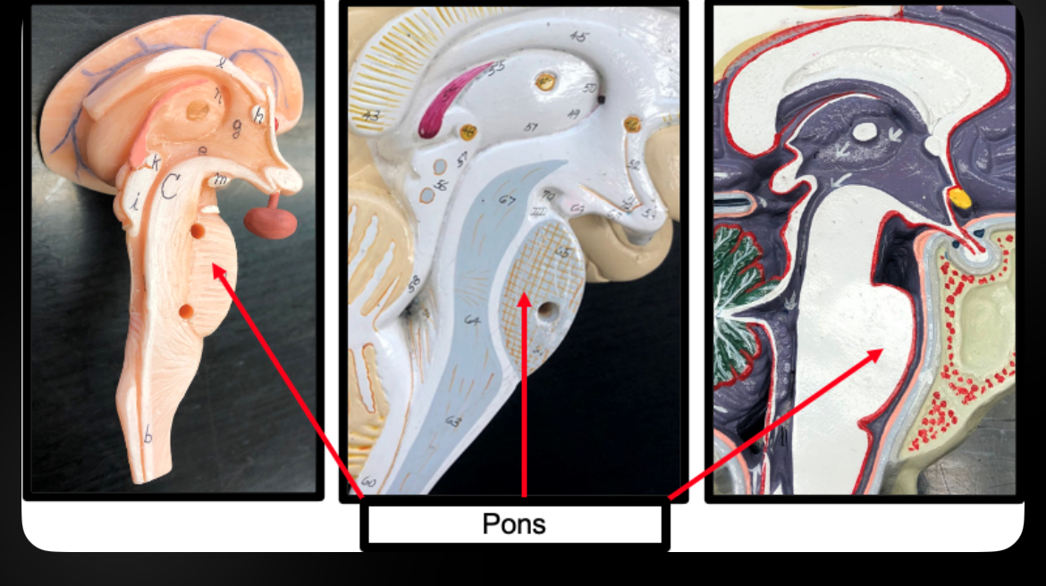
Pons
Relays information between cerebrum and cerebellum; regulates breathing
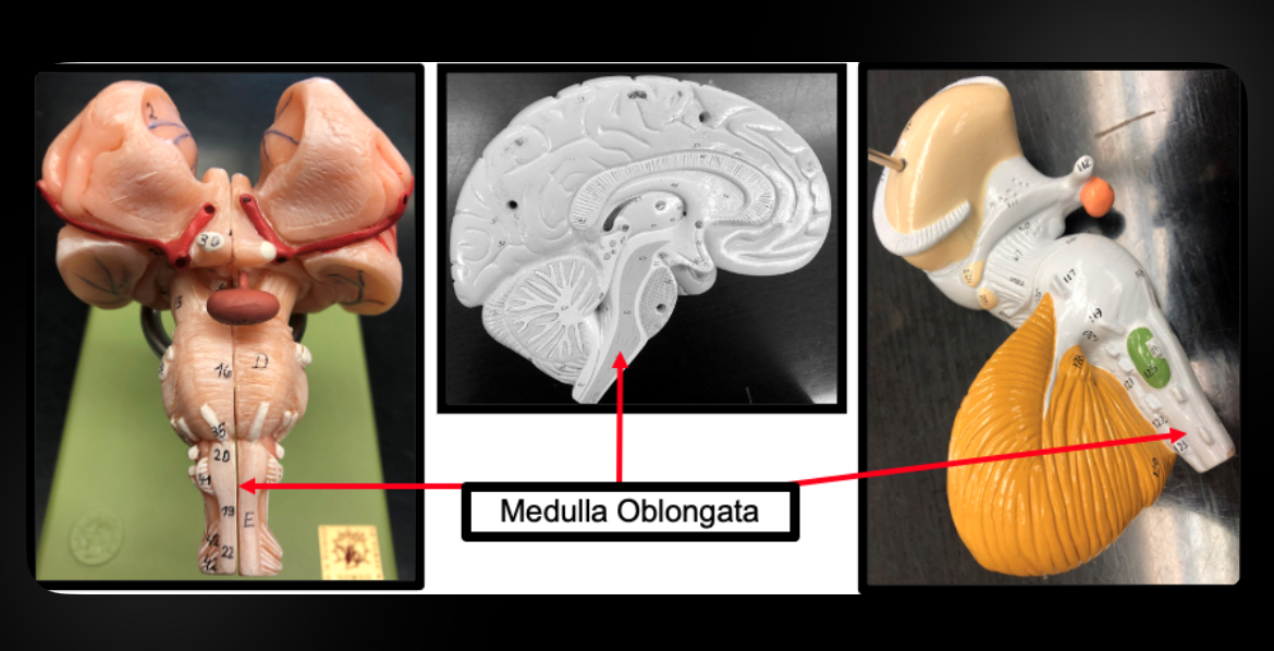
Medulla Oblongata
Controls vital autonomic functions (heart rate, breathing, BP)
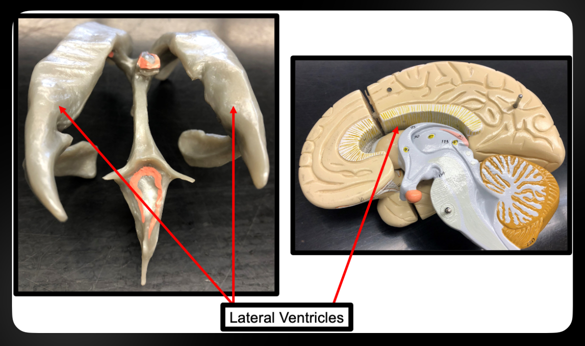
Lateral Ventricles
CSF-filled cavities in cerebral hemispheres
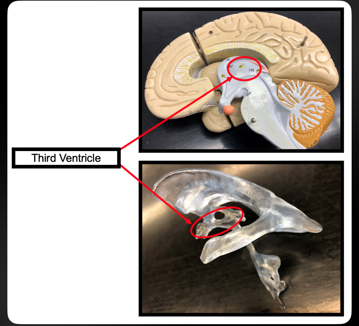
Third Ventricle
CSF cavity in diencephalon
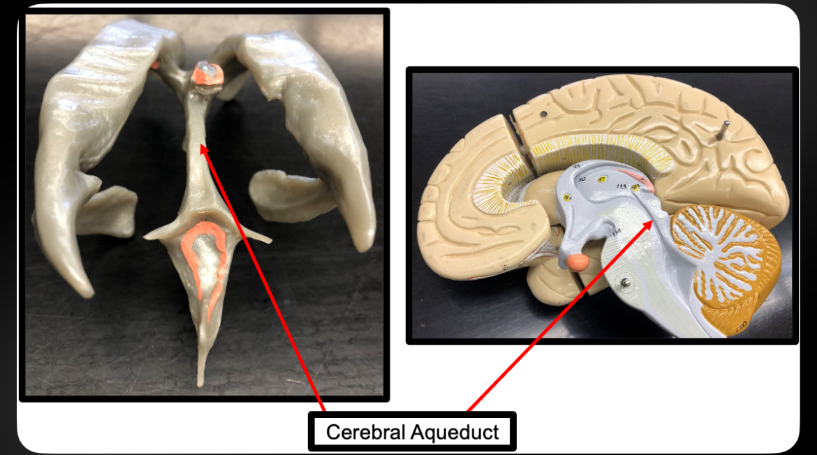
Cerebral Aqueduct
Connects third and fourth ventricles
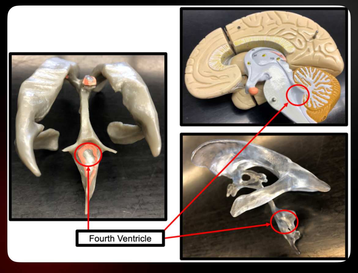
Fourth Ventricle
CSF cavity between pons and cerebellum
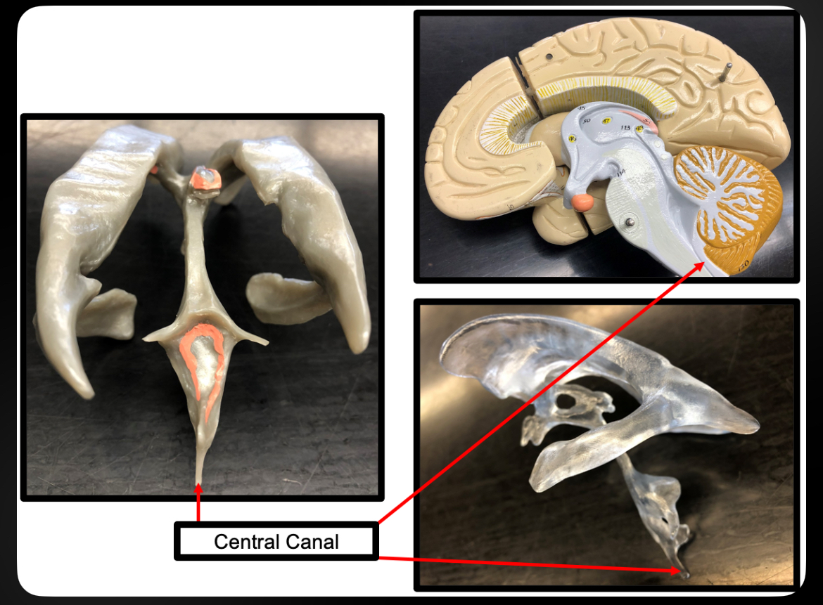
Central Canal
CSF-filled canal in spinal cord
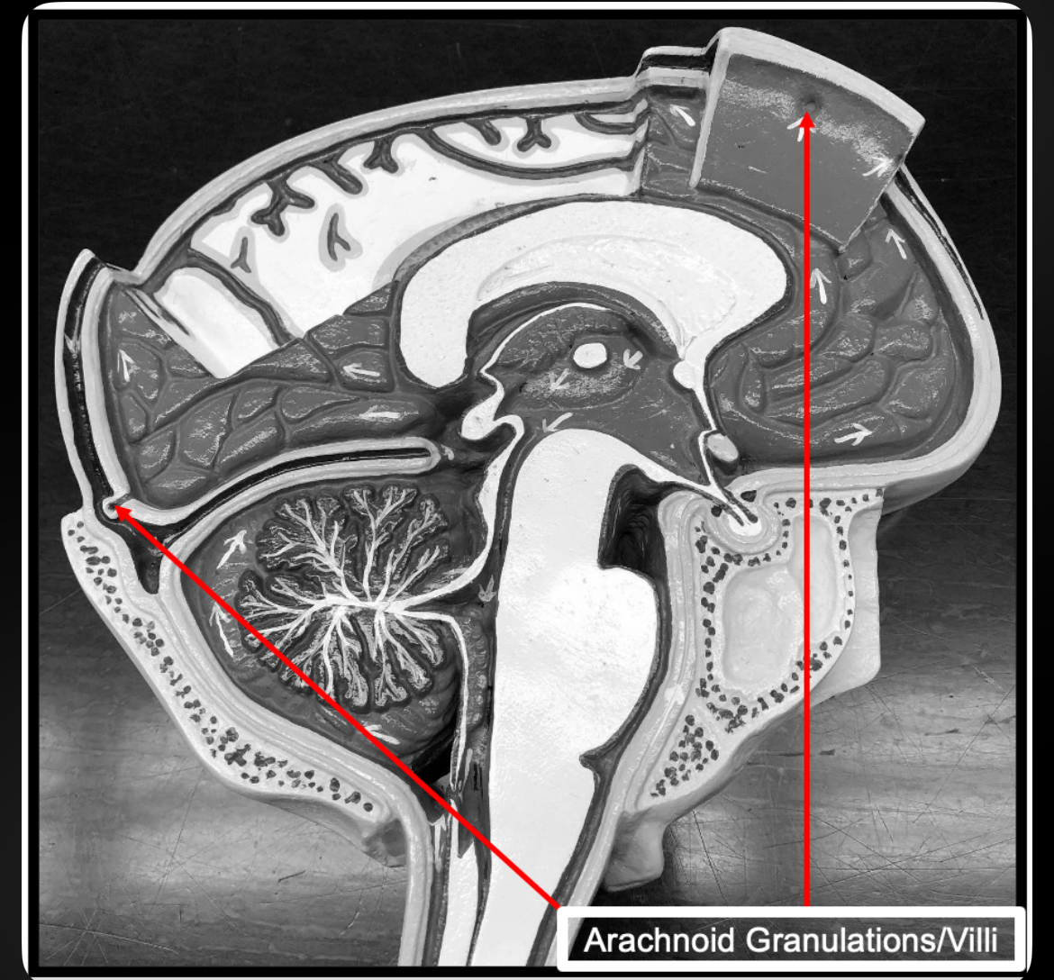
Arachnoid Granulations
Reabsorb CSF into venous blood
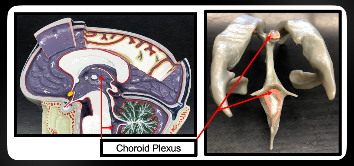
Choroid Plexus
Produces CSF
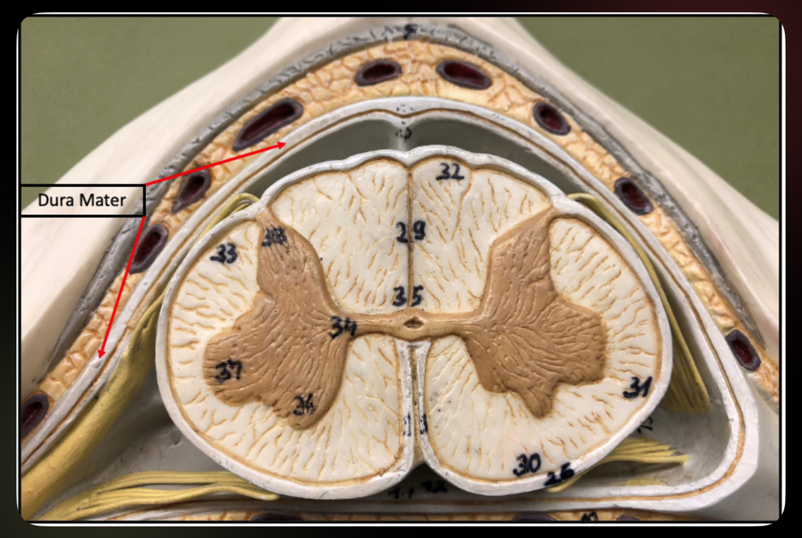
Dura Mater (Spinal Cord)
Outer meningeal layer of spinal cord
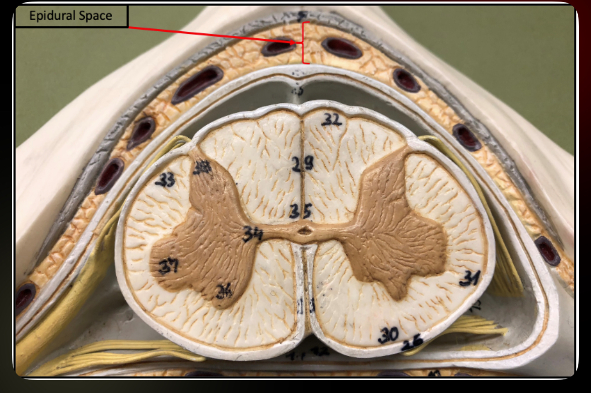
Epidural Space
Space between dura and vertebrae containing fat and vessels
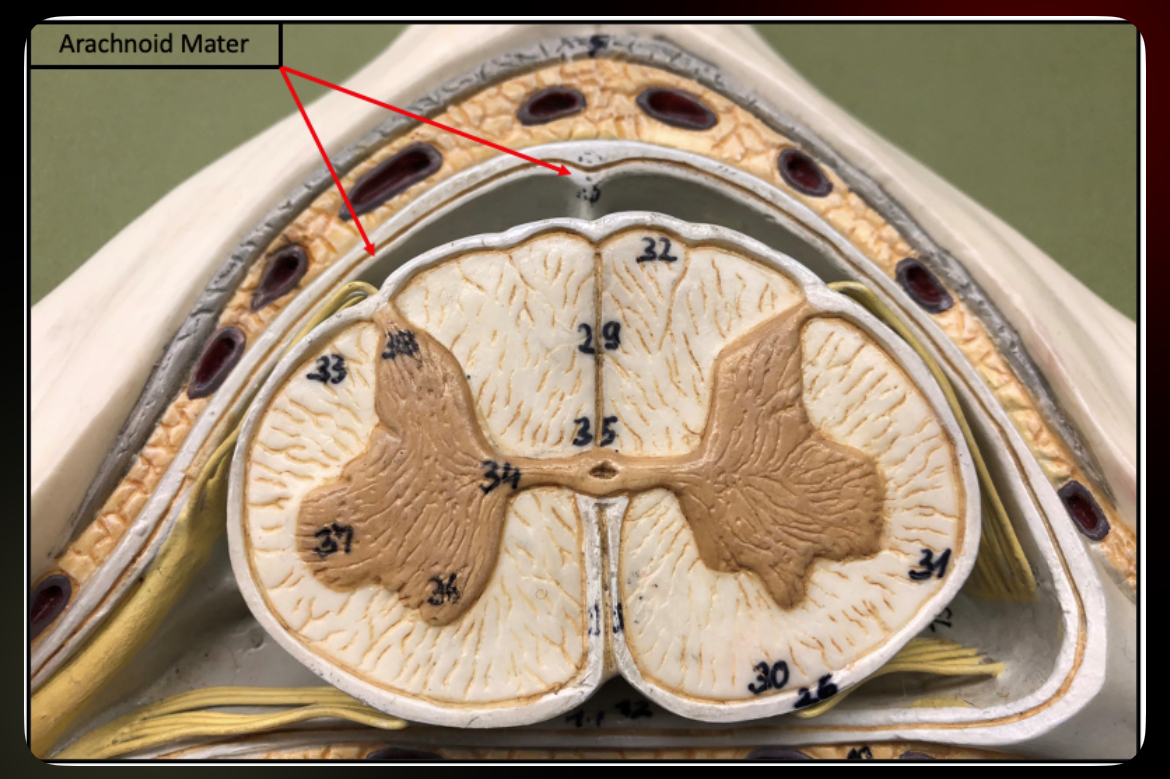
Arachnoid Mater (Spinal Cord)
Middle meningeal layer
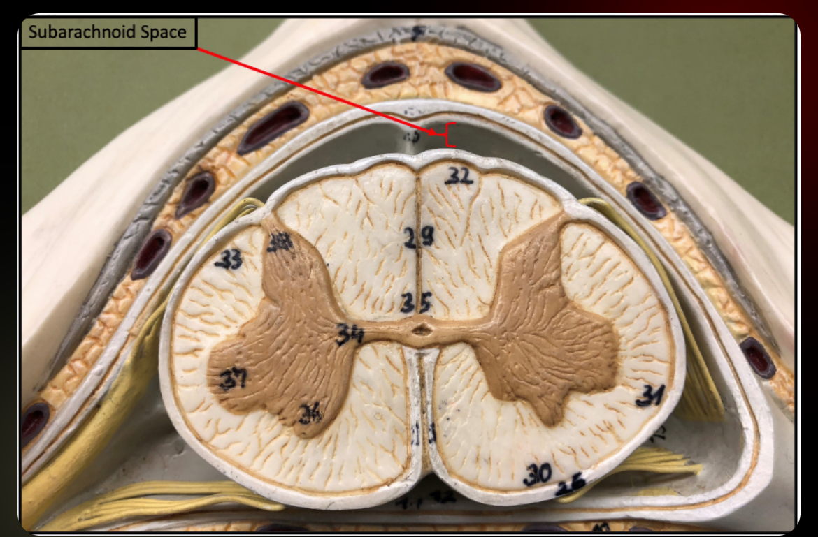
Subarachnoid Space (Spinal Cord)
Contains CSF
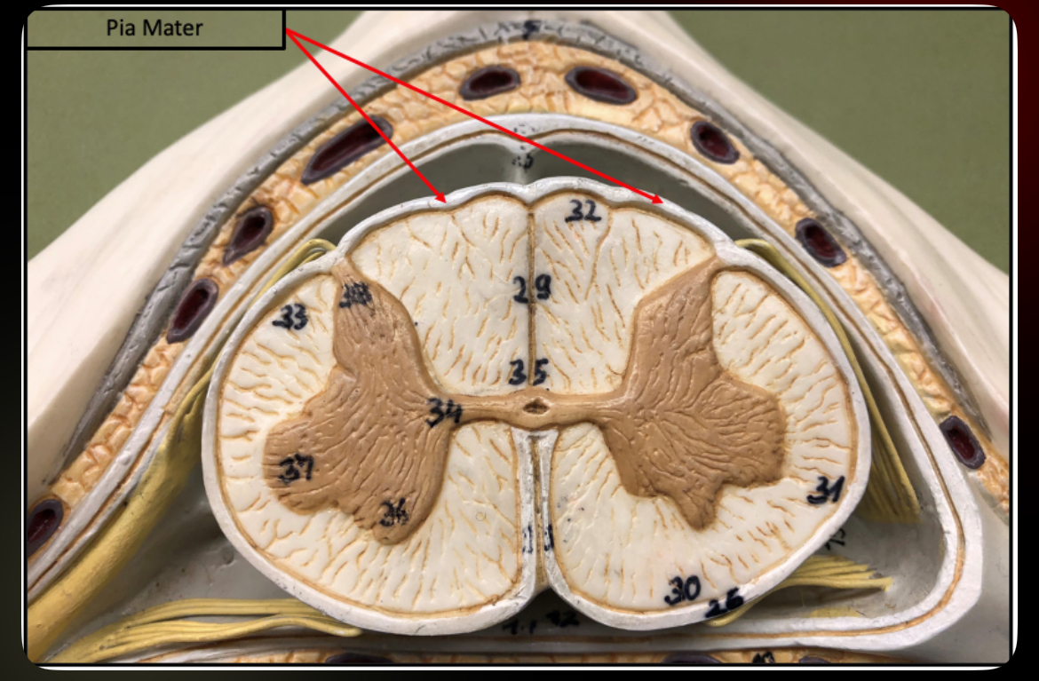
Pia Mater (Spinal Cord)
Inner meningeal layer attached to cord
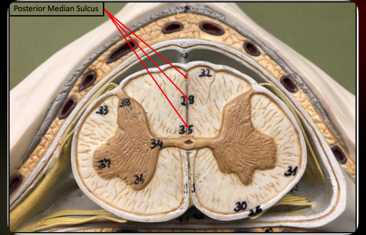
Posterior Median Sulcus
Shallow groove on posterior spinal cord
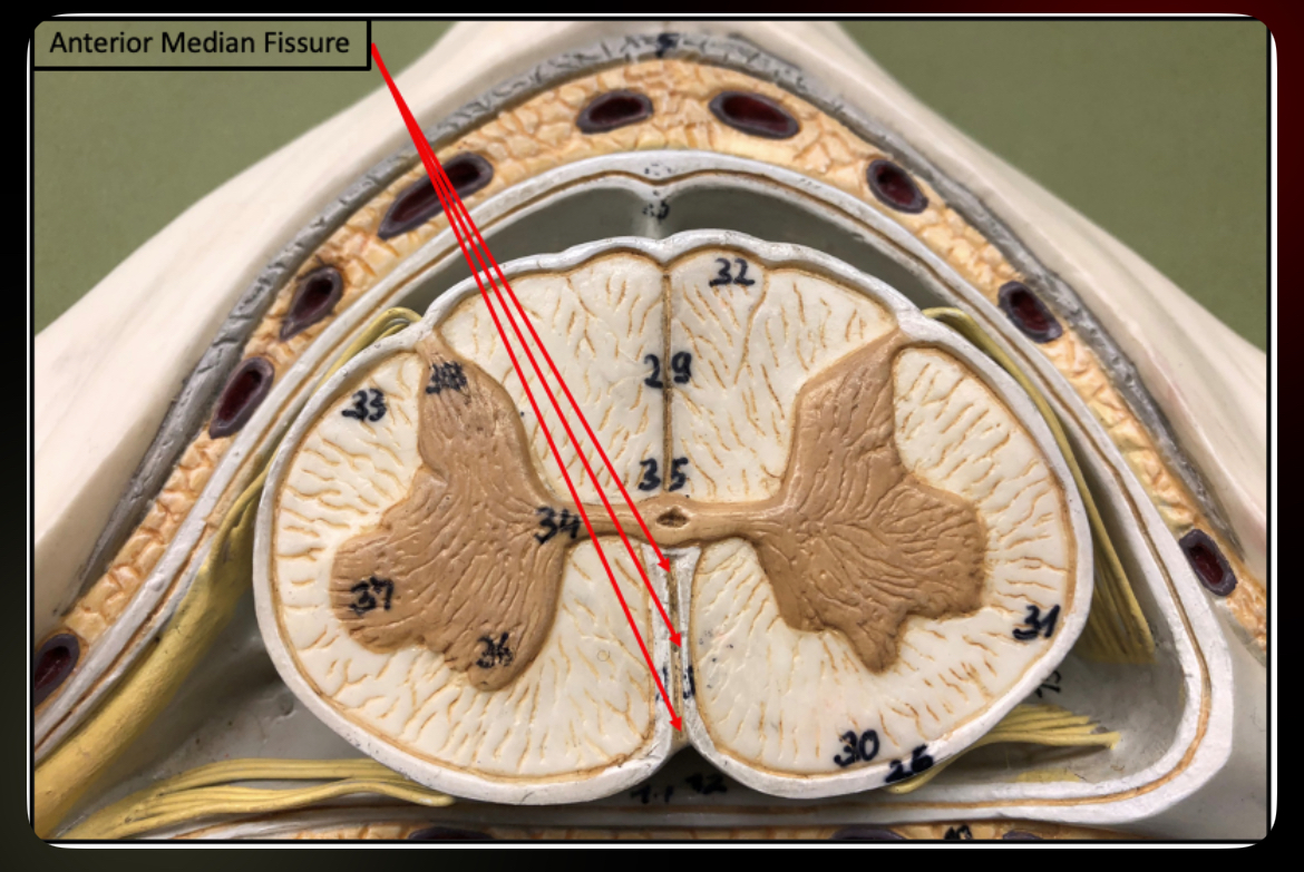
Anterior Median Fissure
Deep groove on anterior spinal cord
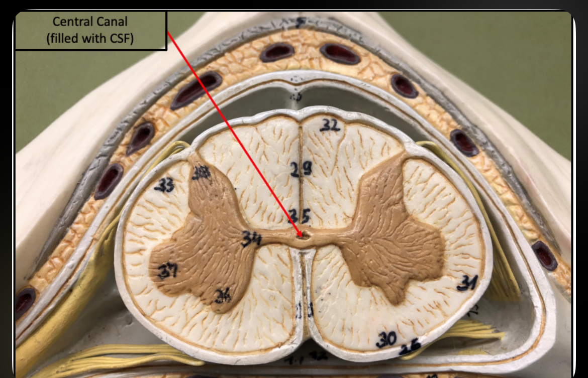
Central Canal (Spinal Cord)
Contains CSF
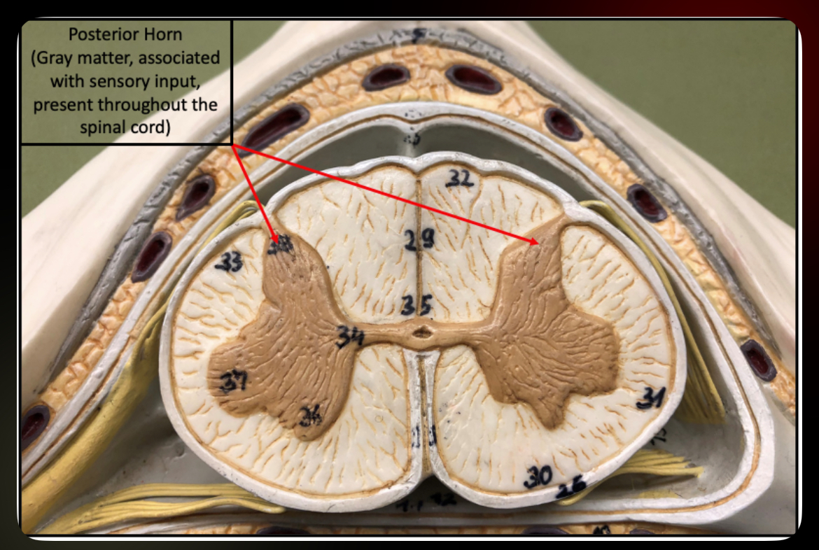
Posterior Horn
Sensory processing region of gray matter
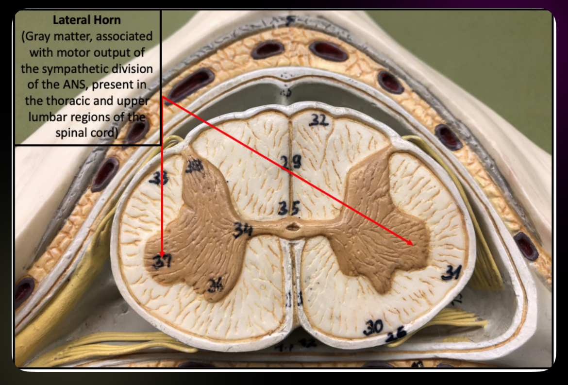
Lateral Horn
Autonomic motor neurons (T1–L2)
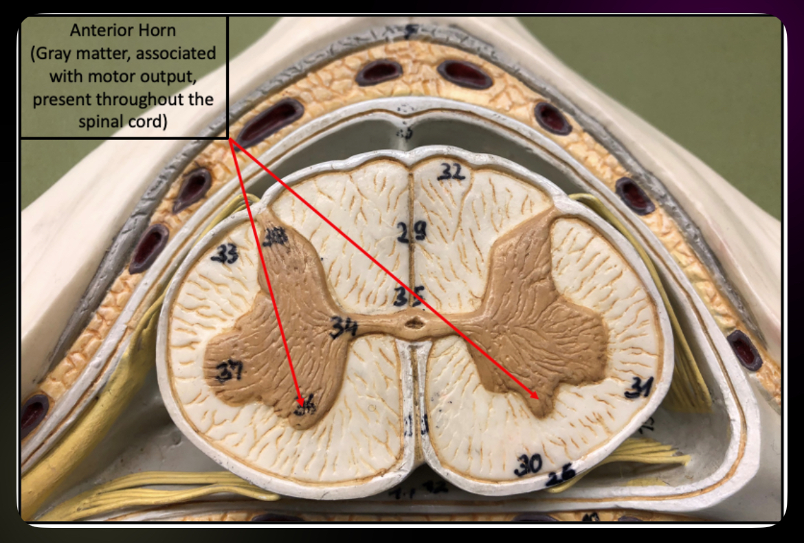
Anterior Horn
Somatic motor neurons
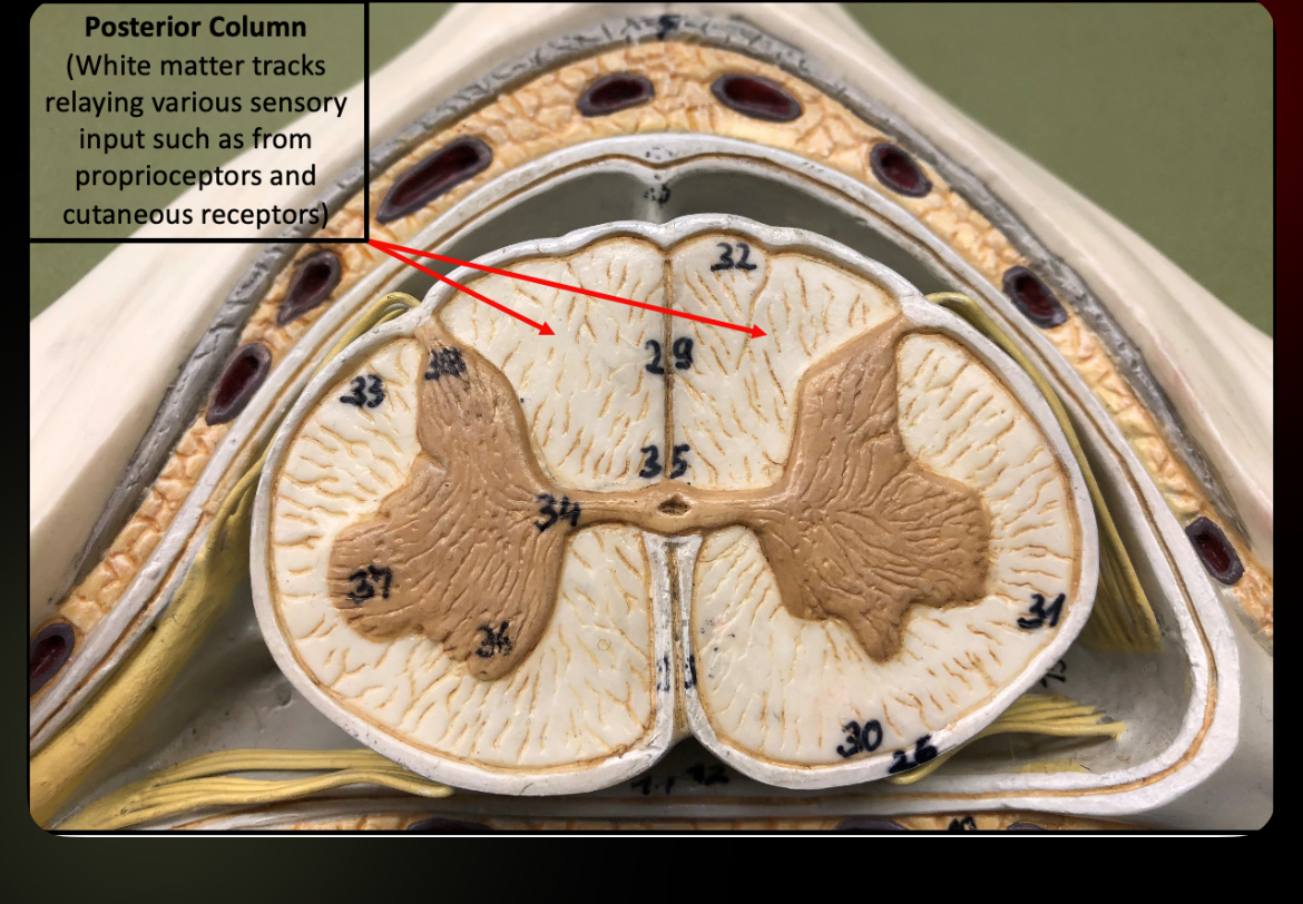
Posterior Column
Sensory ascending tracts
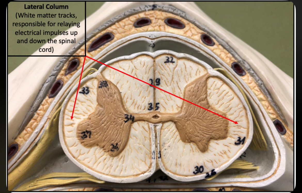
Lateral Column
Ascending and descending tracts
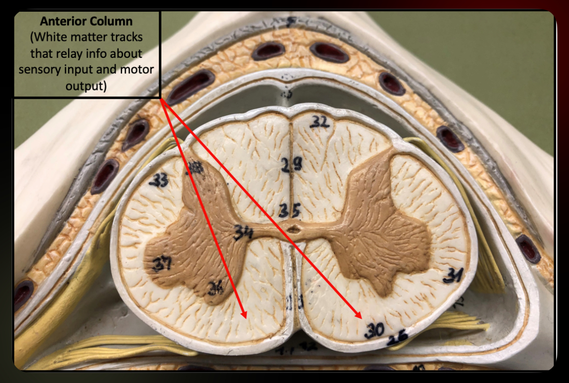
Anterior Column
Motor descending tracts
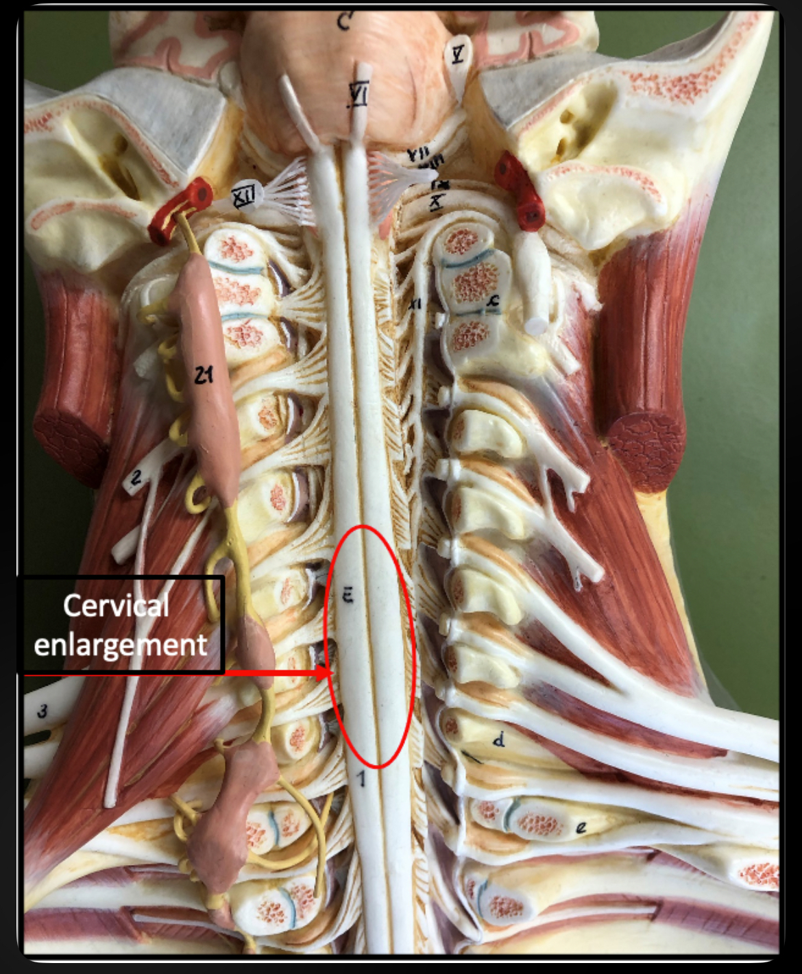
Cervical Enlargement
Supplies nerves to upper limbs
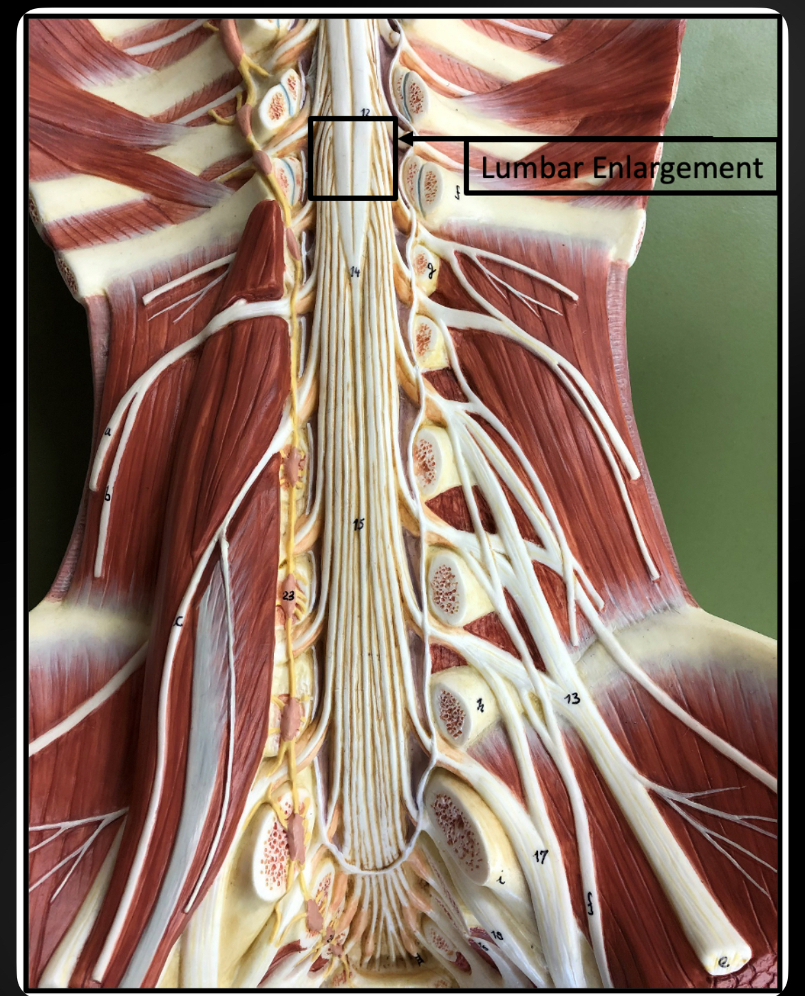
Lumbar Enlargement
Supplies nerves to lower limbs
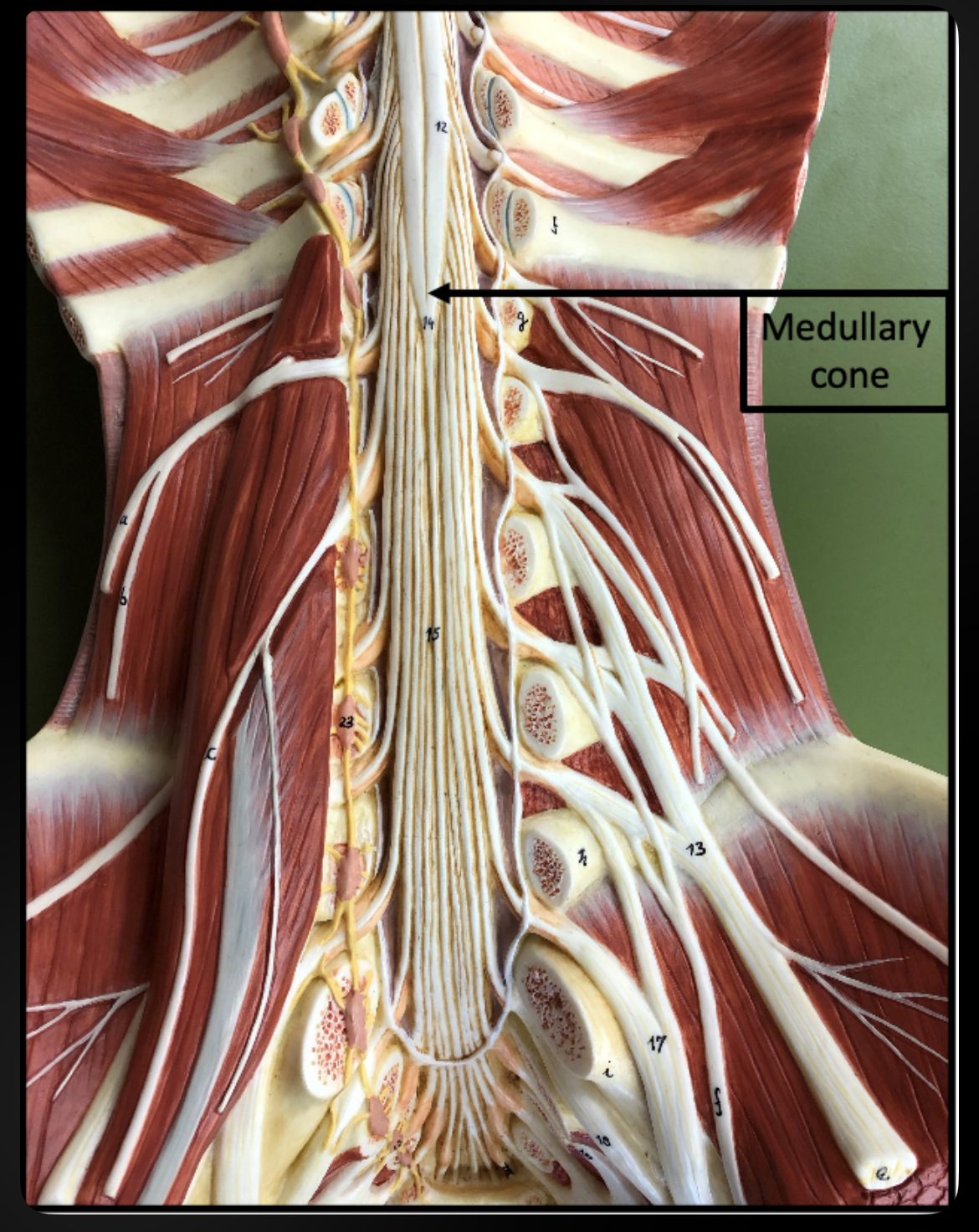
Conus Medullaris
Tapered end of spinal cord
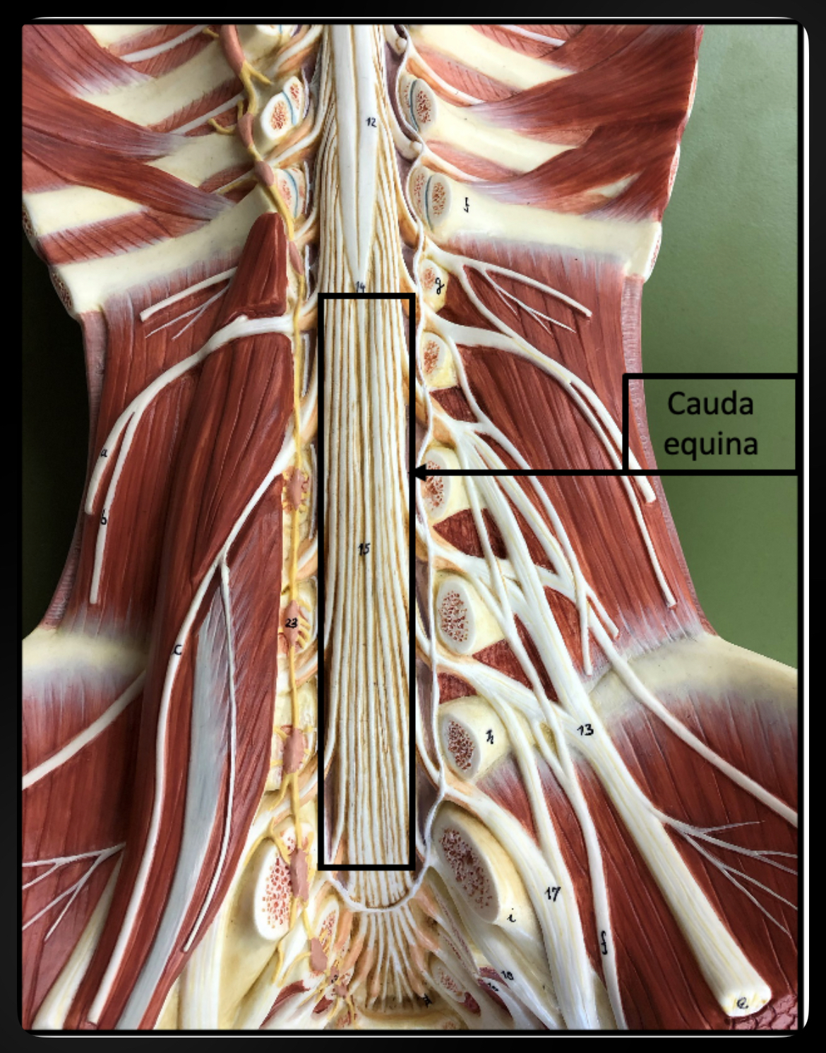
Cauda Equina
Bundle of spinal nerves below conus medullaris
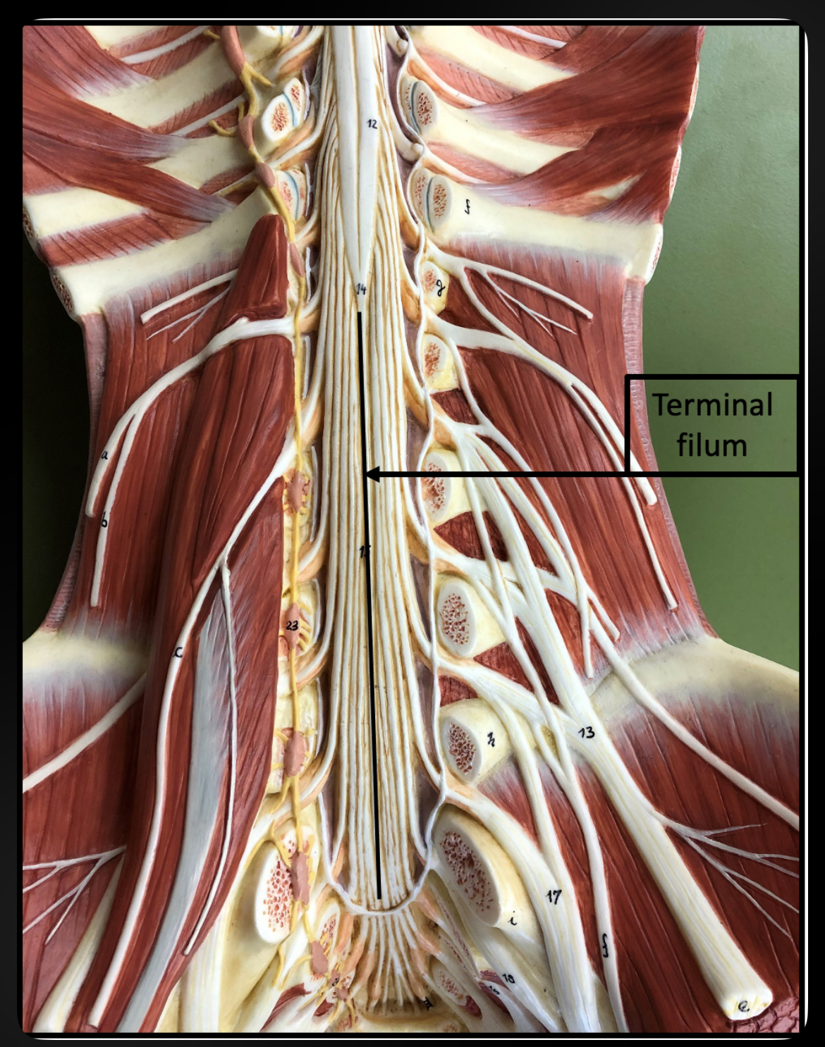
Terminal Filum
Anchors spinal cord to coccyx
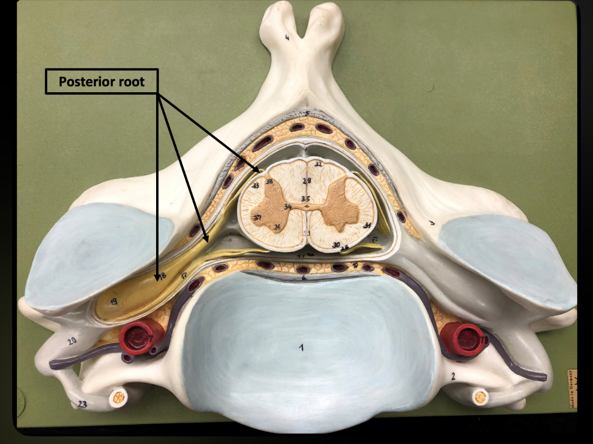
Posterior (Dorsal) Root
Carries sensory input into spinal cord
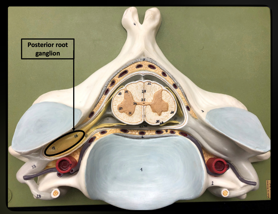
Posterior Root Ganglion
Contains sensory neuron cell bodies
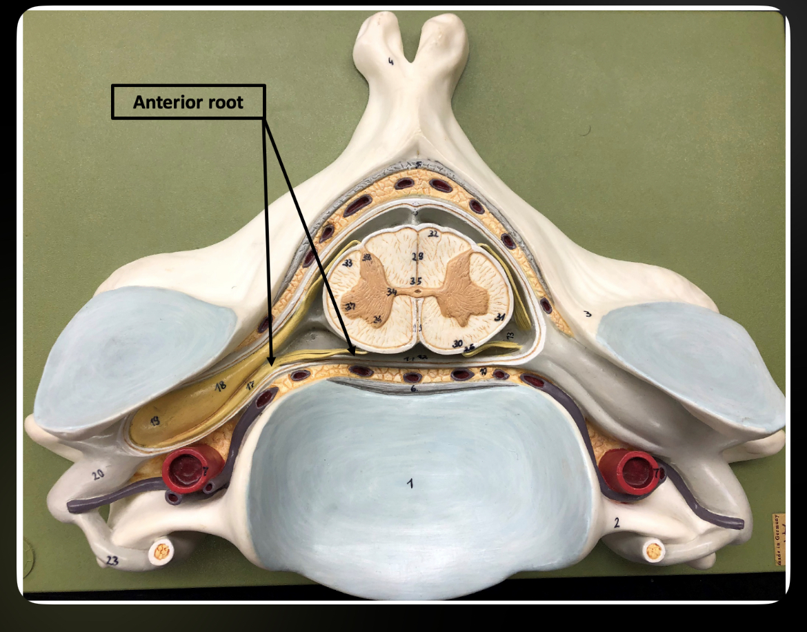
Anterior (Ventral) Root
Carries motor output from spinal cord
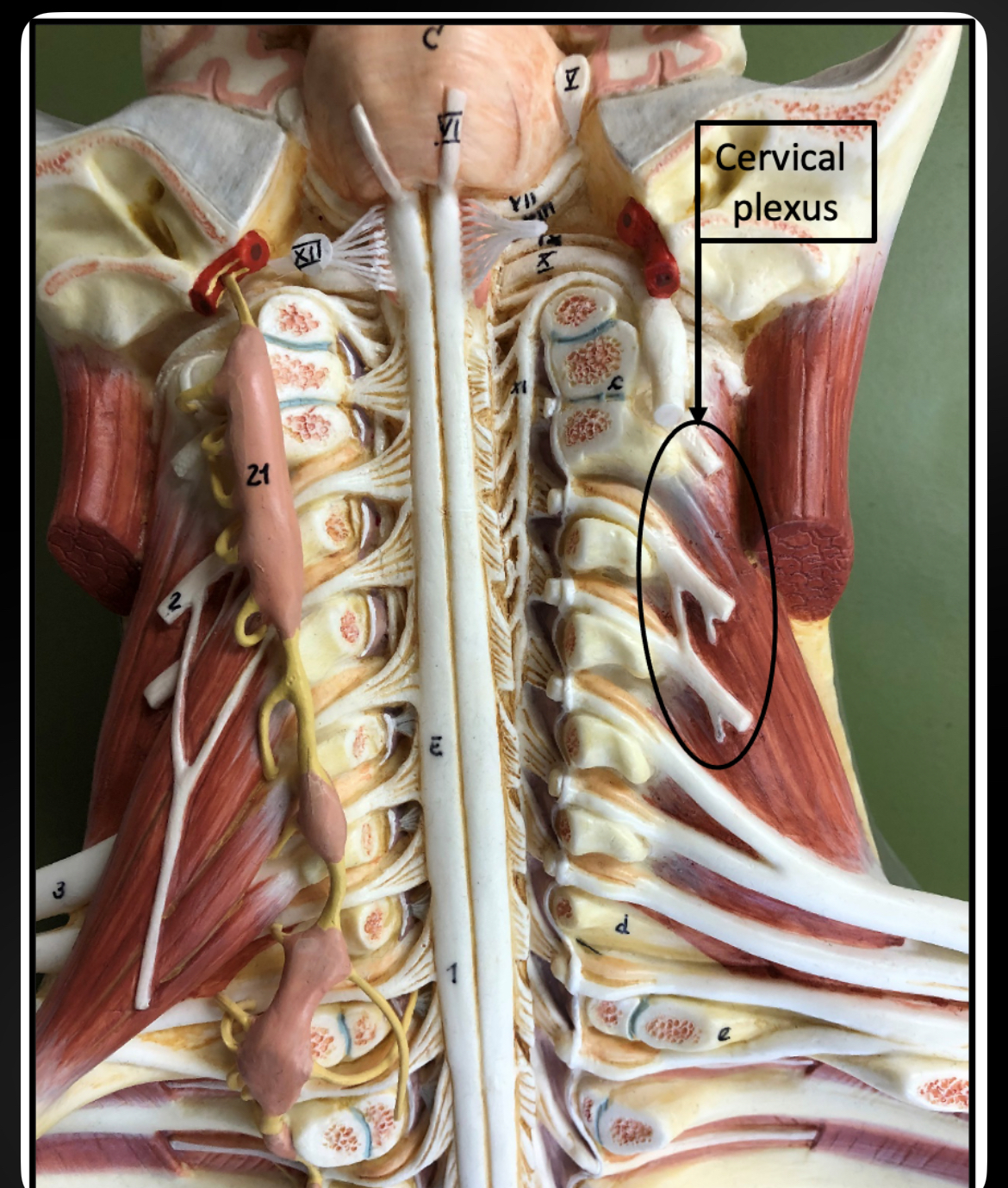
Cervical Plexus
Nerve network supplying neck and diaphragm
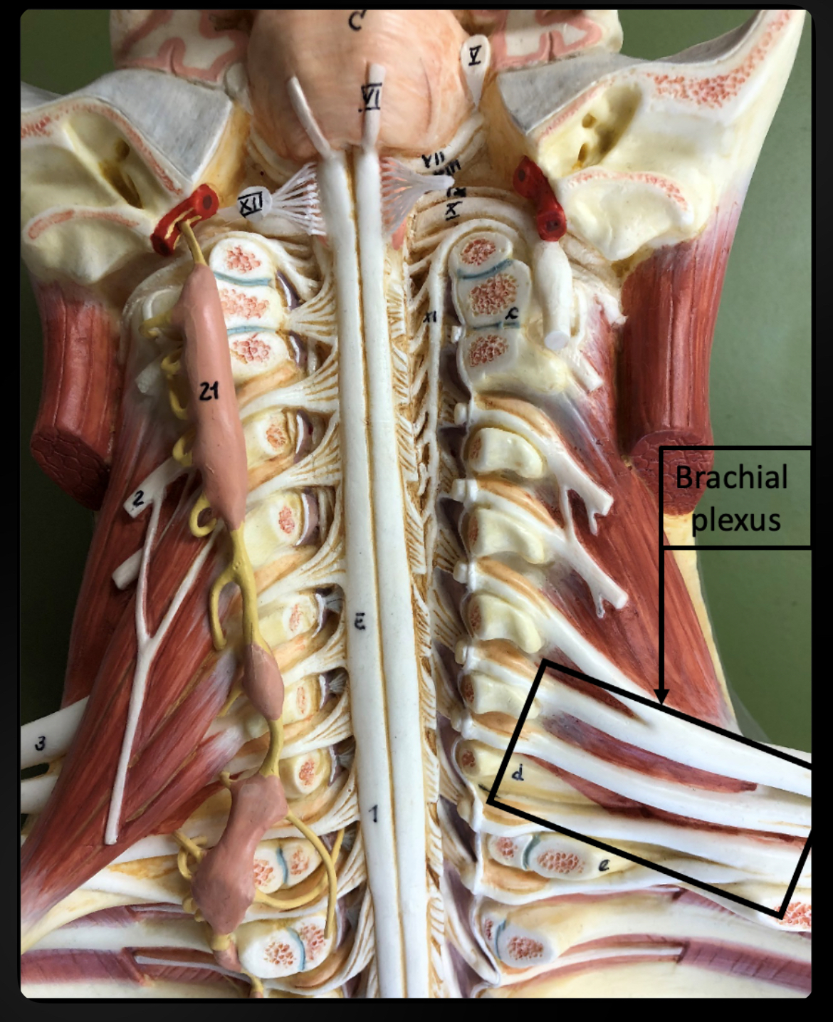
Brachial Plexus
Supplies upper limb
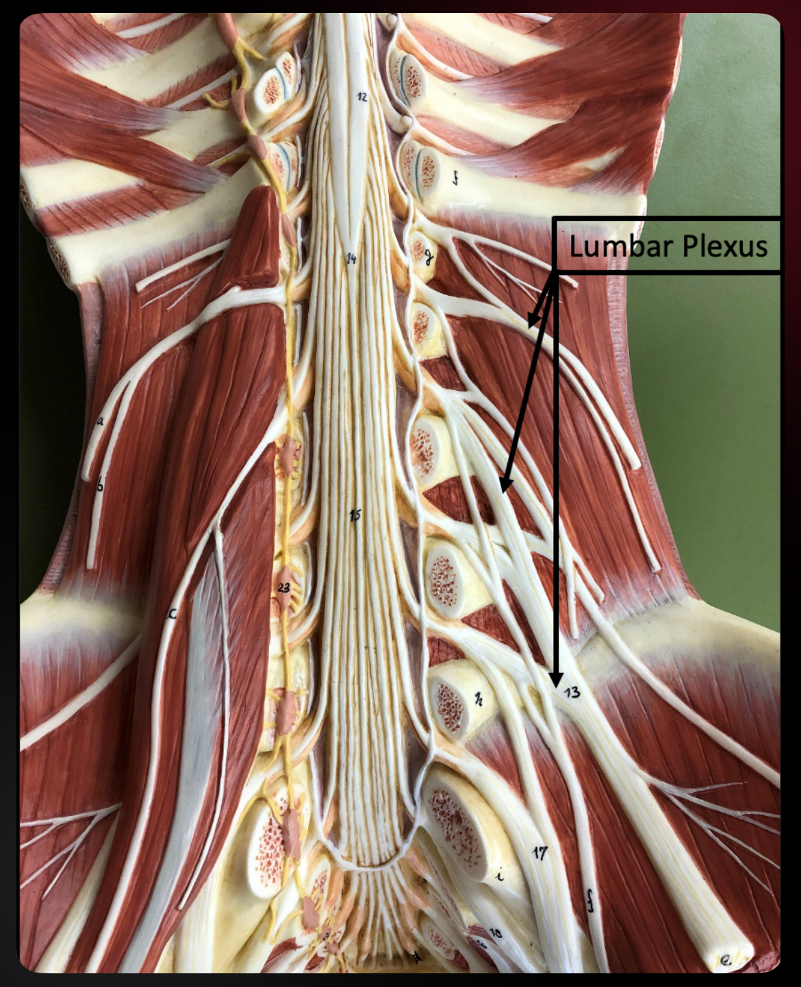
Lumbar Plexus
Supplies anterior thigh
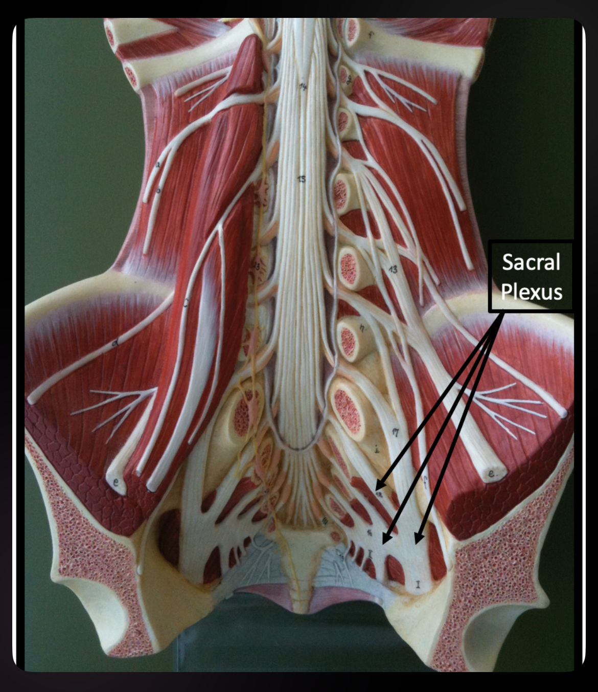
Sacral Plexus
Supplies posterior thigh and lower limb