Lesson 74 Bacterial disease and parasites
1/58
There's no tags or description
Looks like no tags are added yet.
Name | Mastery | Learn | Test | Matching | Spaced | Call with Kai |
|---|
No analytics yet
Send a link to your students to track their progress
59 Terms
metritis
inflammation of uterus
mastitis
inflammation mammary gland
balanoposthitis
inflammation penis or preputial cavity
endometritis
pus or mucopurulent discharge
vaginitis
discharge from vulva
orchitis
inflammation testicles
cryptorchidism
one or both testicles don’t descend from abdominal cavity into scrotum
classification RTI
venereal/ STDs
endogeneous (pathogens in normal biota)
iatrogenic(medical procedures)
brucella abortus
cattle
transmission:veneral, contact, and ingestion
females: abortion with retained placenta, infertility and mastitis
males: orchitis, epididymitis, seminal abnormalities, testicular atrophy, infertility and joint problems
diagnosis of brucella abortus
sample: semen and lymph nodes
cytology: uses Ziehl-neelsen(MZN) stain
serology: serum agglutination test (SAT)
rose bengal plate test-for screening
complement fixation test-for confirmation
PCR
How is B. abortus controlled
Test and slaughter
vaccinat female calves
RB51 strain
Vaccine can cause accidental exposure and cause disease in human
Brucella Canis
dogs (srs problem in breeding kennels)
highly contagious-veneral
males: epididymitis, prostatitis, testicular atrophy, sterility
-carries over into semen
diagnosis of Brucella canis
serology: agglutination test
necropsy: lymph node and spleen
PCR: blood, vag discharge, aborted fetus, semen
how is Brucella canis controlled
testing, euthanizing positive, clean and disinfect
Brucella suis
swine
clinical signs: abortion, sterility, still births, spondylitis (spine arthritis), abscess in organs
Which brucella strain is most significant
B. melitensis
B. melitensis
most serious infection-malta fever or undulant fever zoonotic
transmission: direct contact, milk/cheese
risk group: farm workers, slaughter house, veterinarians
what brucella species is most common in cattle in the USA
B. abortus
Leptospirosis in cows, goats, and sheep
abortion, still birth, weak offspring, infertility, red milk
Leptospirosis in pigs farm and feral
fever, abortion, stillbirth, weak/sick offspring, jaundice, acute kidney failure, loss of appetite
Q fever-Coxiella burnetti
cattle, sheep, goats
ZOONOTIC
transmission: milk, urine, feces. placenta and fluids infective
signs: abortion
risk group: veterinarian, slaughterhouse, dairy workers, farmers, researchers using sheeps
How do you treat Q fever-Coxiella burnetti
treatment: tetracyclines and vaccines available
Why is Q fever a bioterrorism agent
spore like forms that are resistant to heat, drying or common disinfectants
diagnosis of Q fever-Coxiella burnetii
smears of placentra or abortive tissue using Wright Giemsa or Ziehl Neelson
serology
pcr
How is Q fever cases controlled
testing in abortion cases, quarantine, vaccinate
NOTIFIABLE DISEASE
What are the primary clinical sign of Q fever in infected livestock
abortion and stillbirths
Listeria
cattle sheep
non contagious disease. food borne infection
transmission: ingestion, silage (most common source in dairy cows)
signs: abortion, septicemia, head tilt or facial paralysis in sheep
Pathogenesis of Listeria
ingestion→ invasion tissue→ migrate trigeminal nerve→ abscess brain stemm→ meningoencephhalitis→ circling disease
ingestion→ intestine mucosa→ blood→ placental damage→ fetal infection→ abortion, stillbirth
blood→ meninges→ meningitis
diagnosis Listeria
HX: silage feeding
signs: neurological
lesions: perivascular cuffing
culture: brain, blood, milk, placenta, fetus
Types of listeria
L. monocyotogenes: more pathogenic, ZOONOTIC
L. ivanovii: less pathogenic, abortion in sheep
How is Listeria cases handeled
isolation of sick, dispose dead and contaimenated bedding
eliminate source (silage)
ABX in feed
Listeriosis in humans
trasnmission: milk, milk products, cold meat cuts
-mostly from food processing and prep facilities
causes: abortion in women and meningitis in immune compromised adults
What is the main source of Listeria monocytogenes infection in ruminants?
contaminated silage or feed
Posthitis, balanoposthitis
also called pizzle rot or sheath rot
preputial ulcerative dermatitis
Corynebacterium spp
signs: poshthitis-foreskin inflammation
spreads from orifice to mucosa. secondary to bacterial infection
C. Pilosum C. Cystitidis
sheep and goat
-part of prepuce normal biota
predisposing factor: high protein diet→ high urea=increased ammonia
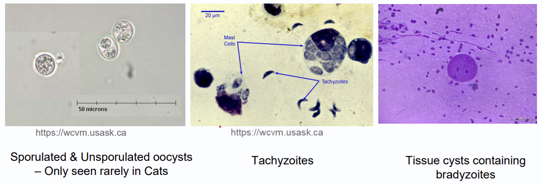
oocysts
resistant stage for environmental transmission
tachyzoites
rapidly dividing tissue stage in vertebrate host
seen in acute infection
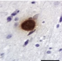
bradyzoites
slowly dividing, encysted tissue stage found in warm blooded vertebrate hosts
seen in chronic infection
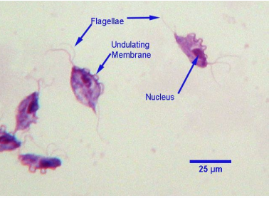
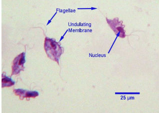
What parasite is this. Sample is from vaginal swabs of aborting cows
Tritrichomonas
definitive host
host where parasite undergoes sexual reproduction
intermediate host
host where parasite undergoes asexual development/ immature stage
paratenic host
host where parasite does not develop further
stage is not required by lifecycle
transport host
host carried parasite, no development
What is the definitive host of toxoplasma gondii
cats
Toxoplasma gondii
sheep, goat, pigs, dogs, cats, humans
zoonotic
trasnmission: spoores of oocysts in contaminated food or water. vertical transmission of tachyzoite, or consumption bradyzoite in infected tissue
risk group: immunocompromised, developing fetus, elderly
signs: abortion, stillbirth, mummification, resorption, weak offspring
-sometimes resp or neuro disorders
Diagnosis of toxoplasma gondii
PCR, ELISA, modified direct agglutination test, Sabin-feldman dye test
How do you treat toxoplasma gondii in dogs with neurological disease
clindamycin, pyrimethamine, supportive care
How do you treat toxoplasma gondii in sheeps
vaccine-toxovax marketed for breeding ewes
How to prevent toxoplasma gondii in cats and humans
avoid raw meat, stop cats from hunting outside, change litter box daily, wahs fruits and vegetables, wear gloves when gardening
How long does it take for toxoplasma gondii in litterbox to become infectious
1-5 days
Neospora caninum
dogs, coyotes, wolves (DH) and cattle sheep goats (IH)
transmission:
-dog:ingestion of infected cow
-cow: horizontal and vertical
signs: major cause abortion in cows, repro disorder, stillbirths, weak births w/ neuro dysfunction
Diagnosis of neospora caninum
cattle-histopath of fetus or placenta, PCR, ELISA
How is neospora caninum cases handeled
NO treatement in cattle
testing, dispose dead and placentas, prevent contact b/w dog and cattle
biosecurity
Tritrichomonos doetus
Bovine
transmission: veneral (folds of prepuce/penis, cervix, and uterus). WORLDWIDE
cows-inflammation reproductive tracts→ infertility, abortion
bulls- no signs but are carriers
How long does it take for cows to naturally clear tritrichomonas foetus infection
after 2-3 estrus cycles
diagnosis tritrichomonas foetus
Hx of decreased calving rate, fertility
vaginal, prepuce, or penis smear, sample, culture, or PCR
How to handle cases of Tritrichomonas foetus
no effective treatment
biosecurity, cull bulls, sexual rest for females
Trichgaurd vaccine-reduce shedding and slightly increase pregnancy and calving rate
AI insemination