Chapter 4 (Face and Neck Development)
1/65
There's no tags or description
Looks like no tags are added yet.
Name | Mastery | Learn | Test | Matching | Spaced | Call with Kai |
|---|
No analytics yet
Send a link to your students to track their progress
66 Terms
The medial nasal processes first fuse internally to form the _____________________ segment that forms the:
a.
b.
c.
The medial nasal processes first fuse internally to form the intermaxillary (premaxillary) segment that forms the:
a. maxillary incisors
b. primary palate
c. nasal septum
Later, the medial nasal processes fuse externally to form:
a.
b.
c.
Later, the medial nasal processes fuse externally to form:
a. middle part of the nose (from root to apex)
b. tubercle of the upper lip
c. philtrum
The lateral nasal processes later form the _________ of the nose.
The lateral nasal processes later form the alae of the nose.
The nares are formed by the fusion of:
a.
b.
c.
The nares are formed by the fusion of:
a. lateral nasal processes
b. maxillary processes
c. nasal processes
The __________________ process develops from the mandibular process.
The maxillary process develops from the mandibular process.
The maxillary processes form the sides of the ____________ lip and fuse with the mandibular process to form the _____________ commissures.
The maxillary processes form the sides of the upper lip and fuse with the mandibular process to form the labial commissures.
The two medial nasal processes form the __________________.
The two medial nasal processes form the philtrum.
Cleft lip and/or cleft palate is caused by ______________________ tissue becoming trapped within the fusing _______________ and ____________________ processes that prevents the ___________________ from forming the structures needed to complete the fusion.
Cleft lip and/or cleft palate is caused by ectodermal tissue becoming trapped within the fusing maxillary and medial nasal processes that prevents the mesenchyme from forming the structures needed to complete the fusion.
The neck is formed from the primitive _______________ and the ____________________ arches.
The neck is formed from the primitive pharynx and the pharyngeal (branchial) arches.
The pharyngeal arches have a central core of ___________________ that forms a bar of _________________.
The pharyngeal arches have a central core of ectomesenchyme that forms a bar of cartilage.
The first arch is the ___________________ arch. It has _____________ cartilage and forms _________________________________________________________________________________________________________ which are innervated by the _______________________ nerve.
The first arch is the mandibular arch. It has Meckel cartilage and forms the mandible, masticatory muscles (and some palatal and suprahyoid muscles), and the lower face (lower lip, mandibular teeth, associated tissues) which are innervated by the 5th cranial (trigeminal) nerve.
The second arch is the _____________ arch. It has ______________ cartilage and forms __________________________________________________________________________________________________________ which are innervated by the ______________________________ nerve.
The second arch is the hyoid arch. It has Reichert cartilage and forms the muscles of facial expression, the middle ear bone and muscles, a process of the temporal bone, and parts of the hyoid bone (including its ligaments and a suprahyoid muscle) which are innervated by the 7th cranial (facial) nerve.
The third arch forms part of the ____________ bone and ________________ muscle which are innervated by the _________________________ nerve.
The third arch forms part of the hyoid bone and pharyngeal muscle which are innervated by the 9th cranial (glossopharyngeal) nerve.
The fourth and sixth arches fuse to form the _________________ cartilages and muscles of the _____________ and ______________which are innervated by the _______________________________ nerve.
The fourth and sixth arches fuse to form the laryngeal cartilages and muscles of the larynx and pharynx which are innervated by the 10th cranial (vagus) nerve.
The first __________________ groove forms the ___________________ membrane (ear drum) and helps to form the mature head and ____________.
The first pharyngeal groove forms the tympanic membrane (ear drum) and helps to form the mature head and neck.
A ________________ cleft cyst forms when parts of the second ___________________ groove do not become ___________________.
A branchial cleft cyst forms when parts of the second pharyngeal (branchial) groove do not become obliterated.
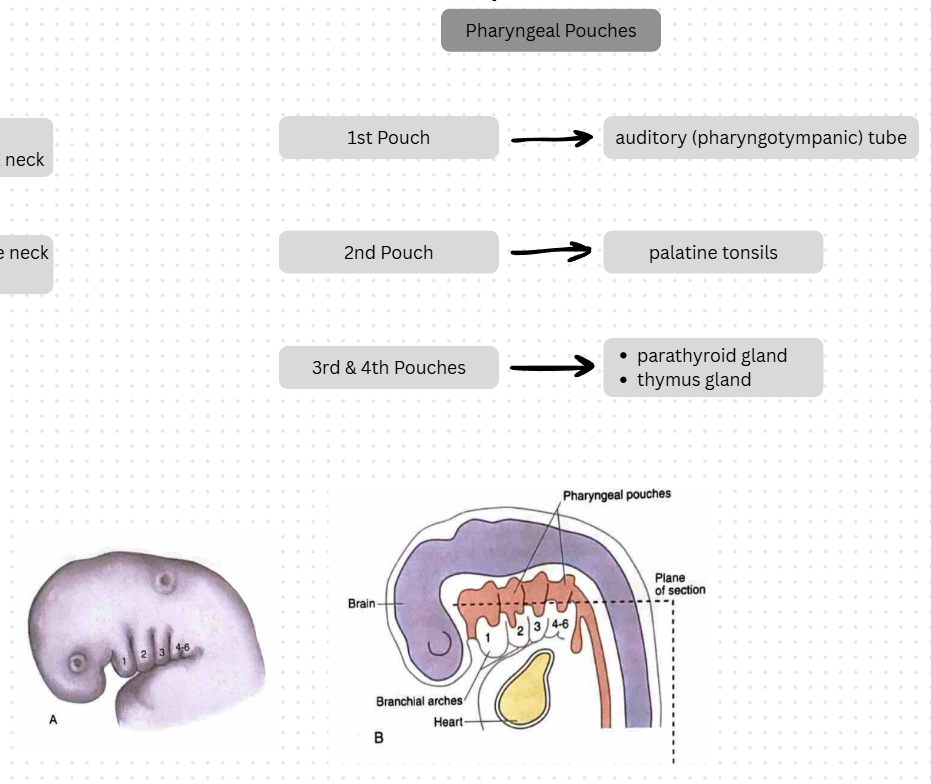
The first pharyngeal pouch forms __________________________________________________________ _________________________________________________________________________________________________________.
The first pharyngeal pouch forms the tympanic membrane, tympanic cavity, mastoid antrum, and auditory (pharyngotympanic) tube.
The second pharyngeal pouch forms _____________________________________________________
_________________________________________________________________________________________________________.
The second pharyngeal pouch forms the crypts and lymphatic nodules of the palatine tonsils.
The third and fourth pharyngeal pouches form _______________________________________ _______________________________________.
The third and fourth pharyngeal pouches form the parathyroid and thymus glands.
1. Which of the following initially primitive structures in the embryo is formed from the stomodeum?
a) Eyes
b) Throat
c) Mouth
d) Nasal cavity
a) Eyes
b) Throat
c) Mouth
d) Nasal cavity
2. Where is the frontonasal process located during development of the face?
a) Cephalic end of the embryo
b) Caudal end of the embryo
c) Surrounding the primitive streak
d) Within the primitive pharynx
a) Cephalic end of the embryo
b) Caudal end of the embryo
c) Surrounding the primitive streak
d) Within the primitive pharynx
3. The olfactory epithelium is responsible for what?
a) smell
b) sight
c) taste
d) hearing
a) smell
b) sight
c) taste
d) hearing
4. Failure of fusion of the maxillary process and what other structure can result in cleft lip?
a) medial nasal process
b) intermaxillary segment
c) nasal septum
d) mandibular arch
a) medial nasal process
b) intermaxillary segment
c) nasal septum
d) mandibular arch
5. Located laterally and posteriorly, what embryonic structure forms pits that create the future internal ear?
a) otic placodes
b) nasal placodes
c) lens placodes
d) stomodeum
a) otic placodes
b) nasal placodes
c) lens placodes
d) stomodeum
6. The pharyngeal or branchial apparatus includes the pharyngeal or branchial arches, the pharyngeal or branchial grooves and membranes, as well as the
a) pharyngeal pouches
b) body of the tongue
c) pharyngeal tonsils
d) buccal developmental depression
a) pharyngeal pouches
b) body of the tongue
c) pharyngeal tonsils
d) buccal developmental depression
7. The intermaxillary segment is a direct growth from which of the following paired processes on the inside of the stomodeum?
a) medial nasal
b) lateral nasal
c) maxillary
d) mandibular
a) medial nasal
b) lateral nasal
c) maxillary
d) mandibular
8. Which numbered pharyngeal or branchial arch is the hyoid arch since it is located inferior to mandibular arch in the embryo?
a) First
b) Second
c) Third
d) Fourth
a) First
b) Second
c) Third
d) Fourth
9. Reichert cartilage is the cartilage within the second pharyngeal or branchial arch that eventually disappears, although parts of it form:
a) a middle ear bone
b) part of the maxilla
c) thyroid cartilage
d) philtrum
a) a middle ear bone
b) part of the maxilla
c) thyroid cartilage
d) philtrum
10. Which of the following is considered a rudimentary and often missing, embryonic pharyngeal or branchial arch?
a) fifth
b) third
c) fourth
d) sixth
a) fifth
b) third
c) fourth
d) sixth
11. Which of the following is considered the first pharyngeal or branchial arch?
a) mandibular arch
b) maxillary arch
c) hyoid bone
d) nasal bones
a) mandibular arch
b) maxillary arch
c) hyoid bone
d) nasal bones
12. Which pharyngeal or branchial arch in the embryo fuses with the fourth pharyngeal or branchial arch?
a) second
b) third
c) fifth
d) sixth
a) second
b) third
c) fifth
d) sixth
13. Where are the pharyngeal pouches located during development of the face and neck?
a) Between the pharyngeal or branchial arches
b) Superior to the maxilla
c) Between the maxilla and mandible
d) Surrounding the future nasal cavity
a) Between the pharyngeal or branchial arches
b) Superior to the maxilla
c) Between the maxilla and mandible
d) Surrounding the future nasal cavity
14. Cervical cysts are developed when which of the following structures do NOT become obliterated upon maturation of the fetus?
a) Branchial or pharyngeal grooves
b) Pharyngeal pouches
c) Nasal pits
d) Mandibular symphysis
a) Branchial or pharyngeal grooves
b) Pharyngeal pouches
c) Nasal pits
d) Mandibular symphysis
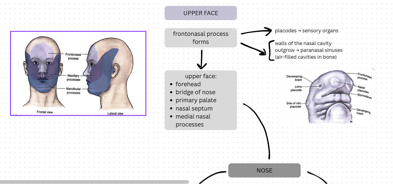
The face starts to develop during week _____ of the ________________ period and is completed by week ______ in the _______________ period.
The face starts to develop during week 4 of the embryonic period and is completed by week 12 in the fetal period.
Mesenchyme is derived from ______________________.
Mesenchyme is derived from mesoderm.
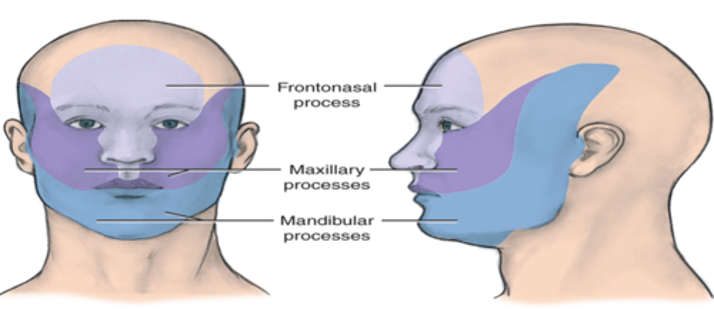
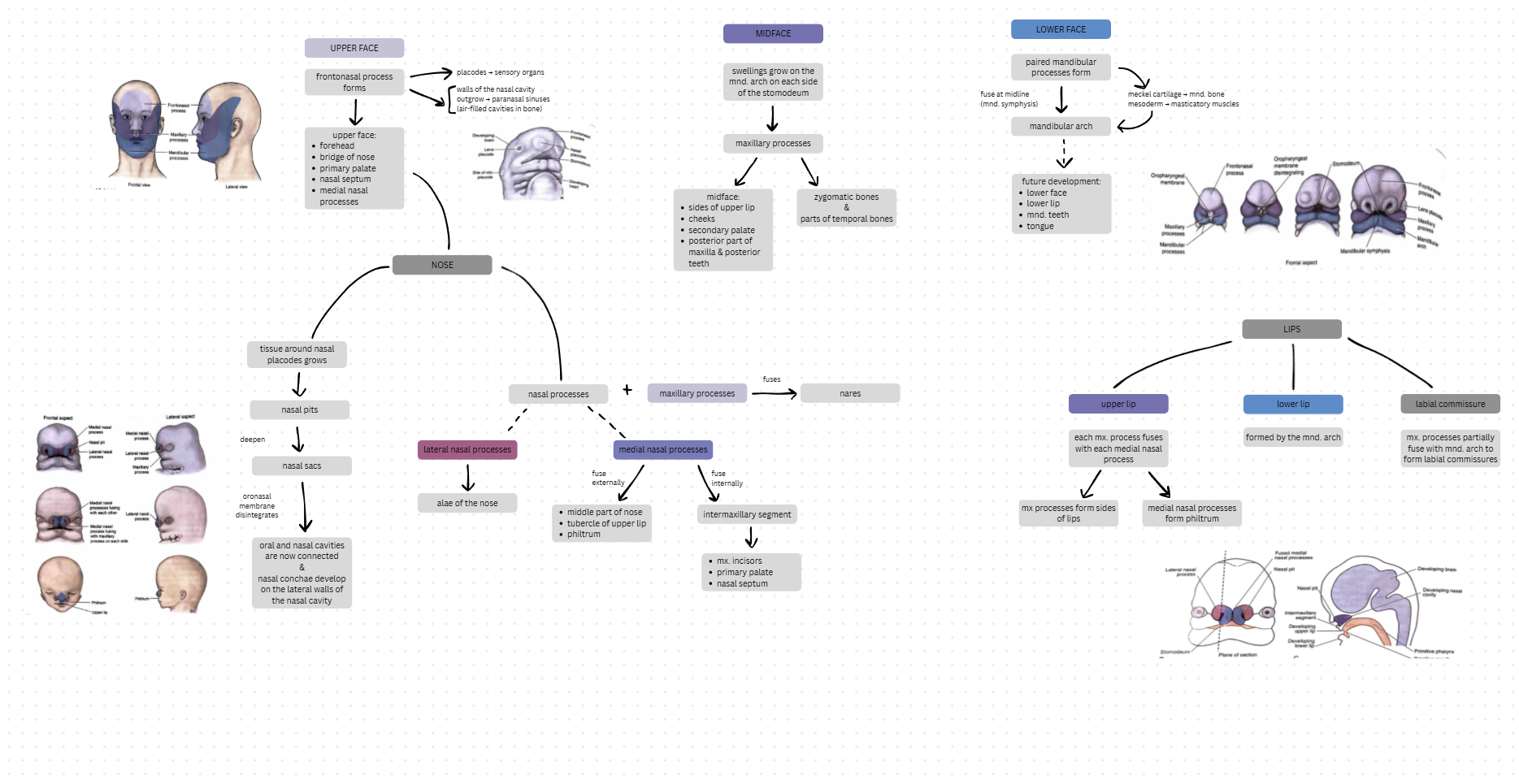
Name the 5 processes (prominences) that form the face and the structures that they develop into:
a.
b.
c.
Name the 5 processes (prominences) that form the face and the structures that they develop into:
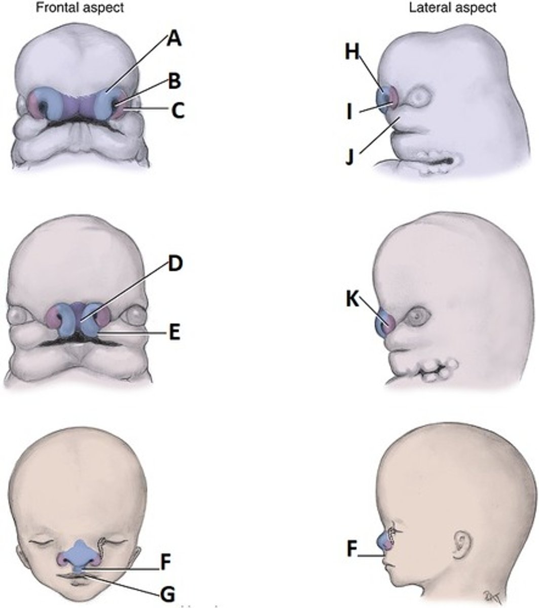
a. FRONTONASAL (single): medial & lateral nasal processes
b. MAXILLARY (paired): midface, upper lip sides, secondary palate, posterior part of maxilla with associated tissue, zygomatic bones, part of temporal bones
c. MANDIBULAR (paired): lower lip, lower face, mandible with associated tissue
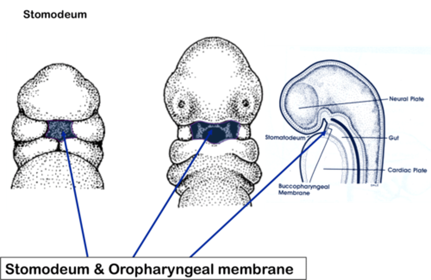
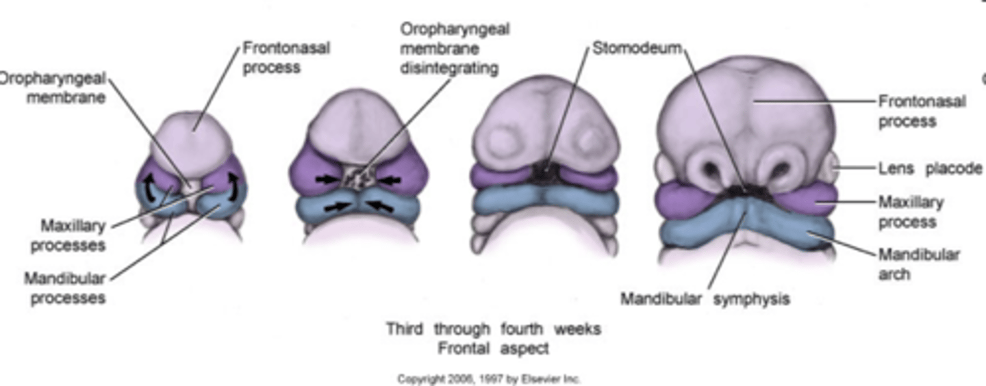
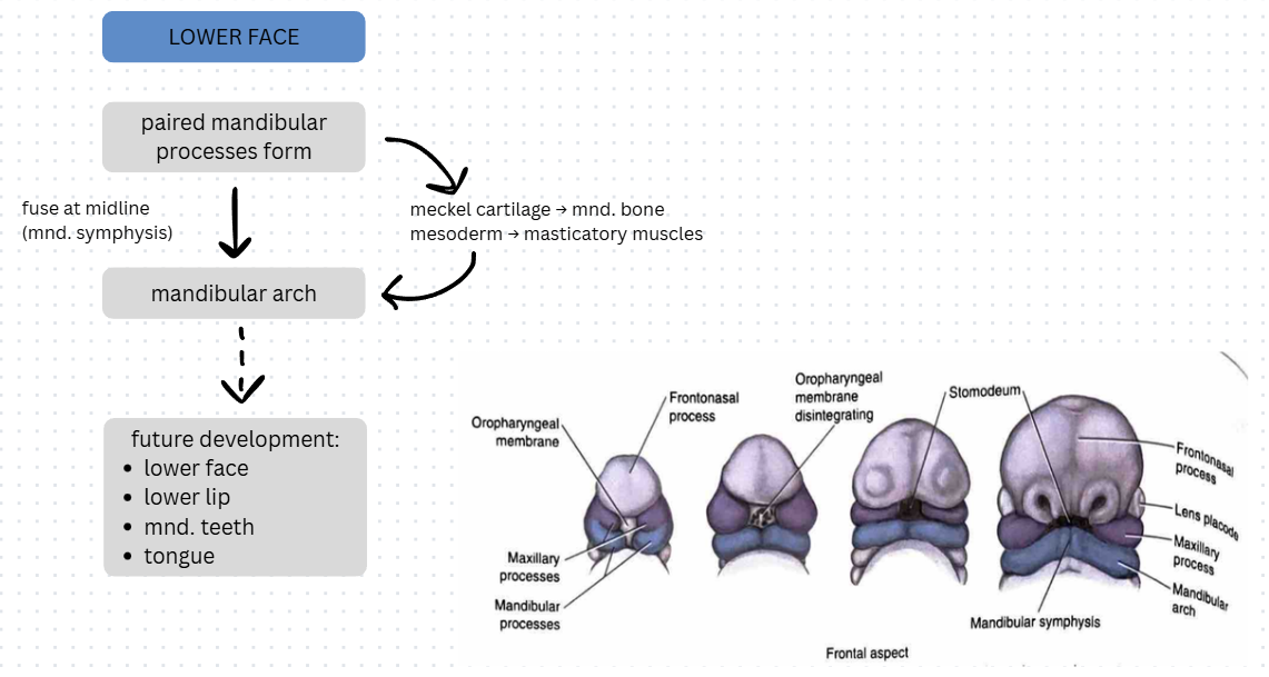
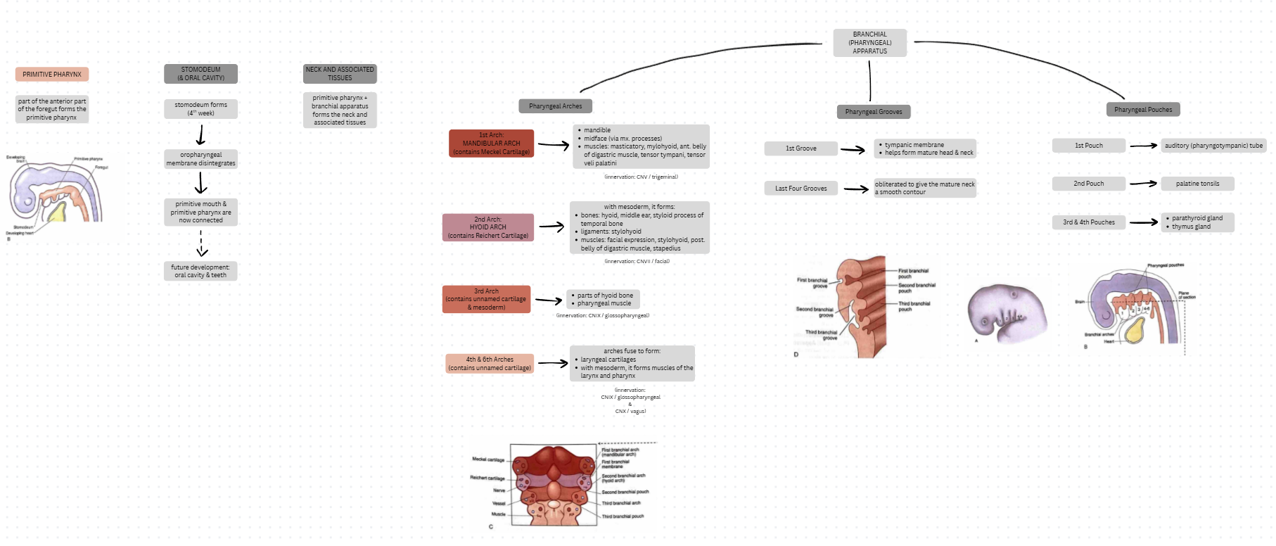
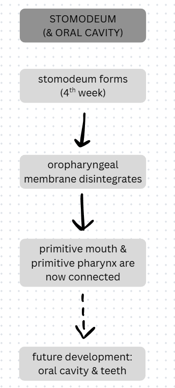
During week 4, a primitive mouth called the ___________________ develops between the _________________ prominence and the _____________ bulge. At the base of the ____________________, the oropharyngeal membrane ________________ which connects the primitive _____________ and the primitive _______________.
During week 4, a primitive mouth called the stomodeum develops between the frontonasal prominence and the cardiac bulge. At the base of the stomodeum, the oropharyngeal membrane disintegrates which connects the primitive mouth and the primitive pharynx.
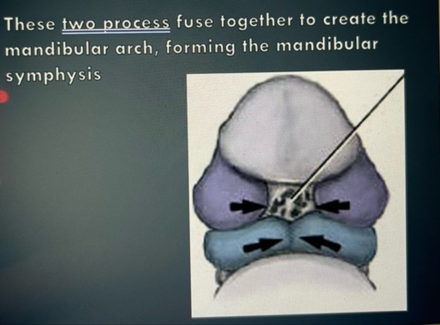
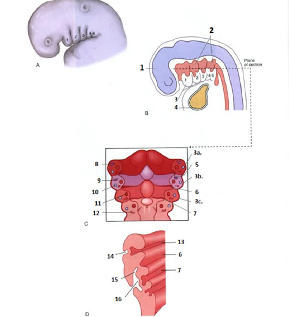
The mandibular processes develop into the mandibular arch which is also known as the first __________________ arch. It has a core of ___________________ and a bar of _____________ cartilage.
The mandibular processes develop into the mandibular arch which is also known as the first pharyngeal arch. It has a core of mesenchyme and a bar of Meckel cartilage.
The mandibular processes fuse at the _______________ to form the mandibular _____________________.
The mandibular processes fuse at the midline to form the mandibular symphysis.
Placodes are areas of thickened _______________ found where ______________ organs are developing.
a. The __________ placodes form the eyes.
b. The __________ placodes form the ears.
c. The __________ placodes form the nose.
Placodes are areas of thickened ectoderm found where sense organs are developing.
a. The lens placodes form the eyes.
b. The otic placodes form the ears.
c. The nasal placodes form the nose.
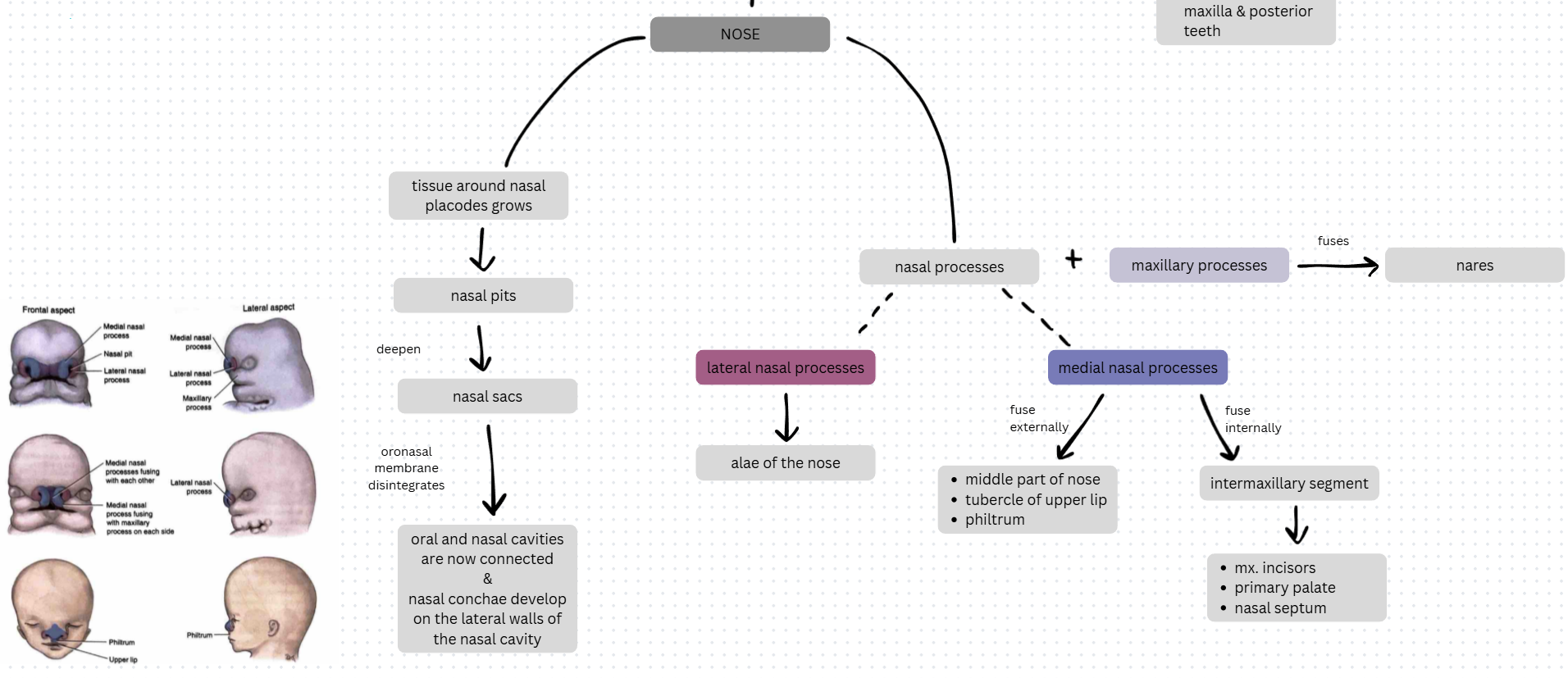
The nasal placodes form nasal __________ that deepen to make the nasal __________ that then grow internally toward the developing _____________. The nasal and oral cavities become connected when the temporary _______________ membrane disintegrates.
The nasal placodes form nasal pits that deepen to make the nasal sacs that then grow internally toward the developing brain. The nasal and oral cavities become connected when the temporary oronasal membrane disintegrates.
The paranasal ______________ form out of the nasal cavity walls and eventually develop four air-filled bony cavities called:
a.
b.
c.
d.
The paranasal sinuses form out of the nasal cavity walls and eventually develop four air-filled bony cavities called:
a. frontal sinuses
b. ethmoid sinuses
c. sphenoid sinuses
d. maxillary sinuses
Describe mesenchyme and its role in head and neck formation.
Mesenchyme: loosely organized embryonic connective tissue cells; derived from mesoderm (middle embryonic layer)
- mesenchyme contains undifferentiated cells that give rise to most tissues (bone, cartilage, blood, lymphatics), including those of the head, neck, and pharyngeal arches
- mesenchyme is also produced by NCCs invading the mesoderm to form ectomesenchyme (ectomesenchyme is involved mostly in the development of the CNS)
What are the divisions of the face?
1. upper face (derived from frontonasal process)
2. midface (derived from maxillary processes)
3. lower face (derived from mandibular processes)
They roughly correspond to the three centers of facial growth during prenatal development.
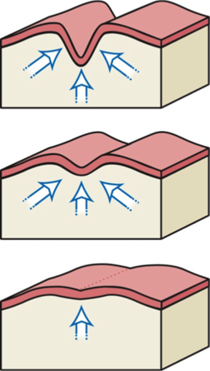
What role does fusion play in facial development?
Two types:
1. fusion of swellings/tissue on the SAME surface
2. fusion of swellings/tissue from DIFFERENT surfaces (occurs for upper lip and palate)
The clefts/furrows between the swellings are eliminated as the underlying mesenchyme migrates into the furrow, making the facial surface smooth. Migration occurs when mesenchyme grows and merges beneath the external ectoderm during maturation of the structure.
Describe the development of the stomodeum and oral cavity.
Stomodeum forms (beginning of 4th week) --> oropharyngeal membrane disintegrates (latter part of 4th week) --> primitive mouth & primitive pharynx are connected --> future development: stomodeum gives rise to oral cavity (lined by oral epithelium derived from ectoderm) and teeth
Describe the development of the lower face.
paired mandibular processes --> fuses at midline to form mandibular arch (1st pharyngeal arch) (mandibular symphysis) --> future development: mandibular arch gives rise to lower face, lower lip, mandibular teeth
- Meckel cartilage --> mandibular bone (intramembranous ossification)
- Mesoderm --> muscles of mastication (5th-10th week, occurs before cartilage ossifies)
Describe the development of the upper face.
frontonasal process --> upper face (forehead, bridge of nose, primary palate, nasal septum, all structures associated with medial nasal processes); placodes --> sense organs
NOSE FORMATION:
- tissue around nasal placodes grows --> forms nasal pits --> deepen to form nasal sacs --> oronasal membrane disintegrates to connect oral and nasal cavities while nasal conchae develop on the lateral walls of the nasal cavity
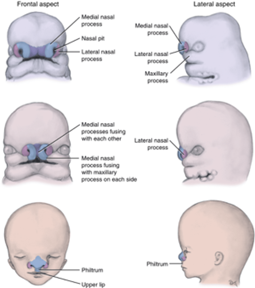
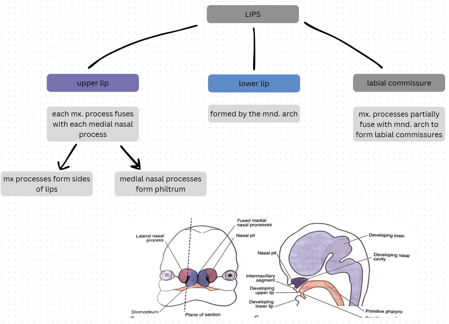
- medial nasal processes --> fuse together externally to form the middle part of the nose (from root to apex), the tubercle of the upper lip, and the philtrum (end of 7th week)
- medial nasal processes --> fuse together internally to form the intermaxillary segment (end of 7th week) --> forms maxillary incisors, primary palate, nasal septum
- lateral nasal processes --> form the alae of the nose
- lateral nasal, maxillary, and medial nasal processes --> fuse to form nares
PARANASAL SINUS FORMATION:
walls of nasal cavity outgrow --> outgrowths become air-filled cavities in bones (sinuses)
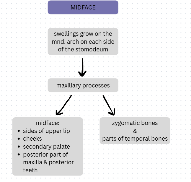
Describe the development of the midface.
tissue grows into swellings on mandibular arch on each side of stomodeum (4th week) --> maxillary processes --> midface (sides of upper lip, cheeks, secondary palate, posterior part of maxilla and its posterior teeth) and zygomatic bones and parts of temporal bones
Describe the formation of the upper and lower lips.
UPPER LIP FORMATION:
- each maxillary process fuses with each medial nasal process (6th week) --> maxillary processes form sides of lip and medial nasal processes form philtrum (fusion completed during 7th week)
- maxillary processes partially fuse with mandibular arch --> labial commissures
LOWER LIP FORMATION:
formed by mandibular arch
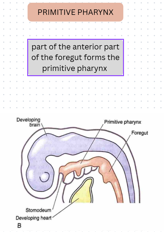
Describe the formation of the primitive pharynx.
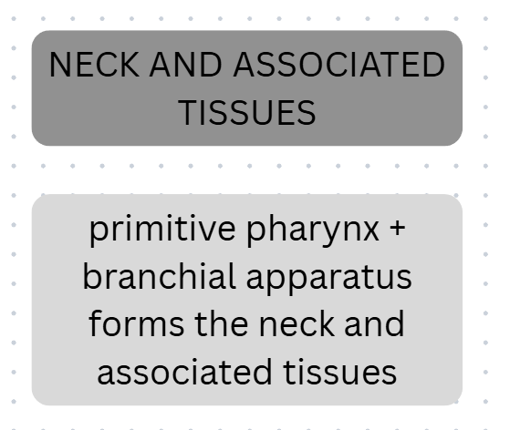
primitive pharynx + branchial (pharyngeal) apparatus --> neck and associated tissues
primitive pharynx:
anterior part of foregut --> primitive pharynx --> joins primitive mouth

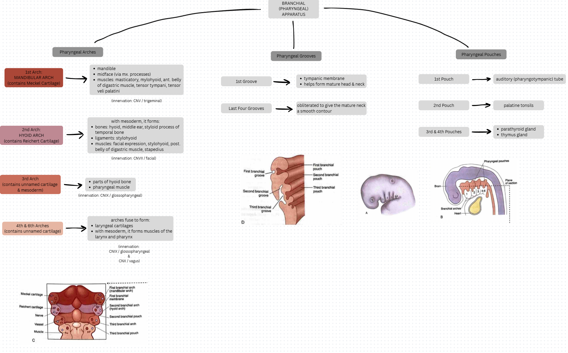
Describe the formation of the pharyngeal (branchial) apparatus.
branchial (pharyngeal) apparatus consists of:
1. pharyngeal arches
2. pharyngeal grooves
3. pharyngeal pouches
Pharyngeal Arches:
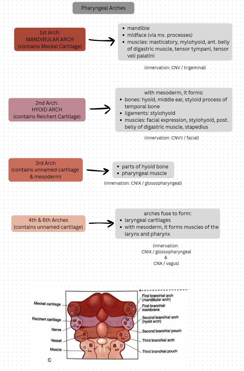
1st arch: mandibular arch (has Meckel cartilage) --> mandible and midface (innervation: 5th cranial trigeminal nerve)
2nd arch: hyoid arch --> lower face (Reichert cartilage and mesoderm forms middle ear bone and muscles, process of temporal bone, parts of hyoid bone with ligaments and suprahyoid muscle, muscles of facial expression) (innervation: 7th cranial facial nerve)
3rd arch: unnamed cartilage and mesoderm forms parts of hyoid bone and pharyngeal muscle (innervation: 9th cranial glossopharyngeal nerve)
4th and 6th arches: unnamed cartilage --> arches fuse --> cartilage becomes laryngeal cartilages and mesoderm becomes muscles of larynx and pharynx (innervation: 9th & 10th vagus cranial nerves)
Pharyngeal Grooves:
1st groove: forms tympanic membrane (ear drum) & helps form the mature head and neck
last 4 grooves: obliterated to give mature neck a smooth contour
Pharyngeal Pouches:
1st pouch: becomes auditory (pharyngotympanic) tube
2nd pouch: becomes palatine tonsils
3rd/4th pouches: parathyroid & thymus glands
FACIAL DEVELOPMENT
Upper Face →
forehead, bridge of nose, primary palate, nasal septum, medial nasal processes
Midface →
midface: sides of upper lip, cheeks, secondary palate, posterior part of maxilla & posterior teeth
zygomatic bones and parts of temporal bones
Lower Face →
mandibular arch → lower face, lower lip, mnd teeth, tongue
Facial Development: UPPER FACE
Frontonasal Process → Upper Face →
forehead, bridge of nose, primary palate, nasal septum, medial nasal processes
Facial Development: Upper Face
NOSE
Frontonasal Process → Upper Face → Nose
Facial Development: MIDFACE
swellings of mnd arch → mx processes →
midface: sides of upper lip, cheeks, secondary palate, posterior part of maxilla, posterior teeth
zygomatic bones & parts of temporal bones
Facial Development: LOWER FACE
paired mandibular processes → mnd arch
mandible bone
masticatory muscles
future development: lower face, lower lip, mnd teeth, tongue
Facial Development: LIPS
each mx process + medial nasal processes = upper lip
mnd arch → lower lip
mx processes + mnd arch = labial commissures
Development of the NECK
Development of the Neck: PRIMITIVE PHARYNX
foregut (anterior part) → primitive pharynx
Development of the Neck: STOMODEUM (& ORAL CAVITY)
Development of the Neck: NECK AND ASSOCIATED TISSUES
primitive pharynx + branchial apparatus = neck and associated tissues
Development of the Neck: BRANCHIAL (PHARYNGEAL) APPARATUS
Development of the Neck: Branchial (Pharyngeal) Apparatus
PHARYNGEAL ARCHES
1st Arch (Mandibular Arch) →
mandible
midface
muscles (masticatory, mylohyoid, ant. belly of digastric, tensor tympani, tensor veli palatini)
2nd Arch (Hyoid) →
bones (hyoid, middle ear, styloid process of temporal bone)
ligaments (stylohyoid)
muscles (facial expression, stylohyoid, post belly of digastric, stapedius)
3rd Arch → parts of hyoid bone, pharyngeal muscle
4th & 6th Arches fuse → laryngeal cartilages, muscles of larynx and pharynx
Development of the Neck: Branchial (Pharyngeal) Apparatus
PHARYNGEAL GROOVES
1st Groove → tympanic membrane, helps form mature head and neck
Last Four Grooves → obliterated to give mature neck a smooth contour

Development of the Neck: Branchial (Pharyngeal) Apparatus
PHARYNGEAL POUCHES
1st Pouch → auditory (pharyngotympanic tube)
2nd Pouch → palatine tonsils
3rd & 4th Pouches → parathyroid gland, thymus gland