RADSCI 313 FINAL (questions are in definition box)
1/153
There's no tags or description
Looks like no tags are added yet.
Name | Mastery | Learn | Test | Matching | Spaced | Call with Kai |
|---|
No analytics yet
Send a link to your students to track their progress
154 Terms
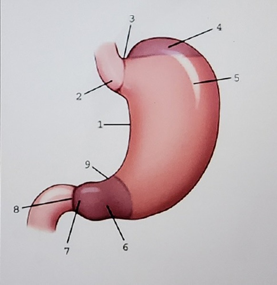
1. Lesser curvature
2. Cardiac antrum
3. Cardiac notch
4. Fundus
5. ?
6. Pylorus/pyloric antrum
7. ?
8. Pyloric sphincter
Identify the anatomy:
Body, pylorus, duodenum
When the patient is in an RAO position, which portion of the stomach and small intestine fills with barium? Name 3 structures.
Body, pylorus, duodenum
When the patient is in a right lateral position, what portion of the stomach and small intestine is filled with barium? Name 3 structures.
Fundus; lies more posterior than the rest of the stomach
When the patient is in a PA recumbent position, what portion of the stomach is filled with air? Why?
B. Small bowel series
Which of the following procedures is considered as a functional study?
A. Air-contrast BE
B. Small bowel series
C. Enteroclysis
D. Barium enema (BE)
A. iliac crests
The majority of AP, PA, and oblique images taken during a barium enema are done on 14" by 17" receptors (IR). Where is the IR centered on sthenic patients for the majority of these projections?
A. iliac crests
B. 2 inches below the iliac crests
C. Costal margin
D. 2 inches above the iliac crests
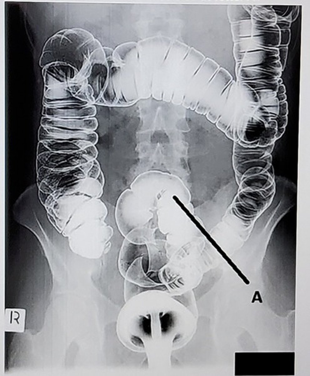
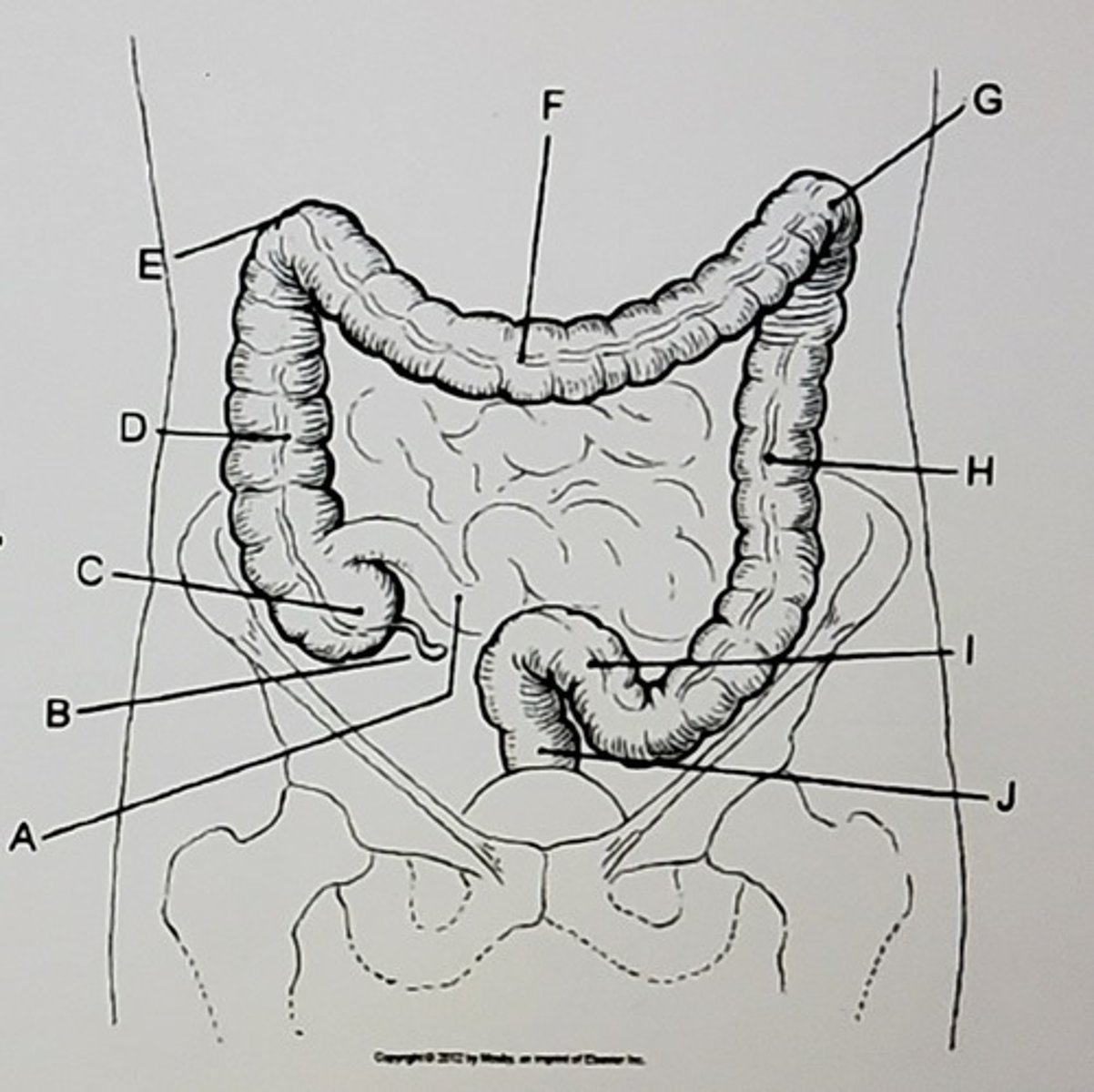
C. Sigmoid colon
What anatomic structure is labeled as letter A?
A. Cecum
B. Rectum
C. Sigmoid colon
D. Descending colon
1, 2, and 3
Preparation of the intestinal tract for examination of the colon includes (select all that apply):
1. Laxatives
2. Clear liquid diet
3. Cleansing enemas or suppositories
C. PA or AP
The entire colon is best demonstrated in which projection?
A. PA axial
B. PA oblique
C. PA or AP
D. Lateral
B. 2 inches above the iliac crests
After ingestion of the barium for small bowel series, where is the IR centered for images taken within the first 15-30 minutes?
A. At the costal margin/L3
B. 2 inches above the iliac crests
C. 1 inch above the iliac crests
D. Level of the iliac crests
A. Small intestine
Which of the following examinations requires the length of time post contrast media administration be marked on the images?
A. Small intestine
B. Esophagram
C. Stomach
D. Large intestine
C. bilbao duodenography tube, past the ligament of Treitz
When conducting a small bowel enteroclysis, what type of tube is used and how far does the radiologist advance it before beginning the administration of contrast?
A. dobhoff, into the third portion of the duodenum
B. nasogastric tube, past the pyloric sphincter
C. bilbao duodenography tube, past the ligament of Treitz
D. salem-sump tube, into the jejunum
1 and 3
Functions of the stomach include (select all that apply):
1. storage of food
2. absorption of food products
3. chemical breakdown of food
A. Duodenal bulb
When positioning a patient for an upper gastrointestinal series, what anatomic structure should be centered to the image receptor for each projection?
A. Duodenal bulb
B. Body of the stomach
C. Fundus of the stomach
D. Jejunum
A. Hypersthenic
For which type of body habits is the stomach almost horizontal?
A. Hypersthenic
B. Hyposthenic
C. Asthenic
D. Sthenic
B. PA oblique, RAO
Which of the following will demonstrate the duodenal bulb and loop in profile?
A. AP oblique, LPO
B. PA oblique, RAO
C. AP oblique, RPO
D. PA
D. Level of the iliac crests
Where is the IR centered for delayed images of the small intestine?
A. 2 inches above the iliac crests
B. 1 inch above the iliac crests
C. At the cost margin/L3
D. Level of the iliac crests
D. Once the contrast media passes the ileocecal valve
When are small bowel series deemed to be complete?
A. Two hours after the ingestion of barium
B. Once the contrast media reaches the rectum
C. Once the contrast media passes the duodenojejunal flexure
D. Once the contrast media passes the ileocecal valve
D. T 5-6
The central ray is directed to what level for all projections and positions of the esophagus?
A. T 9-10
B. mid-sagittal plane
C. C 6-7
D. T 5-6
E. Any of the above
Which of these exercises are used to detect esophageal reflux?
A. Mueller maneuver
B. Valsalva maneuver
C. Compression technique
D. Toe-touch maneuver
E. Any of the above
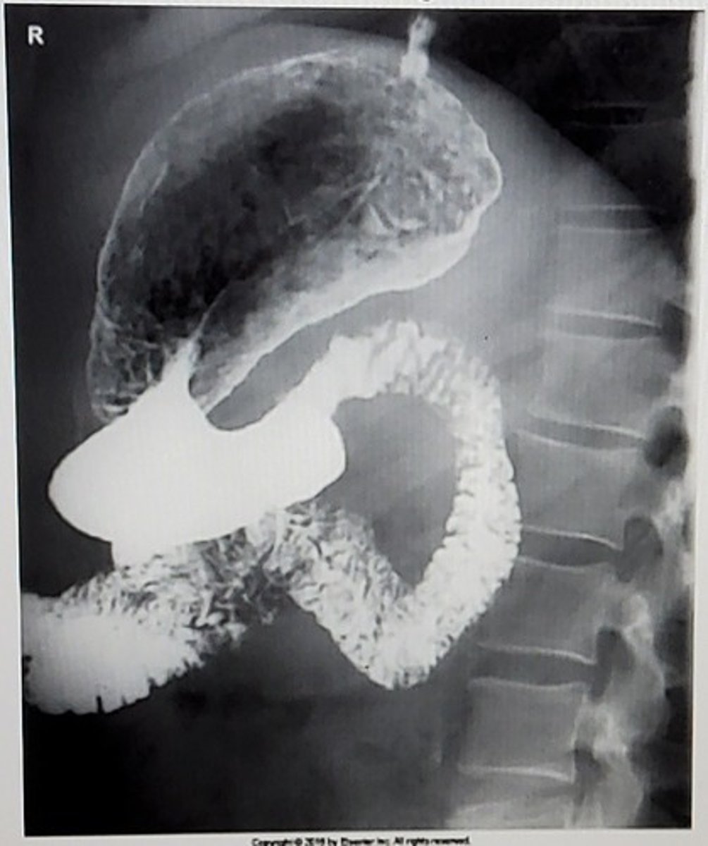
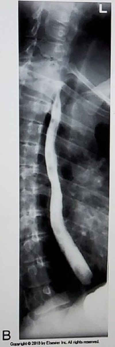
D. Right lateral
What position is this image?
A. RAO
B. AP
C. RPO
D. Right lateral
C. Lateral
Which projection and/or position is most commonly performed during a defogram (evacuative proctogram)?
A. Right posterior oblique (RPO) and left posterior oblique (LPO)
B. AP axial
C. Lateral
D. Anteroposterior (AP) erect
B. Ileocecal valve
The opening between the small intestine and the large intestine is called the:
A. Greater duodenal papilla
B. Ileocecal valve
C. Ampulla of Vater
D. Pyloric valve
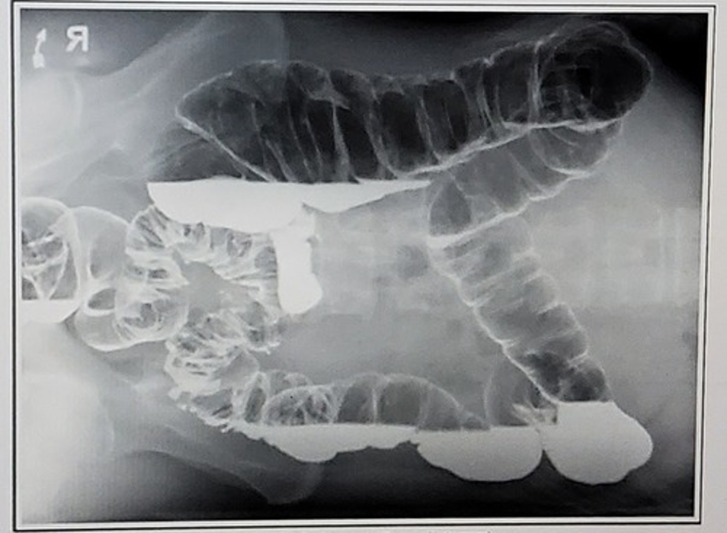
C. Left lateral decubitus
What position is this?
A. Right lateral decubitus
B. AP
C. Left lateral decubitus
D. AP axial
D. Haustra
The large intestine is made up of a series of pouches called the:
A. Taeniae coli
B. Cecum
C. Rugae
D. Haustra
All of the above
Which methods may be used to perform a small bowel enteroclysis study?
A. Double contrast
B. double contrast with CT imaging
C. single contrast
D. All of the above
C. Aortic arch, left mainstem bronchus, left atrium
Luminal indentations of the esophagus are caused by which anatomic features?
A. Cardiac orifice, left atrium, aortic arch
B. Left ventricle, diaphragm, and aortic arch
C. Aortic arch, left mainstem bronchus, left atrium
D. Diaphragm, cardiac antrum, left atrium
A. Double-contrast studies
For select examinations, negative contrast and radiopaque contrast media are used as a pair to outline mucosal linings, joint interspaces, and passageways. These examinations are considered to be:
A. Double-contrast studies
B. Low-contrast studies
C. Extremely dangerous and rarely done
D. Single-contrast studies
D. Between the thoracic spine and heart
A properly positioned lateral or oblique of the esophagus should demonstrate the barium-filled esophagus _________.
A. Overlying the thoracic spine
B. Overlying the heart
C. Just medial to the aortic arch
D. Between the thoracic spine and heart
A. Iliac crests
Where is the IR centered for all decubitus projections of the large intestine?
A. Iliac crests
B. Costal margin
C. 2 inches below the iliac crests
D. 2 inches above the iliac crests
D. AP
When performing an air contrast barium enema, which position would demonstrate the transverse colon filled with air?
A. Lateral
B. AP axial
C. PA
D. AP
B. PA, RAO
Select the projection used to create this image:
A. AP, RPO
B. PA, RAO
C. AP, supine
D. Right lateral
B. Deglutition
The act of swallowing is termed:
A. Aphasia
B. Deglutition
C. Digestion
D. Mastication
A. Suspended on expiration
The respiration phase for all projections of the large intestine is:
A. Suspended on expiration
B. Suspended on full inspiration
C. Suspended respiration, neither inspiration or expiration
D. Slow, shallow breathing
2. 1 and 2
Methods of radiographically examining the colon include:
1. 1 and 3
2. 1 and 2
3. 1, 2, and 3
4. 2 and 3
A.
B.
C. cecum
D.
E.
F.
G.
H.
I.
J.
Identify the anatomy:
B. Suspended respiration on expiration
What is the respiration phase for all radiographic exposures of the stomach and intestines?
A. Suspended after second inspiration
B. Suspended respiration on expiration
C. Slow, shallow breathing
D. Suspended respiration on inspiration
C. Lateral rectum
Which position best demonstrates the rectosigmoid region of the colon?
A. PA
B. Left lateral decubitus
C. Lateral rectum
D. RPO
A. 35 to 40 degrees
What is the degree of body rotation for the PA oblique projection of the esophagus?
A. 35 to 40 degrees
B. 15 degrees
C. 20 to 30 degrees
D. 50 degrees
C. AP oblique, LPO
Which of the following projections will best demonstrate the fundus of the stomach filled with barium?
A. PA oblique, LAO
B. PA
C. AP oblique, LPO
D. PA oblique, RAO
B. for 8 hours
The patient "prep" for an adult who is having a morning stomach examination is food and fluid are withheld:
A. for 12 hours
B. for 8 hours
C. for 24 hours
D. for 4 hours
1 and 4
Which projections will clearly demonstrate the right colic flexure?
1. AP oblique, LPO
2. AP oblique, RPO
3. PA oblique, LAO
4. PA oblique, RAO
5. Right lateral
A. Known or suspected perforation
What is the primary contraindication for the use of barium for any gastrointestinal study?
A. Known or suspected perforation
B. Age
C. History of allergy to barium
D. Risk of aspiration
A. 45 degrees
What is the average degree of body rotation for an AP oblique projection (LPO position) of the stomach and duodenum?
A. 45 degrees
B. 55 degrees
C. 60 degrees
D. 50 degrees
D. 40 to 70 degrees
How much is the body rotated for the PA oblique projection of the stomach and duodenum?
A. 30 to 40 degrees
B. 50 degrees
C. 60 degrees
D. 40 to 70 degrees
1, 2, and 3
Which of the following contrast media are used for examinations of the gastrointestinal tract?
1. air
2. barium sulfate
3. water-soluble iodinated contrast media
4. carbon dioxide
C. 30 to 40 degrees caudad
What is the central-ray angulation for the PA axial projection of the large intestine?
A. 10 to 20 degrees caudad
B. 30 to 40 degrees cephalad
C. 30 to 40 degrees caudad
D. 10 to 20 degrees cephalad
A. 35 to 45
The degree of body rotation for oblique projections of the large intestine is _________ degrees.
A. 35 to 45
B. 20 to 30
C. 30
D. 50
C. AP, left lateral decubitus
Which projection of the colon will best demonstrate the air filled ascending colon and right colic flexure when performing a double contrast enema?
A. AP oblique, LPO
B. AP, right lateral decubitus
C. AP, left lateral decubitus
D. AP oblique, RPO
B. Pyloric sphincter
The muscle controlling the opening between the stomach and the duodenum is termed the:
A. Pyloric antrum
B. Pyloric sphincter
C. Pylorus
D. Ileocecal valve
C. duodenum, jejunum, ileum
In which order does barium flow through the small intestine?
A. ileum, duodenum, jejunum
B. ileum, jejunum, duodenum
C. duodenum, jejunum, ileum
D. jejunum, duodenum, ileum
B. L1/L2
At which level is the IR centered on the sthenic patient for all positions of the stomach and duodenum?
A. L2/L3
B. L1/L2
C. iliac crests
D. L3/L4
A. level of the iliac crests and one inch lateral toward the elevated side of the MSP
When positioning the patient for posterior obliques of the colon, where is the IR centered?
A. level of the iliac crests and one inch lateral toward the elevated side of the MSP
B. level of the iliac crests and one inch lateral toward the dependent (down) side of the MSP
C. level of the iliac crests and along the midsagittal plane (MSP)
D. two inches above the iliac crests and along the MSP
D. 15 minutes
The first small intestine image is typically taken how many minutes after the patient drinks barium?
A. 40 minutes
B. 10 minutes
C. 5 minutes
D. 15 minutes
D. right colic flexure
The ascending portion of the colon joins the transverse colon at the:
A. duodenojejunal flexure
B. left colic flexure
C. sigmoid colon
D. right colic flexure
A. a modified barium swallow only evaluates the oral and pharyngeal phases of swallowing
What is the primary difference between a modified barium swallow and an esophagram?
A. a modified barium swallow only evaluates the oral and pharyngeal phases of swallowing
B. an esophagram is performed to evaluate anatomic structures only
C. the pre-examination prep
D. the typeof contrast media used
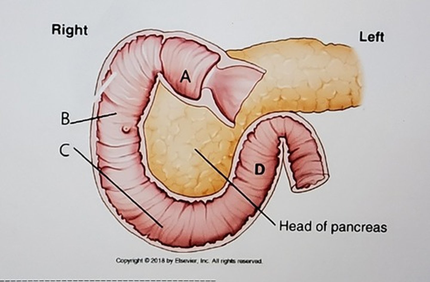
A. Duodenal bulb
B. Second/descending portion
C. Third/horizontal portion
D. Fourth/ascending portion
Identify the specific portions of the structure labeled with letters A, B, C, D.
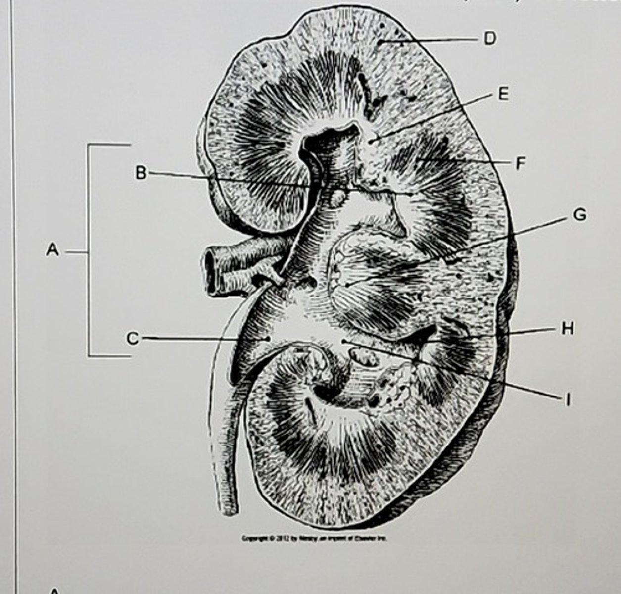
A. Hilum
B.
C. Renal pelvis
D. Renal cortex
E.
F. Renal medulla
G.
H. Minor calyx
I. Major calyx
Identify the letters listed in the picture.
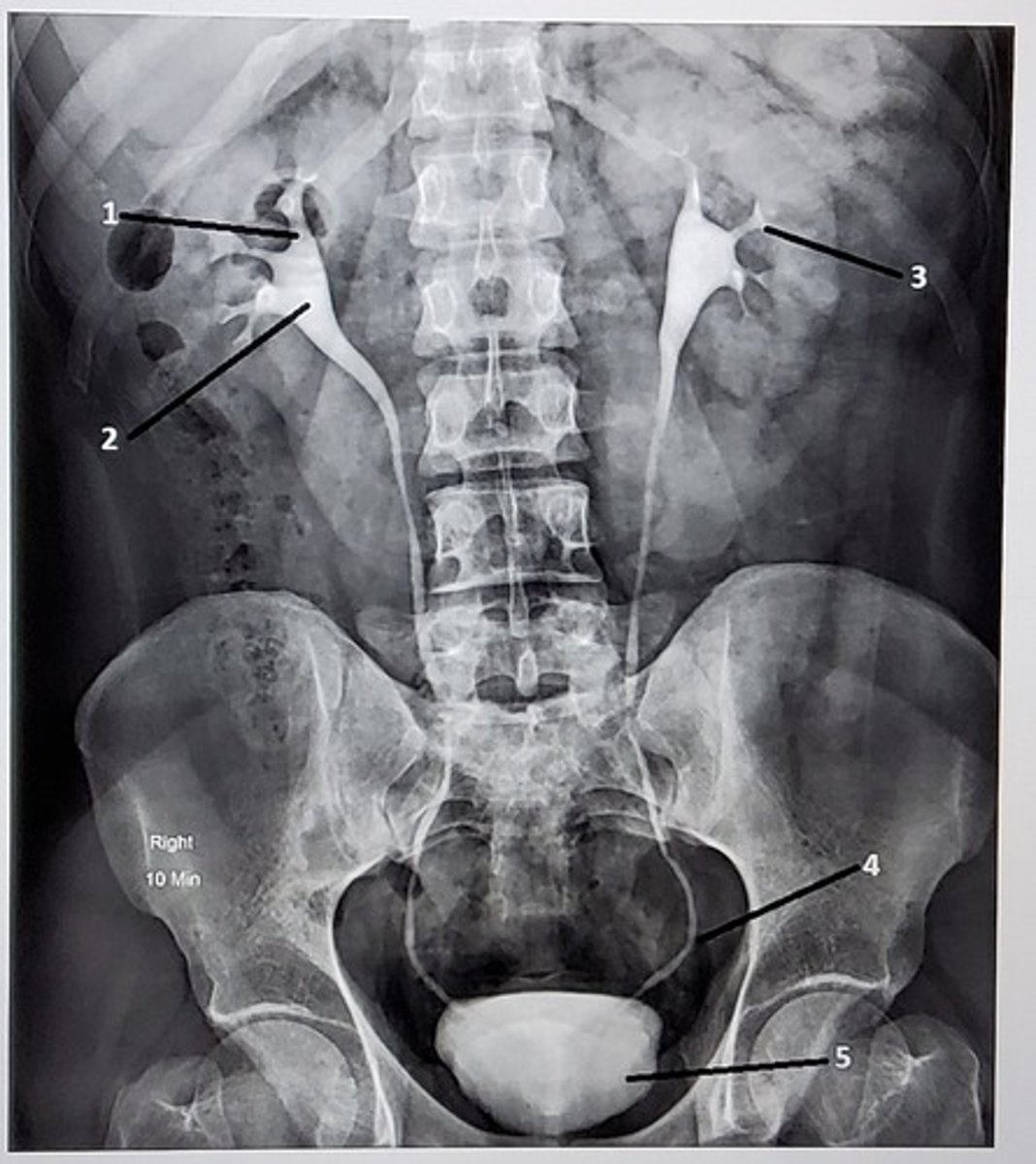
1. Right major calyx
2. Right renal pelvis
3. Left minor calyx
4. Left distal ureter
5. Bladder
Identify the anatomic structures labeled on this image. Your label should be descriptive (proximal/distal, which side of the body, etc).
A. 30° RPO
A male patient comes to radiology for a voiding cystourethrogram. Which of the following projections and/or positions would be performed for this procedure?
A. 30° RPO
B. Recumbent lateral
C. Erect lateral
D. Erect PA
B. Suck on a lemon wedge
How do you get the patients salivary ducts to open when conducting a sialogram?
A. Think about food
B. Suck on a lemon wedge
C. IM injection of glucagon
D. Drink a large glass of water
C. Where the iliac blood vessels cross over the ureters
One of the three constricted points along each ureter is the pelvic brim. The location of this constricted point is:
A. Where the distal ureters connect to the bladder
B. Where the bladder meets the symphysis pubic
C. Where the iliac blood vessels cross over the ureters
D. at the level of the ischial spines
D. the nephron
The structural and functional unit of the kidney is:
A. the glomerulus
B. the calyx
C. Bowman capsule
D. the nephron
A. Direct instillation into the common bile duct
During an endoscopic retrograde cholangiopancreatogram, how does the contrast medium reach the biliary tree?
A. Direct instillation into the common bile duct
B. Orally, by swallowing the contrast
C. Injection into the cystic duct during surgery
D. IV injection
A. starch
Saliva contains certain enzymes to begin the digestion of:
A. starch
B. lipids
C. proteins
D. minerals
Any of these positions
During an operative cholangiogram, the surgeon injects the contrast medium directly into the biliary system. According to Bontrager's which of the following positions may the patient be placed into during the injection of the contrast media?
1. AP
2. AP oblique, RPO
3. AP oblique, LPO
1, 2, 5
The diaphragm has openings in it to allow certain anatomy to pass from the thoracic cavity into the abdominal cavity. Which of the following are diaphragmatic openings?
1. Esophageal hiatus
2. Aortic aperture
3. Cardiac hiatus
4. Fundal aperture
5. Caval hiatus
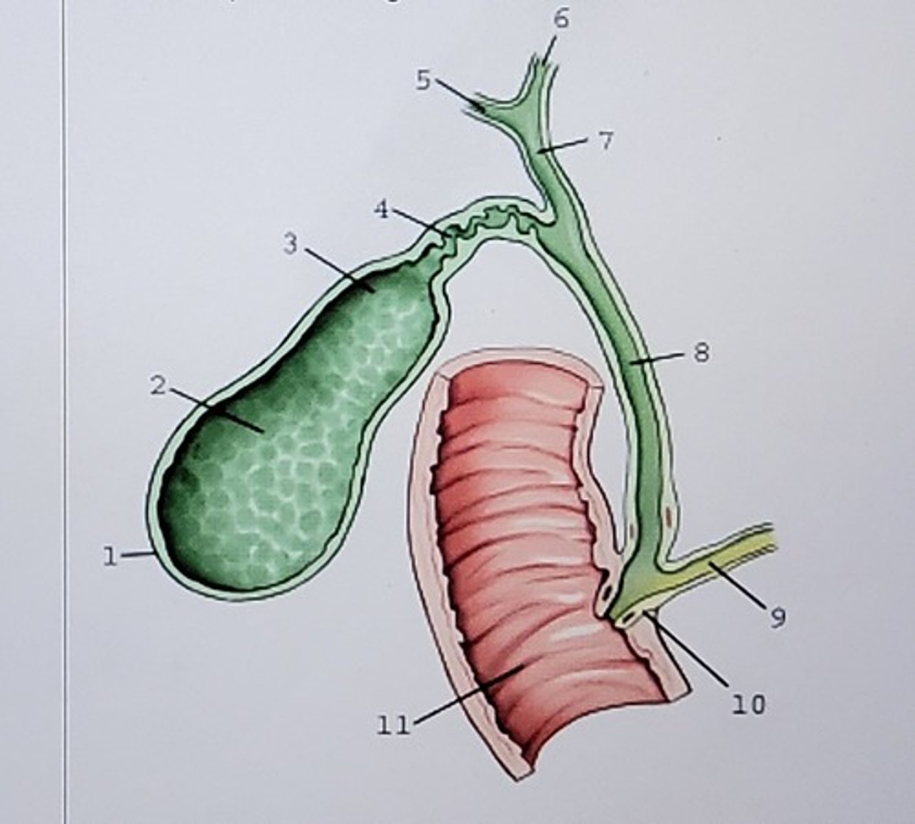
1. Fundus
2.
3.
4. Cystic duct
5.
6.
7.
8.
9.
10. Hepatopancreatic ampulla
11.
Label this picture:
D. All of these are salivary glands
Which of the following structures is not one of the salivary glands?
A. Sublingual
B. Parotid
C. Submandibular
D. All of these are salivary glands
B. Evaluate the patient for a paralyzed hemidiaphragm
Diaphragm fluoroscopy is performed for which of the following reasons?
A. Investigate the reason for shortness of breath
B. Evaluate the patient for a paralyzed hemidiaphragm
C. Evaluate for abducens nerve palsy
D. It is part of the post-stroke evaluation
C. sialography
Radiographic examination of the salivary glands using contrast medium is called:
A. angiography
B. parotitis
C. sialography
D. sialostenosis
B. Gravity flow through a catheter
How is contrast media normally introduced during a retrograde cystogram?
A. With an injection through the urethra with a large syringe and catheter
B. Gravity flow through a catheter
C. Intravenously
D. With a cystoscope
A. 350 to 500 mL
What is the total capacity of the average adult bladder?
A. 350 to 500 mL
B. 100 to 250 mL
C. 500 to 750 mL
D. 1000 to 1200 mL
1 and 2
Functions of the kidney include:
1. Removal of waste products from the blood
2. Maintaining fluid and electrolyte balance
3. Recycling of red blood cells
A. endoscopic retrograde cholangiopancreatiography (ERCP)
Which of the following uses a fiber optic scope to diagnose and treat hepatobilliary disorders?
A. endoscopic retrograde cholangiopancreatiography (ERCP)
B. Percutaneous transhepatic cholangiography (PTC)
C. T-tube cholangriography
D. Operative cholangiography
A.
B.
C. common bile duct
Label the picture:

D. Cholangiography
Radiographic examination of just the biliary ducts is termed:
A. Cholelithiasis
B. Cholecystography
C. Cholecystocholangiography
D. Cholangiography
D. emulsify fats
The main function of bile is to:
A. break down cholesterol
B. begin the digestion of complex sugars
C. begin the digestion of proteins
D. emulsify fats
C. stimulates the gallbladder to contract
What is the primary function of cholecystokinin?
A. stimulates the production of bile
B. inhibits the formation of gallstones
C. stimulates the gallbladder to contract
D. serves as an enzyme to break down certain food nutrients
3, 4, and 5
As a member of the team performing a bronchoscopy, what is your roles as the radiologic technologist?
1. assist the pulmonologist by capturing and preserving tissue samples
2. monitor the patients vital signs
3. utilize the c-arm as directed by the pulmonologist, centering over the bronchoscope tip
4. ensure the entire team is wearing appropriate radiation protection apparel
5. perform a post-procedure mobile CXR if ordered
1 only
What type of contrast media is used for imaging the urinary and hepatobiliary systems?
1. water soluble nonionic contrast media
2. barium sulfate
3. oral, water soluble iodinated contrast media
4. oil-based iodinated contrast media
A. retrograde urography
Which of these examinations is usually performed by a physician in an operating room?
A. retrograde urography
B. voiding cystourethrogram
C. IVU
D. Cystography
D. bile
"chole-" is a prefix for terms pertaining to the:
A. Liver
B. ducts
C. gallbladder
D. bile
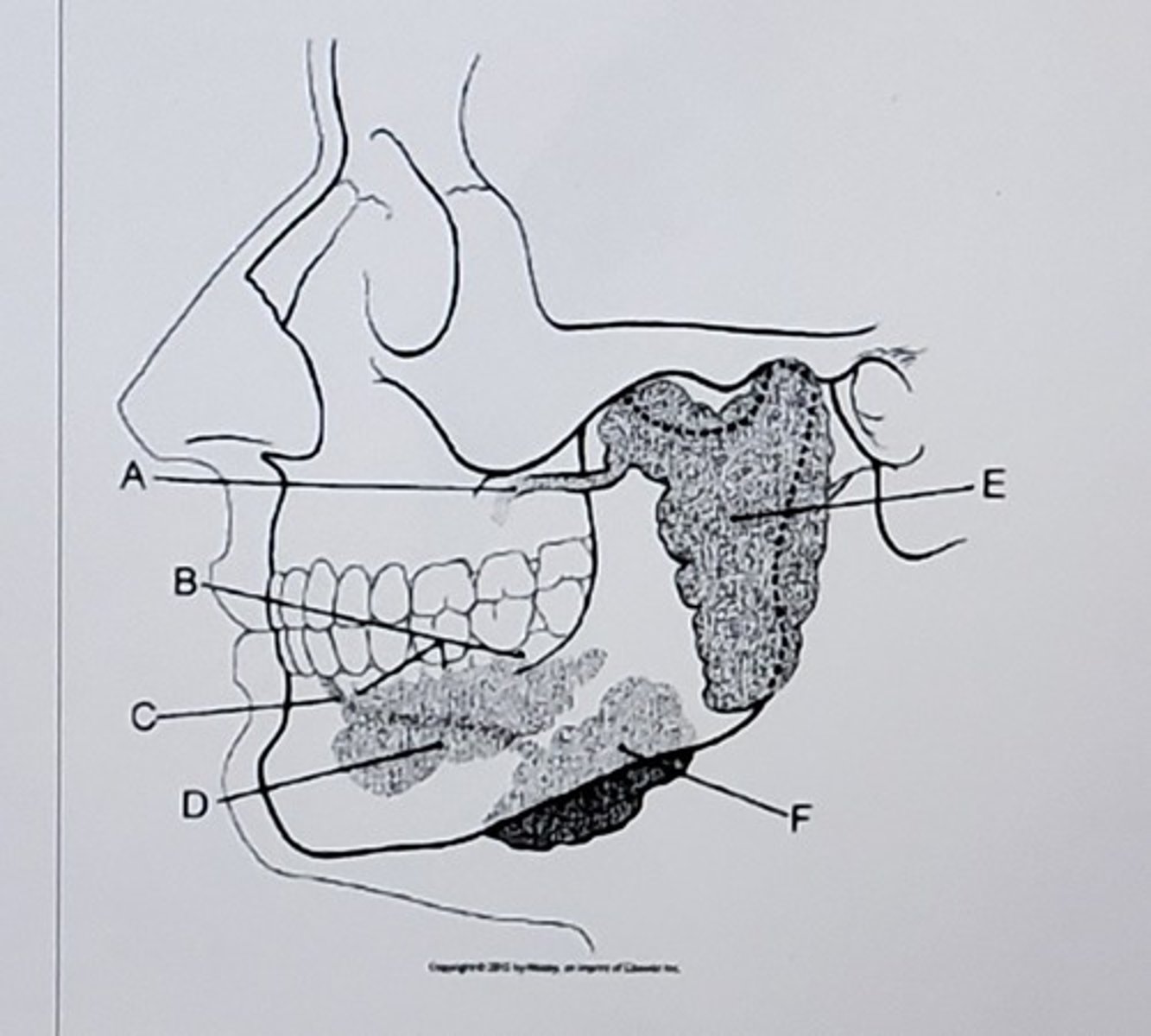
A.
B.
C.
D.
E. Parotid gland
Label the picture:
A. ensure there are no air bubbles in the syringe, needle, or tubing as air bubbles mimic stones
When preparing the contrast media for any cholangiography study, what precaution should always be followed?
A. ensure there are no air bubbles in the syringe, needle, or tubing as air bubbles mimic stones
B. have an extra supply of contrast media ready should the radiologist need to inject more than the usual amount
C. Always use the highest concentration of injectable contrast media your department has
D. Dilute the contrast media with 75% sterile saline to 25% contrast
B. 45-60
How many degrees should the patient be rotated for AP oblique projections of the urinary bladder?
A. 16-30
B. 45-60
C. 10-15
D. 30-39
D. Stay with the patient for 15 minutes
You have just injected 50cc of radiopaque. IV contrast into your patient for an intravenous urogram. You have paperwork to do and want to step out of the room to do it. Should you go? If not, how long do you stay with the patient to watch for an adverse reaction?
A. Stay with the patient for 25 minutes
B. Stay with the patient for 30 minutes
C. Stay with the patient for 5 minutes
D. Stay with the patient for 15 minutes
A. suspended on expiration
The respiration phase for exams pertaining to the urinary system is made on:
A. suspended on expiration
B. suspended on inspiration
C. shallow breathing
D. suspended respiration
D. liver
Where is bile formed?
A. duodenal mucosa
B. gallbladder
C. pancreas
D. liver
All of the above (the radiologist performs #4, not the patient)
During a diaphragm fluoroscopy exam, what activities does the radiologist ask the patient to perform?
1. Take short, quick breaths
2. Breath normally
3. take deep breaths
4. measure the distance the diaphragm moves between inspiration and expiration
D. upright
Which position best demonstrates mobility of the kidneys?
A. Supine
B. lateral decubitus
C. RPO
D. upright
D. intravenous urography
Which examination best demonstrates the functioning ability of the urinary system?
A. cystography
B. retrograde urography
C. voiding cystourethrography
D. intravenous urography
A. 2 inches above the symphysis pubis
The central ray for AP and oblique projections of the urinary bladder when performed as part of a cystogram should be directed:
A. 2 inches above the symphysis pubis
B. to the iliac crests
C. to the symphysis pubic
D. to the ASIS
A. iliac crest
The central fay for scout radiographs of the urinary system is centered to the level of the:
A. iliac crest
B. ASIS
C. Xiphoid process
D. lower rib margin
C. Nonionic iodine-based contrast
When setting up for a aialogram, which type of contrast media will you obtain for the radiologist to use?
A. Gastrografin
B. Barium
C. Nonionic iodine-based contrast
D. Carbon dioxide crystals
B. four
The liver is divided into ______ major and minor lobes.
A. two
B. four
C. three
D. six
B. sublingual
Which salivary gland has multiple small ducts which open into the floor of the mouth?
A. submandibular
B. sublingual
C. parotid
D. periramal
C. decrease body rotation
A radiograph of a right posterior oblique (RPO) position taken during an IVU reveals that the left kidney is foreshortened and superimposed over the spine. What must the technologist do to correct this error during the repeat exposure?
A. increase CR angulation
B. place the patient in the LPO position to better demonstrate the right kidney
C. decrease body rotation
D. increase body rotation
A. cystourethrography/retrograde urethrogram
Radiography of the urethra is called _____________.
A. cystourethrography/retrograde urethrogram
B. cystography/cystogram
C. cystoureterography
D. retrograde urography
B. the left kidney is longer and narrower than the right
Which of the following is true regarding the kidneys?
A. the right kidney is longer and narrower than the left
B. the left kidney is longer and narrower than the right
C. the left kidney is lower than the right
D. both kidneys are the same width and length