Neurons and synaptic transmission
1/13
There's no tags or description
Looks like no tags are added yet.
Name | Mastery | Learn | Test | Matching | Spaced | Call with Kai |
|---|
No analytics yet
Send a link to your students to track their progress
14 Terms
Motor neurons
Form synapses with muscles and control their contractions
Neurotransmitter
Chemical substances that play an important role in the workings of the nervous system by transmitting nerve impulses across a synapse. They can be classified as either excitatory or inhibitory in their action
Relay neurons
These neurons are the most common type of neuron in the CNS. They allow sensory and motor neurons to communicate with each other
Sensory neurons
Carry nerve impulses from sensory receptors to the spinal cord and the brain
Synapse
The conjunction of the end of the axon of one neuron and the dendrite or cell body of another
Synaptic transmission
Refers to the process by which a nerve impulse passes across the synaptic cleft from one neuron (the presynaptic neuron) to another (the postsynaptic neuron)
Explain the structure and function of neurons
100 billion neurons in nervous system, 80% in brain
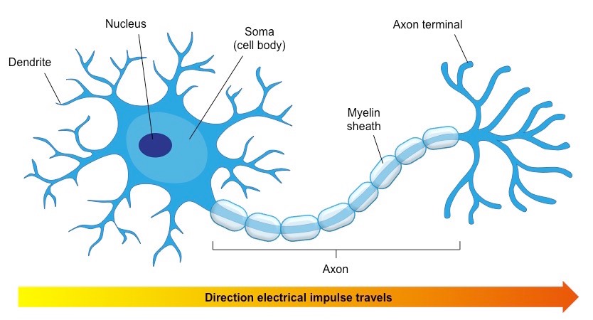
Specialised cells that carry neural information throughout the body
3 types: Sensory, motor and relay
Consists of a cell body, dendrites and an axon
Dendrites at one end of a neuron receive information/signals from other neurons or sensory receptors
They are connected to the cell body (neuron’s control centre)
From there the impulse is carried along the axon, where it terminates at the axon terminal
In many nerves there’s an insulating layer around the axon called the myelin sheath which allows the nerve impulses to transmit more rapidly along the axon - if damaged they slow down
Length of axon varies from a few mms to 1m

Explain what action potential is
neurons must transmit information both within the neuron and from one neuron to the next
When a neuron is not sending a signal it is in a resting state and the inside is more negatively charged than the outside
When a neurotransmitter binds with the receptor of a neuron it becomes positively charged and if that charge reaches a certain potential action potential is fired
The dendrites of neurons receive information from the sensory receptors or other neurons
This information is then passed down to the cell body and on to the axon
Once the information has arrived at the axon, it travels down its length in the form of an electrical signal known as an action potential
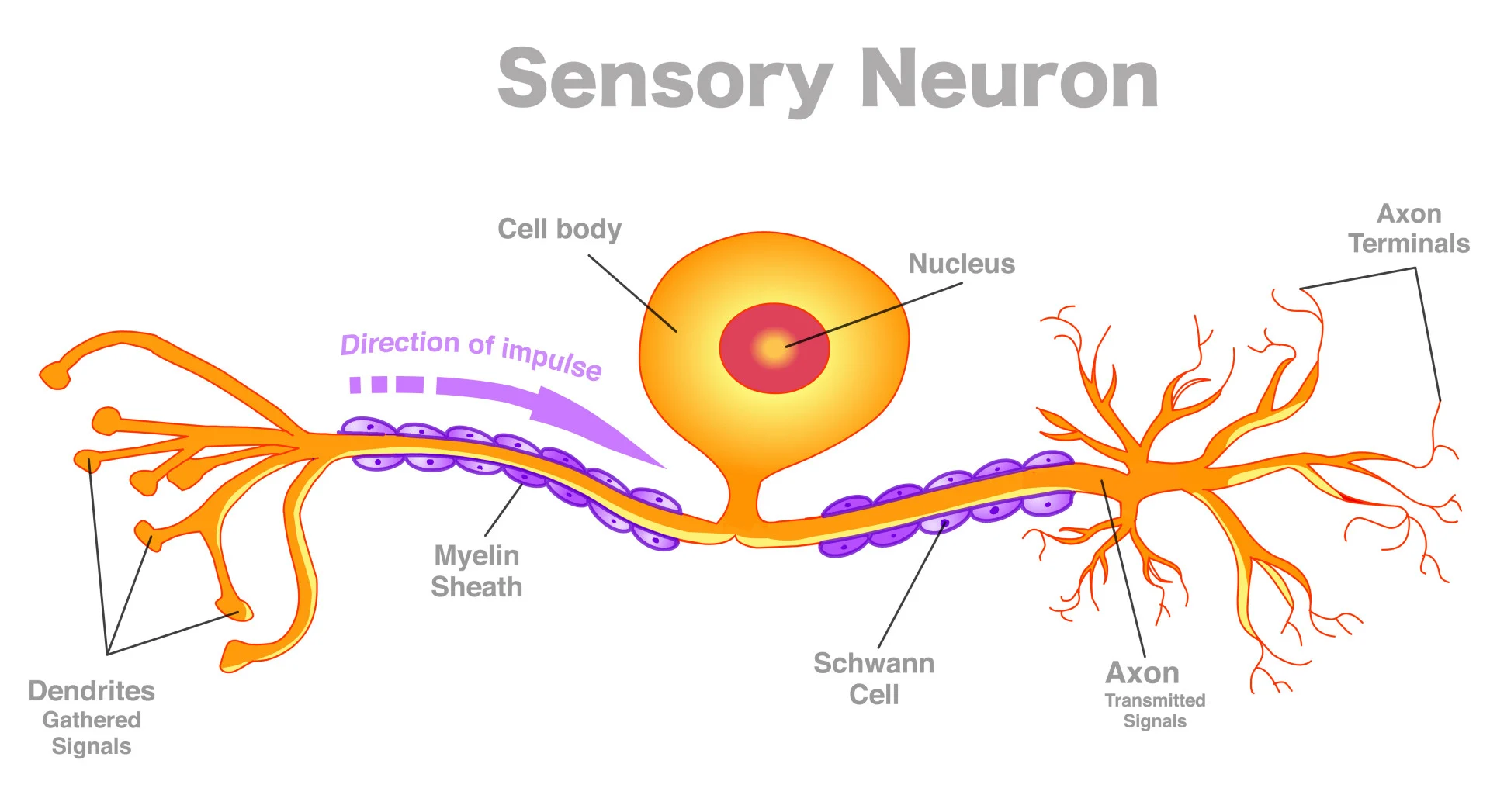
Explain the structure and function of sensory neurons
carry nerve impulses from sensory receptors (PNS) to the CNS (brain + spinal cord)
Sensory receptors found in various locations in the body
Sensory neurons convert information from these receptors into neural impulses
When these impulses reach the brain, they’re translated into sensations so that the organism can react appropriately
Some of the neurons terminate in the spinal cord which allows reflex actions to occur quickly without the delay of sending impulse to the brain
Structure: Cell body in the middle off to one side, long dendrites and axons
Explain the structure and function of relay neurons (interneurons)
allow sensory and motor neurons to communicate with each other
Lie wholly between brain and spinal cord (in CNS)
Structure: Short dendrites and axons, often don’t have a myelin sheath
Explain the structure and function of motor neurons
conduct signals from CNS to effector organs like muscles and glands
Form synapses with muscles and control their contractions
When stimulated, they release neurotransmitters that bind to receptors on the muscle and trigger a response that leads to muscle movement
When the axon fires, the muscle with which it has formed synapses with contracts
The strength of the muscle contraction depends on the rate of firing of the actions of the motor neurons that control it
Muscle relaxation is caused by inhibition of the motor neuron
Structure: Cell bodies are in the CNS, but they have long axons which form part of the CNS, short dendrites
Where are neurotransmitters released?
Released by synapses and pass over synaptic cleft to another connecting neuron
Explain the process of synaptic transmission
neurons communicate with each other via neural networks and each neuron is separated from the next by a tiny gap called the synapse (in between the presynaptic and postsynaptic neurons)
Synapse includes the end of the presynaptic neuron, the membrane of the postsynaptic neuron and the gap in between (the synaptic gap/cleft)
Once an action potential has arrived at the terminal button at the end of the axon, it needs to be transferred to another neuron or to tissue
At the end of the axon there are a number of sacs (synaptic vesicles) that contain chemical messengers that assist in the transfer of the impulse (the neurotransmitters)
As the action potential reaches the synaptic vesicles it causes them to release their contents through a process called exocytosis
The released neurotransmitter diffuses across the synaptic gap where it binds to specialised receptors on the surface of the cell that recognise and are activated by it - each neurotransmitter has its own specific molecular structure that fits perfectly to the receptor site, like a lock and key
Once they have been activated the receptor molecule produces either excitatory or inhibitory effects on the postsynaptic neuron
Whole process takes only a fraction of a second
The effects are terminated at most synapses by a process called ‘re-uptake’: The neurotransmitter is taken up again by the presynaptic neuron, where it is stored and made available for later release (a sort of recycling process)
How quickly the presynaptic neuron takes back the neurotransmitter from the synaptic cleft determines how prolonged its effects will be (quicker = shorter effects on postsynaptic neuron)
Some antidepressants prolong action of neurotransmitter by inhibiting the re-uptake process
Neurotransmitters can also be ‘turned off’ after they have stimulated the postsynaptic neuron through the action of enzymes produced by the body (which make the neurotransmitters ineffective)
