Canine and Ruminant Feet (Week 2, Mod 7)
1/11
Earn XP
Description and Tags
Name | Mastery | Learn | Test | Matching | Spaced | Call with Kai |
|---|
No analytics yet
Send a link to your students to track their progress
12 Terms
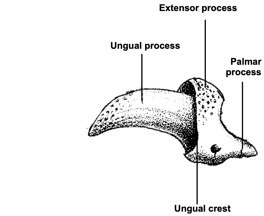
Describe the distal phalanx of the carnivore… what are the 4 main components?
1) Ungual Process - supports claw
2) Ungual Crest - vascular root of claw
3) Extensor Process - extensor tendon insertion point
4) Palmar Process - flexor tendon insertion point
Describe the digital pads of the canine foot… what do they look like, and how are they placed in relation to the skeleton?
Digital pads (n=4):
overlies DIP (distal interphalangeal) joint
Metacarpal / metatarsal pad (n=1):
heart shaped
overlies MCP / MTP joints
Carpal / stopper pad (n=1):
Forelimb only
Protects accessory carpal bone when hyper-extended during a hard break
What are some functions of the digital footpad? Think of a few… how do they protect the metocarpo/metatarso joints in particular?
Functions of footpads:
Allow weightbearing over entire digit
Support digit & metacarpo / metatarso-phalangeal joints
Protect deep structures
Anti-concussion / shock absorption
Resistance to trauma / wear & tear
Traction – prevent slipping
Metacarpo/metatarso - phalangeal joint support
Interosseus muscle for each digit
(cf suspensory ligament in horse)
Support from metacarpal / metatarsal foot pad
Describe the epithelium of the foot pad… how is it modified for its function?
Epidermis
Keratinised stratified squamous epithelium
Thick for protection
Maintained by increased turnover of cells
Rough surface (conical papillae) for traction
Sweat glands
heat loss for thermal regulation… can only do that via sweating from paw pads or panting
Variable amounts pigmentation
NO BLOOD OR NERVE SUPPLY TO THE EPIDERMIS
Describe the contents of the digital cushion… what would happen if this gets cut?
Contains:
Fibrous and elastic tissue
Fat
Vascular channels
Is HIGHLY vascularized; acts as a sponge
Function: Shock absorption
Clinical significance:
Would be VERY messy and bloody
Since its made of elastic tissue, would need some suturing
Keep in mind when bandaging feet, paw pads have sweat glands, so need extra padding for the sweat and need to change bandage often
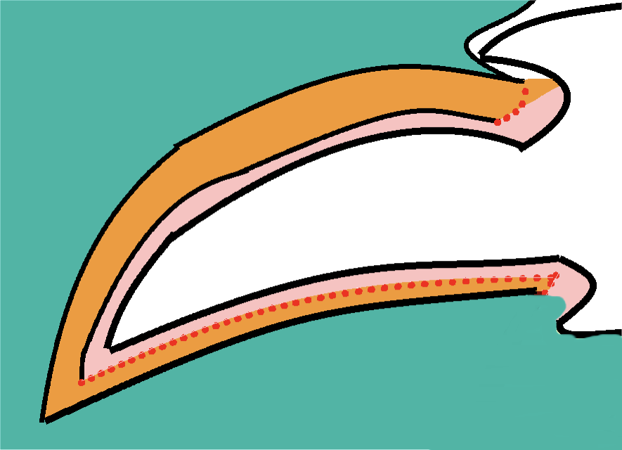
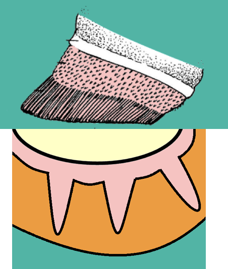
What are the 3 main components of the carnivore claw? Describe its structure
1) Dermis
Fused to the PERIOSTEUM of the ungual process
Sensitive; bleeds
2) Germinative layer
Active in 2 areas of the claw (red dotted lines on image)
Recess at ungual crest
Forms the dorsal and lateral surfaces of the claw
Palmar surface of ungual process
Forms the UNDERSIDE of claw
This is how the claw grows
3) Epidermis
Heavily keratinized (horn ± pigment)
How does the canine claw remain in one stabilized position?
Is a balance of forces between the dorsal elastic ligaments (pink), common digital extensor, and the deep digital flexor tendon
Allows the tip of the claw to touch the ground, keeping it from either hyperextension or hyperflexion
How does the feline claw RETRACT? (essentially, what ligaments are at play here?)
To be retracted:
Has a VERY strong dorsal elastic ligament, keeps the claws in its “neutral position” (= distal interphalangeal joint hyperEXTENSION)
Distal phalanges rotate dorsally
To be exposed:
Deep digital flexor tendon FLEXES the DIP
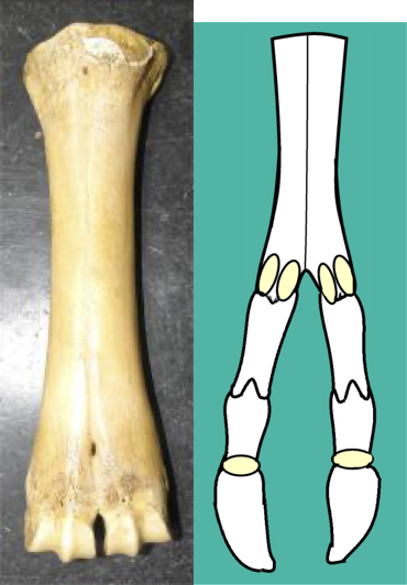
Describe the overall structure of the ruminant foot… How is it different from the carnivore, starting at the metacarpals.
The two metacarpals they have are metacarpals 3 and 4
Are FUSED TOGETHER; looks like a mega-cannon bone of a horse
At the distal end, has grooves where the proximal phalanges would fit… are also for 2 pairs of proximal sesamoids
Digits 3 & 4:
All bones present
Weight bearing
2 x proximal interphangeal joint
2 x distal interphalangeal joint
2 x distal sesamoid bones
** Digits 2 and 5…
Present as dewclaws / little hooves on the palmar aspect of the metacarpo/metatarso-phalangeal joint
1st digit is absent
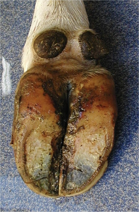
Describe the overall visible components of the ruminant hoof…
Components:
Cloven hoof
Medial and lateral claws
Interdigital cleft
Sole
Heel
Wall:
Lateral - convex
Continuous with heel
Medial - vertical
Stops at parapedal groove (about 2/3s down the digit; on the inside)
Transition hard to soft horn = prone to damage
White line - between the hoof wall and the sole
Describe ruminant hoof growth (may need to look at equine hoof notecards for better explanation)
Starts from the coronary band
Only distal part of dermis covered by laminae
Sensitive laminae
Insensitive laminae
Primary laminae ONLY
No secondary
What are 4 clinical issues that could occur to the ruminant hoof?
1) Horn overgrowth:
Due to soft ground
FL = inner claw overgrowth
HL = outer claw overgrowth
2) Solar ulcers:
especially at parapedal groove
Due to abrasive flooring
3) Interdigital dermatitis
4) Secondary infection
can track up DDFT sheath