ALL practicals - parasitology
1/83
Earn XP
Description and Tags
Remember: schizogony and merogony is the same!. Includes finished: Practical 1, Practical 2, Practical 3, Practical 4, practical 5, Practical 6, Practical 7, Practical 8, Practical 9.
Name | Mastery | Learn | Test | Matching | Spaced |
|---|
No study sessions yet.
84 Terms
Parasite is defined as?
A weaker organism that gets food and shelter from another organism and gets the benefits from this association.
Parasitism: intimate + obligatory relationship bw. two heterospecific organisms which the parasite, usually the smaller one of the two partners, is metabolically dependent on the host. One partner benefits, while the other is harmed.
Definition of obligatory parasite and facultative parasite?
Obligatory parasite → completely relies on its host for part or all of its life cycle.
Facultative parasite → does not completely dependent on the parasitic way of life. It can live without a host, but also adapt to be in such a relationship.
Definition of permanent parasite and temporary parasite?
Permanent parasite → Lives its whole life in or on a host animal (ex. tapeworms, lice)
Temporary parasite → Only lives part of its life in or on a host animal, during the feeding of mosquitoes, ticks etc.
Definition of definitive host?
Organism that is the place for the adult or sexual stage of the parasite, where parasite reaches sexual maturity.
Definition of an intermediate host?
Host that is a temporary enviroment for the parasite, necessary for its development, but where the parasite does not reach sexual maturity. But it may undergo asexual reproduction in this host.
Definition of paratenic host?
This host is one in which the parasite does not undergo development, but one in which it remain alive and infective to another host.
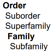
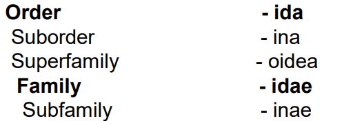
Write the endings of the different categories of organisms (levels - taxa). What is the ending for Diseases?
- ida for order
- ina for suborder
- oidea for superfamily
- idae for family
-inae for subfamily
-osis for disease
In fecal examination, it is important to immediately look at the fecal samples after collection, why is that?
Fecal examination - most common
Collecting fecal samples immediately after defecation is crucial because it helps preserve the integrity of the sample and prevents the degradation of any parasitic organisms or eggs present. If the sample sits for too long, environmental factors like heat and moisture can affect the viability of the pathogens, making the diagnostic results less reliable.
if not seen within few hours → refrigerated until testing, if needed to be sent to another lab, should be packed with cold packs. Helminths eggs may be preserved with equal vol. of 5-10% buffered formaline. Never for lung worms!
collection of fresh materal: can be directly from rectum of large animals or indirectly, immediately after defecation in small animals.
Other diagnostic methods:
coprological
urine, skin
haemotological
histopathological
immunological
molecular biological
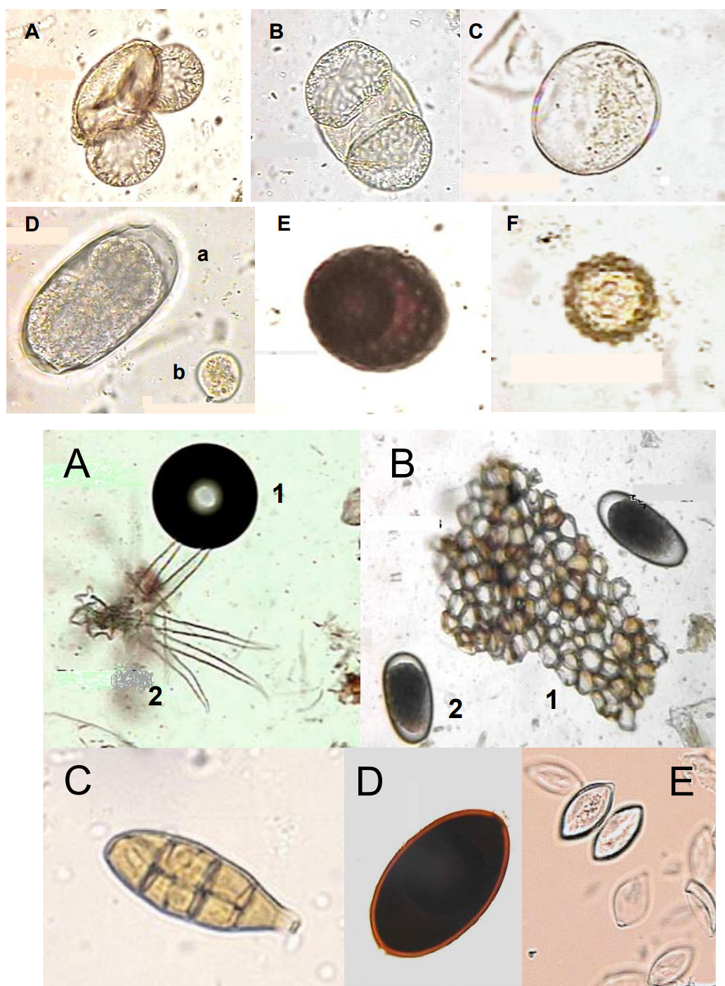
Pseudoparasites are?
Organisms (organic/anorganic substances) that may resemble parasites but do not rely on a host for their life cycle, nor are they actual parasites.
similar to the propagation stages of parasites (egg, cyst, larvae)
non-parasitic substances: plant fibre, hairs, pollen grain (AF), mucosal cells, cells of plants (B1), air bubble (A1)
non-specific parasitic objects:
monocystis sp. - protozoa from earthworms
eggs + larvae of parasites that came from another host after ingestion of feces. Like for instance pig ingesting faces of poultry or dogs eating ruminant faeces.
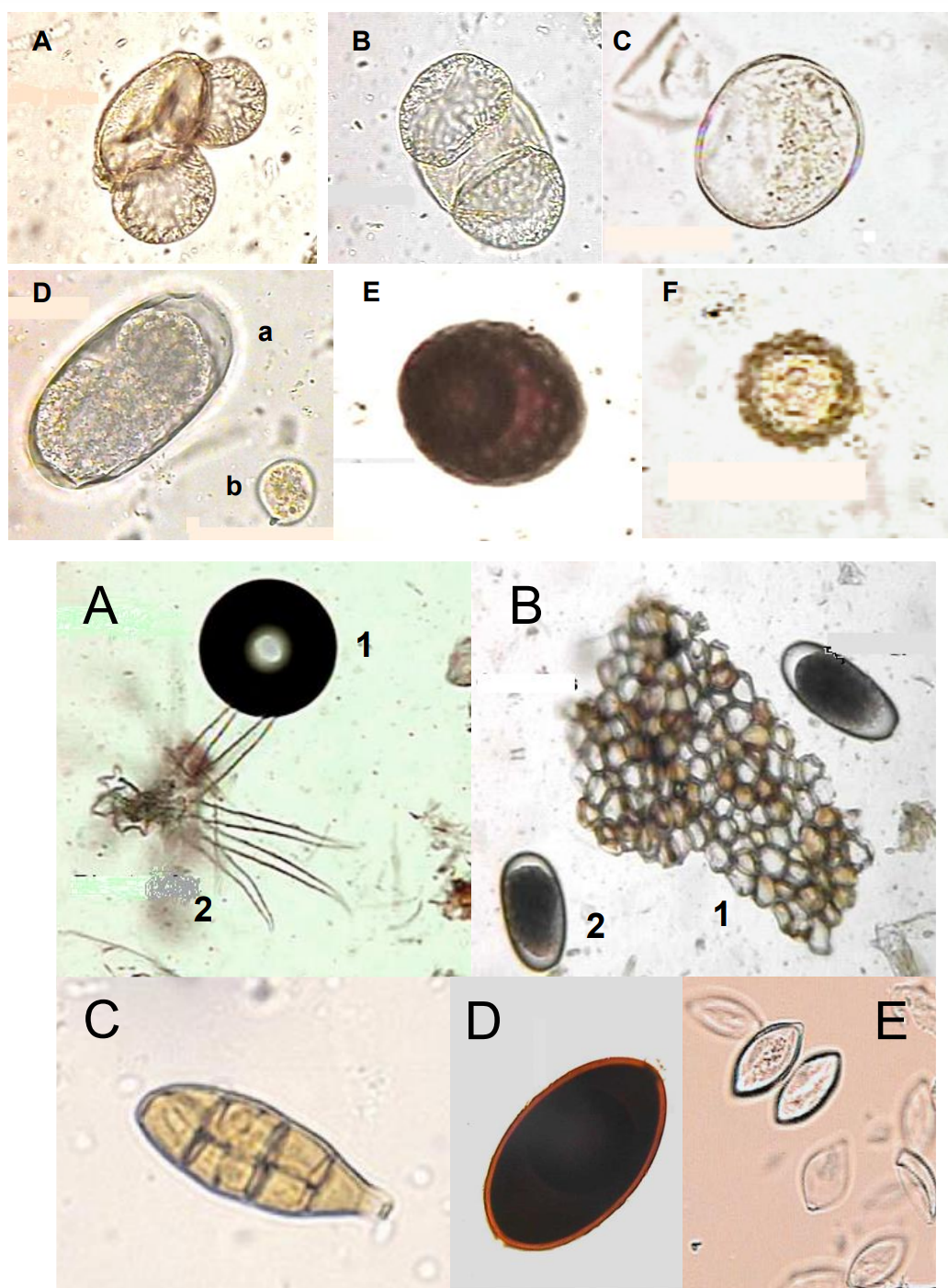
How do we describe the morphology of parasitic objects/eggs?
Base it upon 5 basic criteria:
size
shape (ex. oval, symmetrical, oval-symmetrical, lemon-shaped)
structure of shells
trematode - 2 thin shells
cestode - pseudophylidia (two thin shells with operculum at one pole and a small button on the other), and cyclophyllidea (three layers).
nematode - three layers, protein layer, chitin layer and vitelline layer internally.
acantocephalan - three layers
internal structure (unembryonated vs. embryonated)
trematode eggs: unembryonated ex. fasciola. Embryonated ex. paramphistomum.
cestode eggs: pseyudophylidia unembryonated, cyclophyllidea embryonated.
nematode eggs:
unembryonated
embryonated
Acantocephalan eggs: embryoanted (acantor)
colour (brown, reddish-brown, dark brown, light brown/grey, yellow, transparent)
The direct smear of faeces is done by? Flotation method is?
Direct smear - suitable for rapid examination. Small amount of feces is placesd on a slide, mixed with drop of water, spread out covered with a slip and examined directly.
Flotation method: depending on mixing of fecal sample with liquid whose specific gravity is greater than the parasite. - using “flotation fluids”.
sample preparation: mixing faeces with water → semisolid suspension
filter the suspension by tea strainer
centrifugation
add flotation solution, filling 1/3
resuspend the sediment by metallic stick
centrifugation
microscopy: take parasitological loop to take 3 drops from surface of tube, put on glass slide.
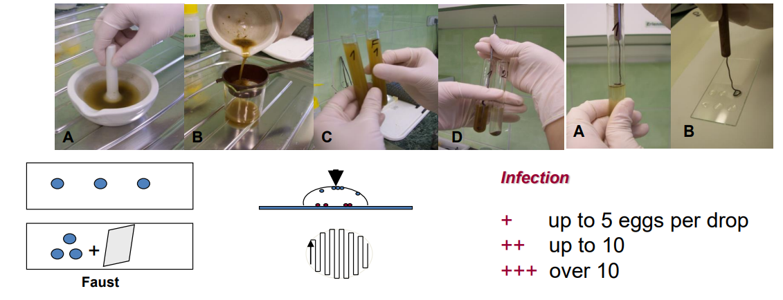
What are the 3 flotation fluids to know? Which animals are they used for?
FAUST (33% ZnSO4): gravity 1.18 – used for protozoan of carnivores
KOZÁK-MÁGRA: gravity 1.24 – used for cestode and nematode eggs of carnivores, and feaces of poultry
BREZA: gravity 1.3 – used for cestode and nematode eggs of ruminants, horses, pigs and rodents
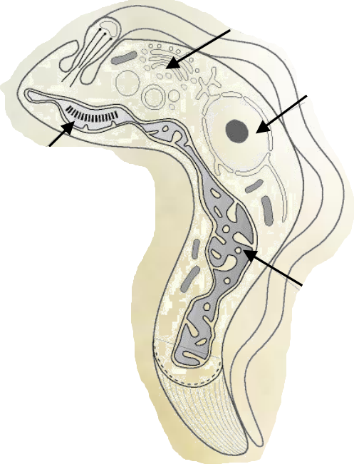
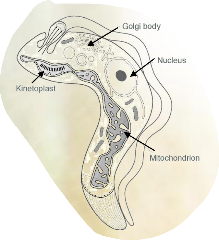
Which parasite is this?
Protozoa - Genus: Trypanosoma
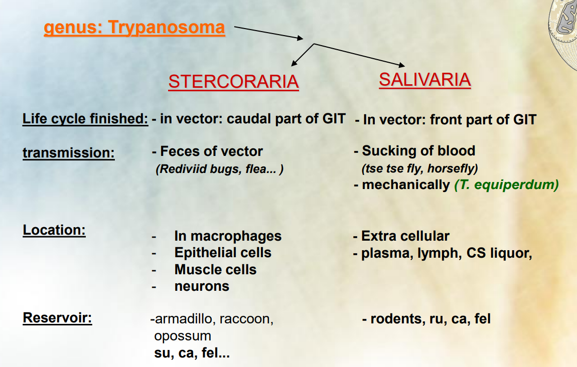
The difference of the subdivisions Stercoraria and Salivaria of the genus Trypanosoma? What are an example of species under them?
Stercoraria: species transmitted by the feces of their vector, typically Rediviid bug. Stercoraria species, such as trypanosoma cruzi, are associated with Chagas disease and are transmitted when a vector defecates near a bite wound. Life cycle done in vector, caudal part of GIT.
Reservoir: pig, dog and cat
Location: extra cellular, plasma, lymph and CS liquor.
Salivaria: species transmitted through the saliva of their vector, such as the tsetse fly. Salivary species like trypanosoma brucei, cause sleeping sickness and are transmitted by the bite of an infected tsetse fly. Life cycle done in vector, front part of GIT.
Though, T. equiperdum transmits mechanically.
Reservoir: Rodents, ru, dog and cat
Located in macrophages, epithelial cells, muscle cells and neurons
Trypanosoma cruzi cause?
Chagas disease
Latin america, in man, domestic and wild animals
Which trypanosoma can cause chronic sleeping sickness?
Trypanosma brucei - gambiense
Trypanosomiasis - in africa, man monkey, dog, pig/Glossina spp.
Which trypanosoma can cause acute sleeping sickness?
Trypanosoma brucei - rhodensiense
Trypanosomiasis - Africa, man, animal/Glossina spp.
Which trypanosoma does not require a vector?
Trypanosoma equiperdum
What are the vectors of trypanosoma evansi?
Vectors: Tabanus spp. (horse flies), stomoxys spp. (stable flies)
Vampire bats, SURRA eating on blood are also vectors, non-cyclic.
occurs in horses, ruminants and dogs
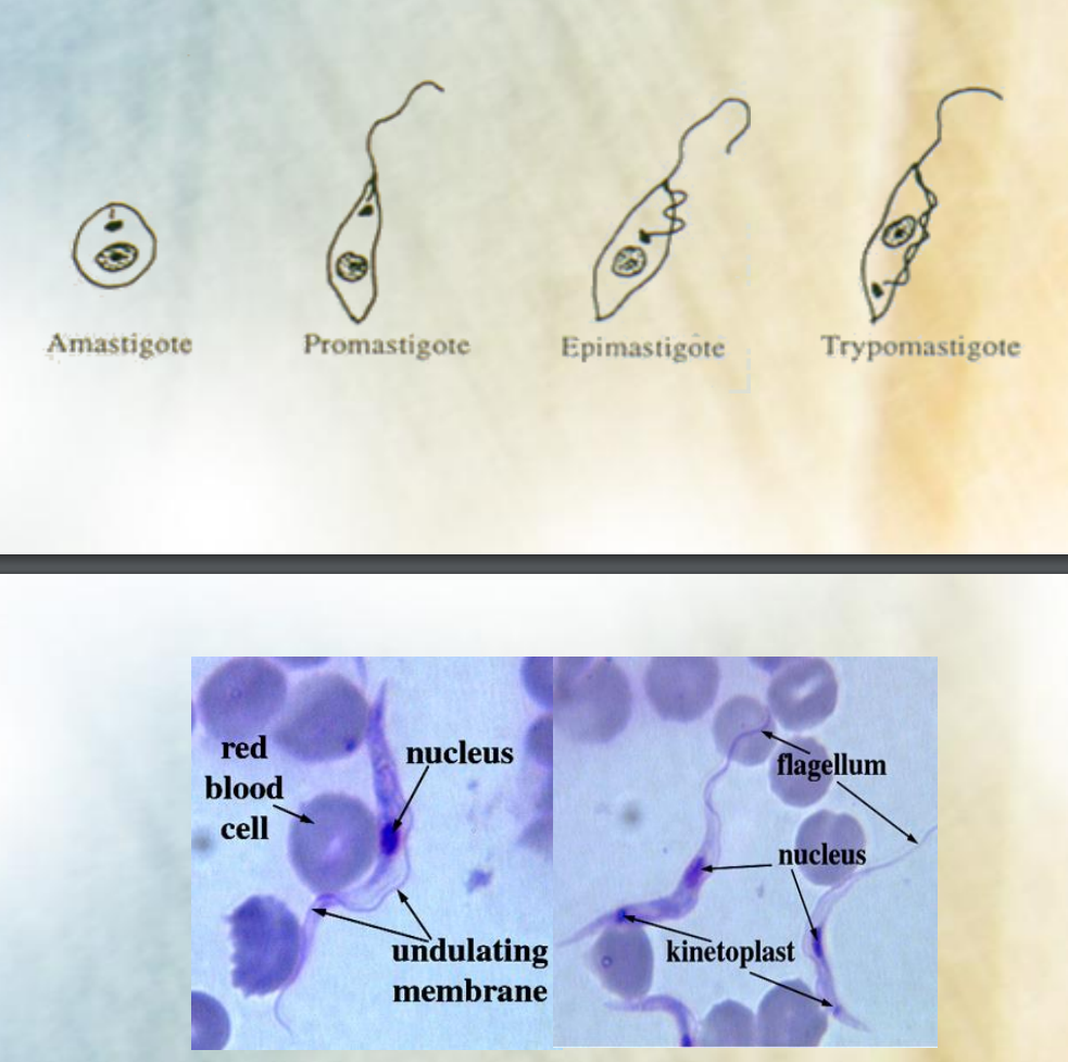
What are the 4 life cycle stages of parasite (Trypanosoma)?
AMASTIGOTE - oval, no flagellum
PROMASTIGOTE - elongated, kinetoplast in front of nucleus, flagellum
EPIMASTIGOTE - undulating membrane formed
TRYPOMASTIGOTE - kinetoplast goes behind nucleus, still has undulating membrane and flagellum
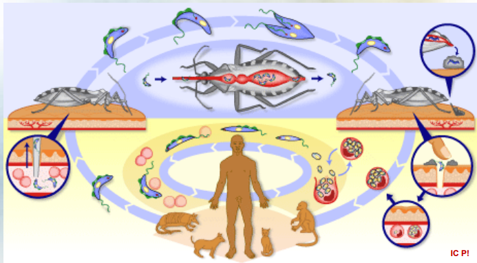
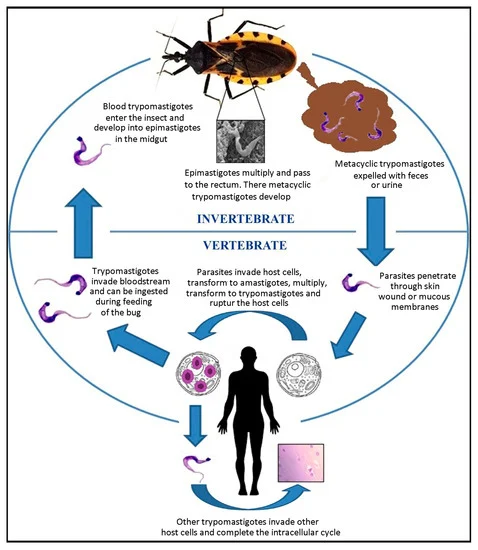
What parasite has this type of lifecycle?
Subdivision of trypanosoma: STERCORARIA (T. Cruzi)
Trypomastigotes: The cycle starts with trypomastigotes present in the blood of an infected host. These are the infectious form of the parasite.
Reduviid bugs: The Trypomastigotes are ingested by Reduviid bugs (the vector) during a blood meal.
Epimastigotes: Inside the bug, the Trypomastigotes transform into Epimastigotes and reproduce through binary fission over 8-10 days.
Trypomastigotes in feces: The Epimastigotes eventually transform back into Trypomastigotes and are excreted in the bug's feces.
Skin lesion: The Trypomastigotes can enter a new host through a skin, wound, during scratching of the bite site.
invasion of cells: Once inside the host, Trypomastigotes invade host cells. (macrophages)
Amastigotes: Inside the macrophages, they transform into Amastigotes, where they multiply.
Lymph nodes (lnn.): From the macrophages, the Amastigotes can spread to the lymph nodes.
Blood: Eventually, they return to the bloodstream in their Trypomastigote form, continuing the cycle → epimastigote → trypomastigote etc.
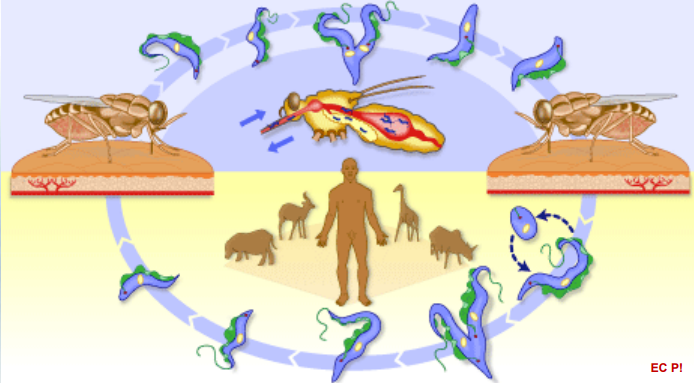
To which parasite does this lifecycle belong to?
Subdivision of Trypanosoma: SALIVARIA (T.bruzei)
Trypomastigotes in Blood of infectected host
Tse Tse Fly: The tse tse fly (the vector) ingests blood during a meal, getting the trypomastigotes.
Binary Fission in the Fly: Inside the tse tse fly, the Trypomastigotes undergo binary fission, multiplying in number.
Salivary Gland: The parasites migrate to the salivary glands of the tse tse fly.
Epimastigotes: In the salivary glands, they transform into Epimastigotes. This form continues to reproduce through binary fission.
Trypomastigotes Formation: They revert to the Trypomastigote form, ready to infect a new host.
Inoculation to the Host: During a blood meal, the tse tse fly inoculates/injects Trypomastigotes into the host's bloodstream.
Binary Fission in Blood and CNS: Once in the host, Trypomastigotes multiply through binary fission in the bloodstream and can invade the central nervous system (CNS).
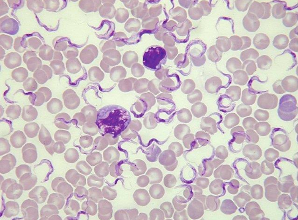
which parasite is this?
Trypanosoma
Characterize the Genus Leishmania.
intracellular parasite in macrophages
vector: sandflies
damage of CNS, hyperplasia of cells, skin, mucosa + internal organ damage
can be skin form (L. tropica minor et major) or visceral form (L. donovani)
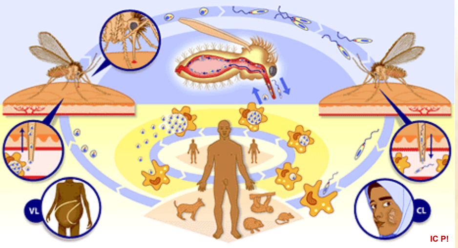
Which parasite does this life cycle belong to?
Leishmania.
complex life cycle, involving two main hosts, the sandfly vector and the mammalian host (ex. humans)
begins when sandfly vector feeds on the blood of an infected mammal, it will ingest the amastigotes from it.
These amastigotes will transform into promastigotes in the midgut of sandfly, and multiply by binary fission. These will then migrate to the sandfly`s salivary glands (pharynx of vector)
During another blood meal, the sandfly will inoculate/inject the promastigotes into the skin of a new mammalian host, where they invade macrophages and transform back into amastigotes.
Inside the macrophages, they multiply by binary fission → rupture of the affected cell and subsequent infection of new macrophages.
The infection can cause agglomeration → spread to visceral organs, skin and mucosa.
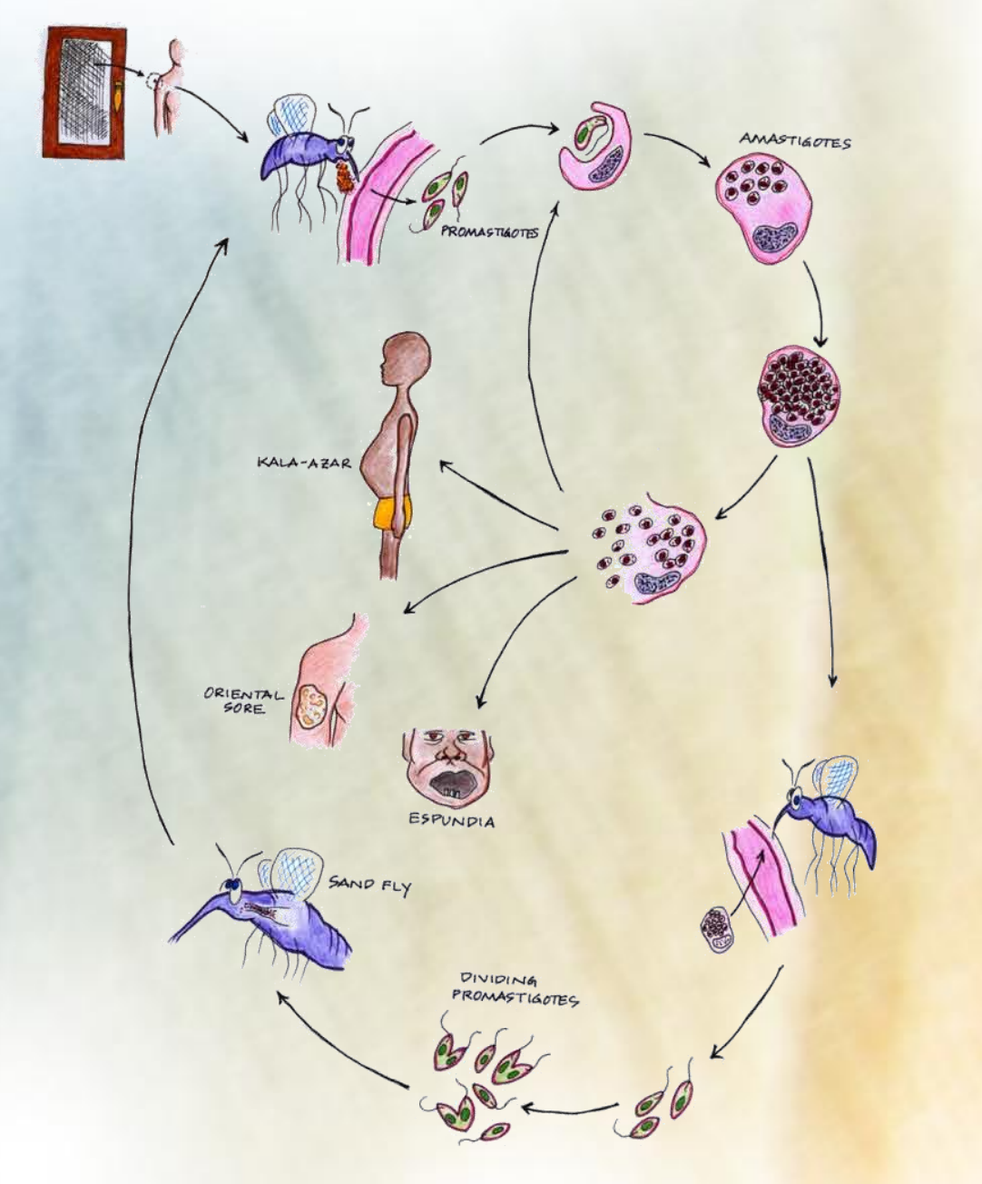
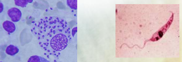
These shows the stages?
Amastigote stage and promastigote stage (Leishmania)
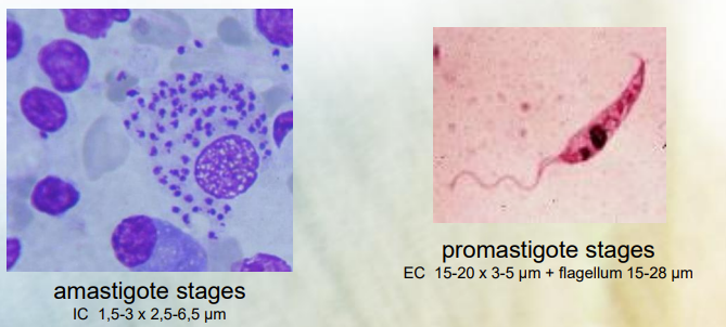
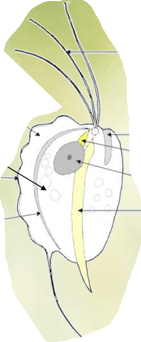
Which parasite is this?
Protozoa - Trichomonas
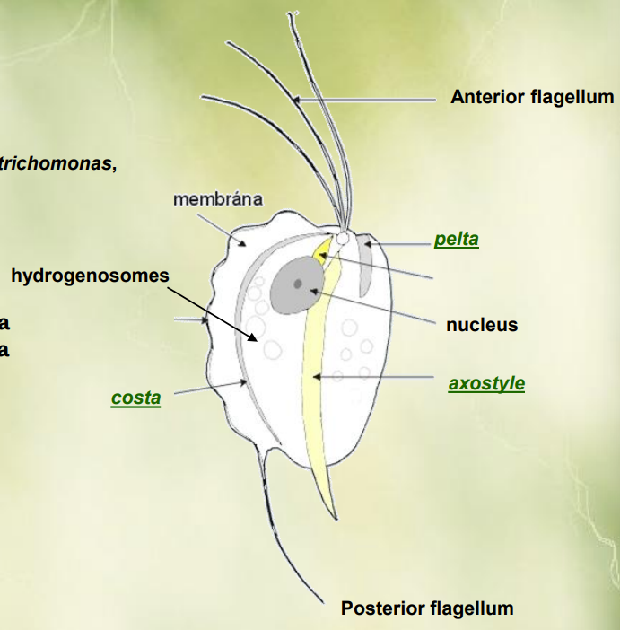
How many flagella do the trichomonas have?
4 anterior flagella, and 1 posterior
While pentatrichomonas (Pt) → 5 anterior flagella
Tritrichomonas (Tt) - 3 anterior flagella
Ditrichomonas (Dt) - 2 anterior flagella
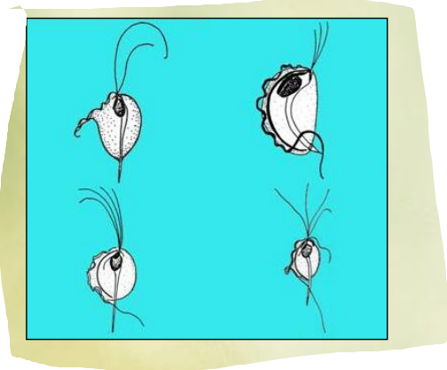
Trichomonas reprodude by?
Binary fission
Asexual reproductive process, one organism divides into 2 identical daughter cells. The process begins by replication of the organism`s genetic material, followed by the elongation of the cell. After that, the cell membrane pinches inwards, dividing the cytoplasm → 2 separate cells, each a copy of the original DNA.

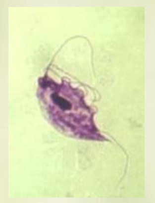
Describe the tritrichomonas foetus.
Protozoan parasite, affecting cattle
flagellated, with 3 anterior flagella and 1 posterior flageullum
causes reproductive issues such as abortion.
Drop shape, 20×30um, short axostyl. Reproduction by binary fission, rarely makes pseudocysts. Transmission by intercourse or artificial insemination.
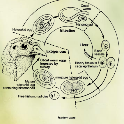
This is the life cycle of?
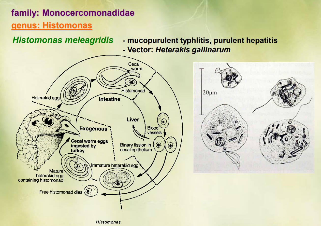
Genus Histomonas of the family monocercomonadidae.
Histomonas meleagridis
cycle begins with heterakid eggs containing histomonas, which are ingested by turkeys.
Inside the turkey`s intestine, the eggs hatch, releasing immature heterakid worms (cecal worms)
the histomonas enter the cecal epithelium, multiplying by binary fission
as they multiply, the invade blood vessels → spreads
histomonads continue to grow, eventually leading to death of free histomonads.
mature heterakid eggs containing the histomonas can now be released in the turkey`s feces.
These cecal worm eggs can then be ingested by another turkey → continuation of cycle.
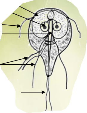
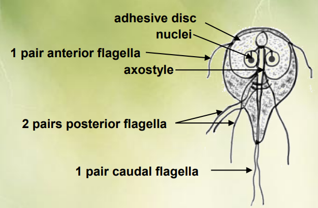
This is of?
Genus: Giardia of family Hexamitidae
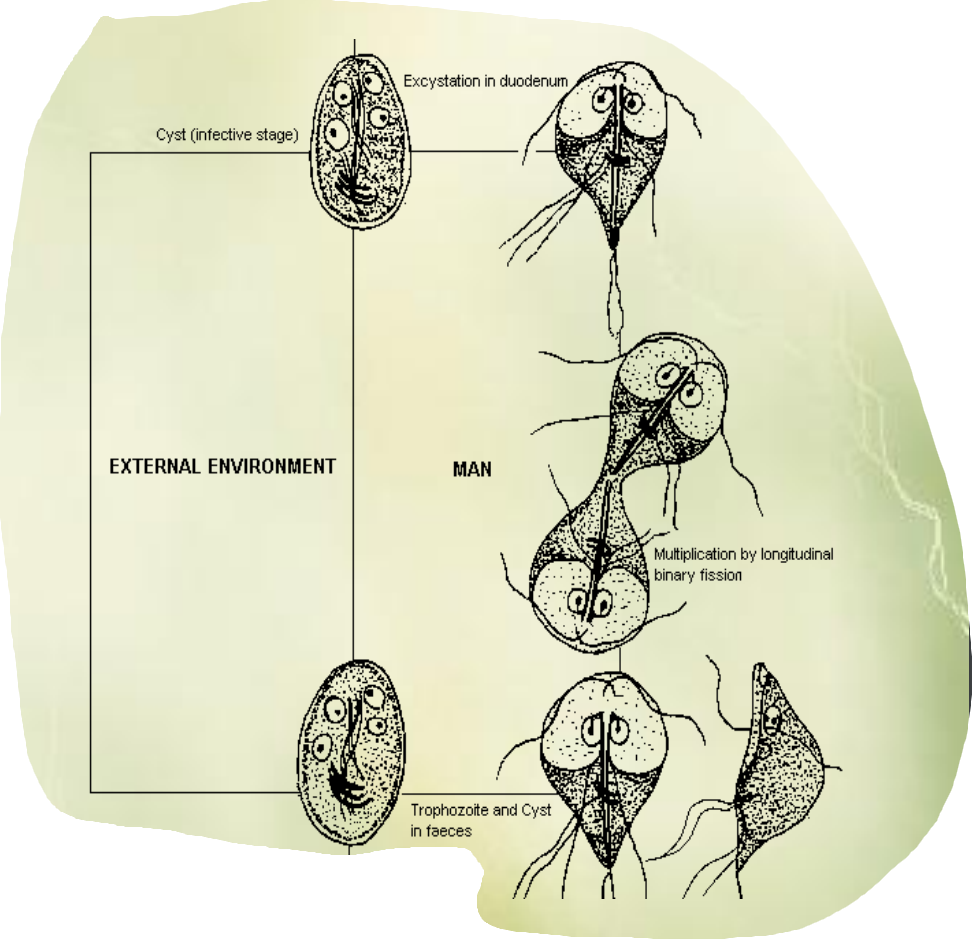
This is the life cycle of?
Life cycle of Giardia Intestinalis
ingestion: cysts are ingested by the host through contaminated water or food
excystation: in the intestines, cysts are exposed to stomach acid → activating the release of trophozoites.
trophozoite stage: multiplication by binary fission, attaching to intestinal wall, GIT symptoms.
encystation: trophozoites transform back into cysts
excretion: cysts are excreted in the host`s feces, where they can contaminate water or soil, completing the life cycle.
Way of transmission: water, food (with cysts)
occurrence: spring autumn.
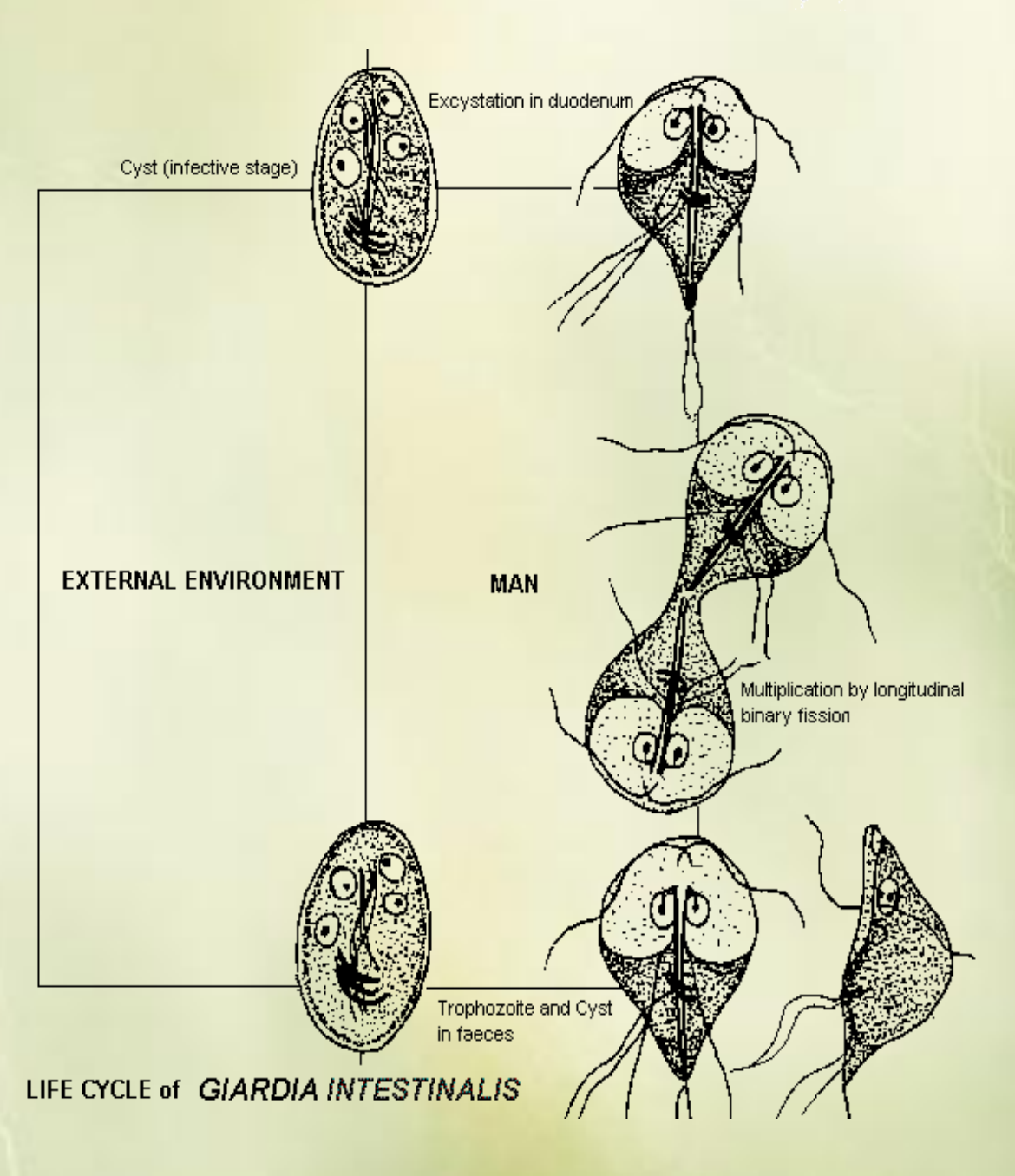
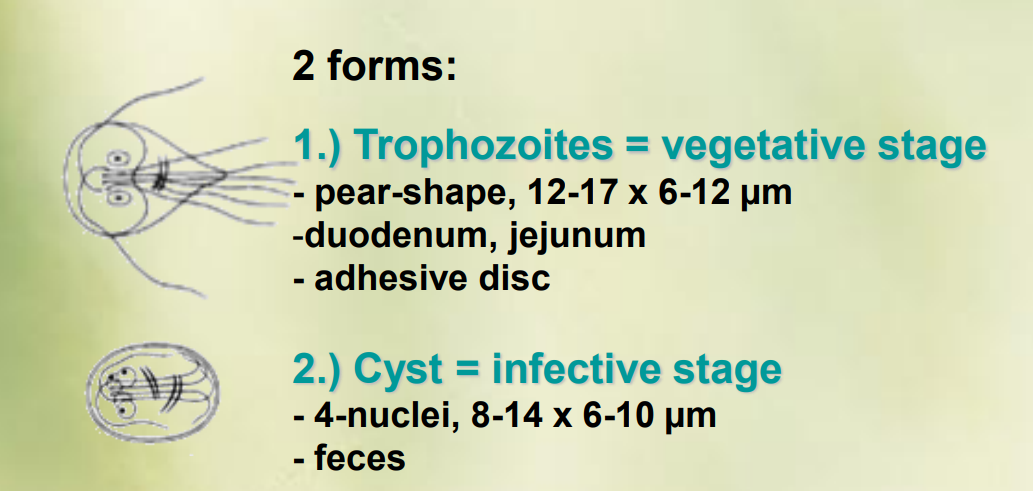
The 2 forms of giardia is?
Trophozoites = vegetative stage
pear-shape, 12-17-6-12 um
duodenum, jejunum
adhesive disc
cyst = infective stage
4-nuclei, 8-14 × 6-10 um
feces
The two forms are present in species of giardia, like giardia duodenalis, intestinalis, agilis and muris.

This is?
Apical complex → apicomplexa (syn. sporozoa)
apical complex: structure in certain protozoa, specifically in the group apicomplexa that consist of specialized organelles located at the anterior end of the organism. It helps in the invasion of host cells.
Apicomplexa, also known as sporozoa, is a subhylum of Alveolata (phylum) of protozoa. Characterized by the presence of apical complex. Members of this group include for ex. Plasmodium (causing malaria) and Toxoplasma.
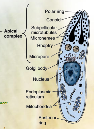
The apical complex consist of which 5 things?
Polar ring
conoid
preconoidal rings
subpelicular microtubules
Micronemes
Rhoptry
Dense granules

this is picture of?
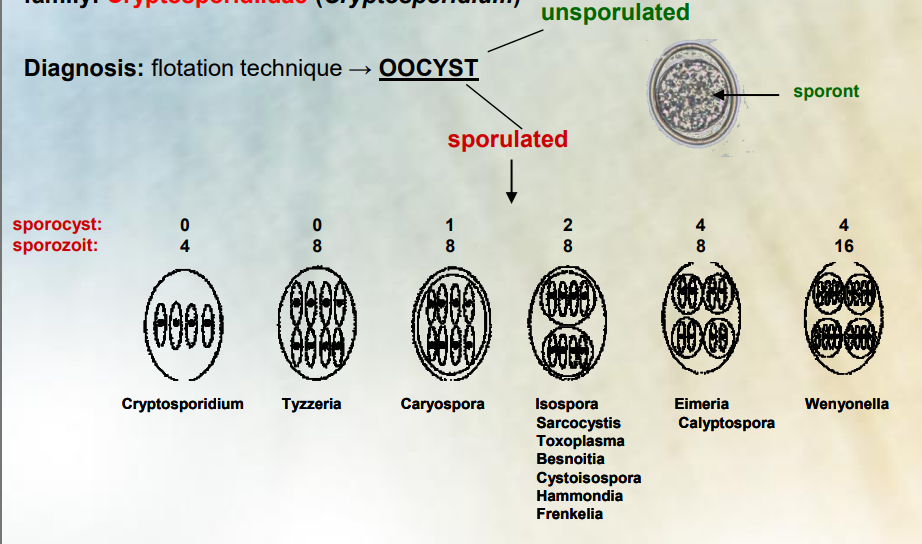
Sporylated oocysts examples.
An oocyst can be categorized as unsporulated (immature and not yet capable of reproduction) and as sporulated (mature and capable of infection)
These are drawings of the sporulated oocyst in the different species (orders) like:
cryptosporidum having 4 sporozoits but no sporozyst.
Tyzzeria (of family Emeriidae) having 8 sporozits, 0 sporozyst.
Caryospora having 8 sporozoits, 1 sporozyst.
Isospora, sarcocystic, toxoplasma, besnoita, cystoisopsora etc. having 8 sporozoits, 2 sporozysts.
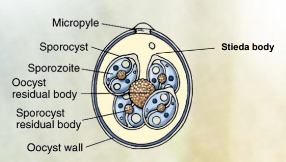
Eimeria, calyptospora having 8 sporozoits, 4 sporozysts.
The diagnosis by flotation technique is done by identifying an oocyst (cystic stage of protozoa), as unsporulated or sporulated.
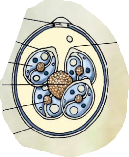
Eimeria is?
Genus within apicomplexa. Order: Eimerida, Family: Eimeriidae
Eimeria: 4 sporozysts with 2 sporozoits in each.
causes coccidiosis
host specificity
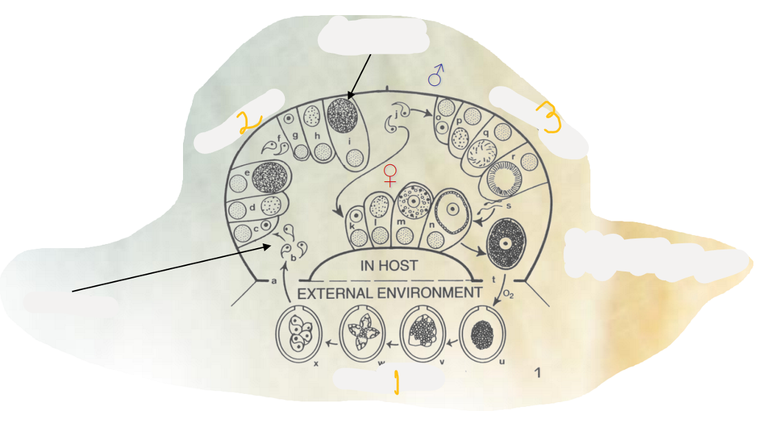
The 3 phases of monoxenous life cycle in general of Eimeria?
exogenous sporogony: formation of sporozoits within the oocyst from single organism outside host environment.
endogenous merogony: Once ingested by new host, sporulated oocyst release sporozoites that invade the cells, inside these, the sporozoites undergo asexual replication → production of merozoites.
endogenous gamegony: production of gametes resulting in sexual reproduction within the host. - some merozoites differentiate into microgametes and macrogametes.
Monoxenous: life cycle in one host - Eimeria.
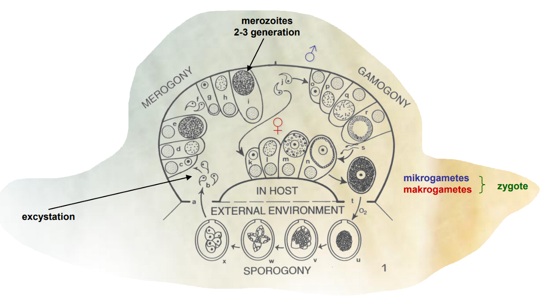
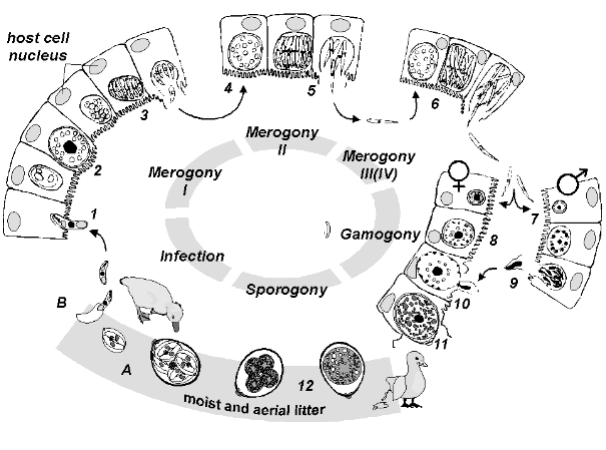
This is the life cycle of?
Eimeria
Exogenous sporogony: Formation of sporozoites within the oocyst from a single organism outside the host environment.
Infection: Sporulated oocysts are ingested by a new host, releasing sporozoites.
Endogenous merogony I: Sporozoites invade host cells and undergo asexual replication to produce merozoites.
Endogenous merogony II: More rounds of asexual replication occur, producing additional merozoites.
Endogenous merogony III: The final round of asexual replication occurs, leading to the production of many merozoites ready to infect new cells.
Endogenous gamogony: Some merozoites differentiate into microgametes and macrogametes, resulting in sexual reproduction within the host.
Eimeria truncata is in which species?
Geese - in the kidney
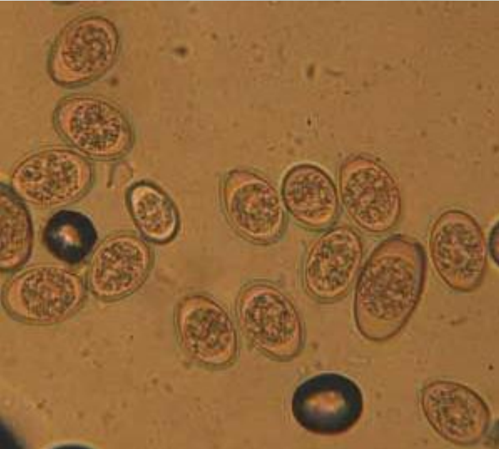
this is?
Unsporulated oocysts of Eimeria spp.

This is what? (4 circles within a circle)
Cryptosporidium (genus) of family: crytosporidiidae.
species ex. C. parvum (intestine), C. muris (intestine), C. baylei, C. meleagridis
no host specificity, no microspyle and sporocyst, oocyst contains 4 FREE sporozoites.
Endogenous sporulation.
Microgametes have no flagellum.
Cryptosporidium meleagridis and C. baylei are in which species?
of birds, mainly in respiratory tract.
The size of cryptospodium is?
5-7 micrometers (um)
Where does these take place?
sporogony
merogony/schizogony
microgametogony
macrogametogony
Sporogony takes place outside the host, in the environment, where oocysts mature and develop into sporozoites.
Merogony or schizogony takes place within the host's cells, where asexual reproduction occurs and merozoites are produced.
Microgametogony takes place within the host, typically in the intestinal lumen, where male gametes (microgametes) are produced.
Macrogametogony takes place within the host, usually in the intestinal lumen, where female gametes (macrogametes) are formed.
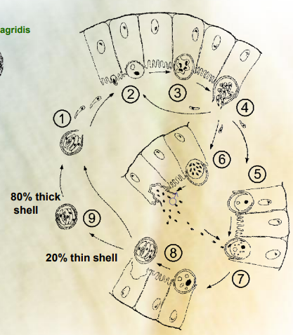
This is the life cycle of?
Cryptosporidium (genus) - of family cryptosporidiidae.
Ingestion: The cycle begins when a host ingests oocysts containing sporozoites from contaminated water or food.
Excystation: Inside the gastrointestinal tract, the oocysts are exposed to stomach acids, activating them to release sporozoites.
Sporozoite: The released sporozoites then invade the epithelial cells of the intestines.
Trophozoite formation: Within these cells, the sporozoites develop into trophozoites, which are the active feeding forms.
Merogony: The trophozoites then undergo asexual reproduction known as merogony, resulting in the production of merozoites.
Merozoite release: These merozoites are released back into the intestinal lumen, where they can infect new epithelial cells or develop into gametes.
Gamogony: Some merozoites differentiate into male and female gametes, ready for sexual reproduction.
Fertilization: Male gametes (microgametes) fertilize female gametes (macrogametes), forming a zygote.
Oocyst formation: The zygote develops into an oocyst, which can either sporulate inside the host to become infectious or be excreted in the feces of the host, thus reinitiating the cycle when ingested by another host.
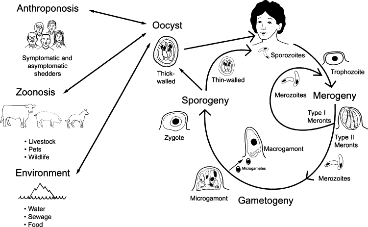
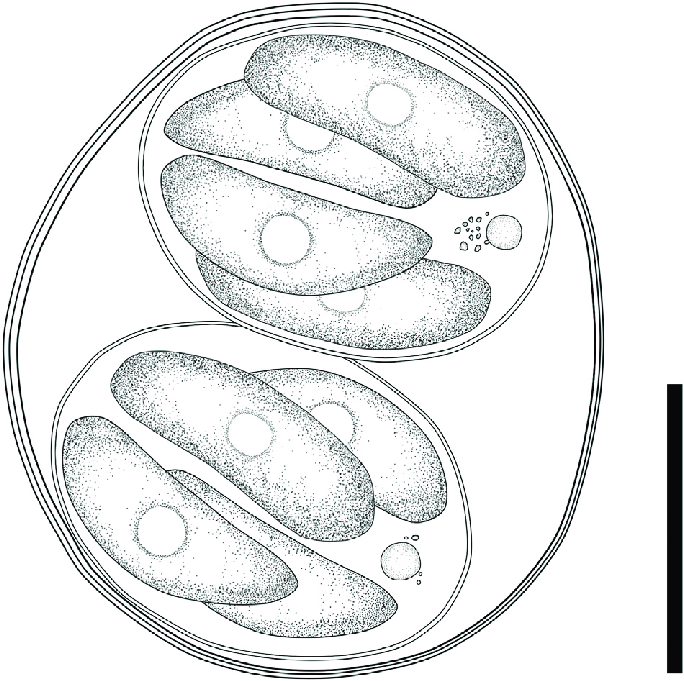
which species of parasite is this?
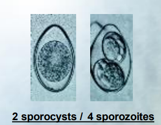
Cystoisosporosis - containing 2 sporocysts with 4 sporozoites inside each.
n pigs: Cystoisospora suis, C. almaataenis, C. neyrai
in dogs and cats: C. felis, C. rivolta, canis
Sporogony - exogenous, Merogony + gamegony happening in the epithelial cells of small intestine.
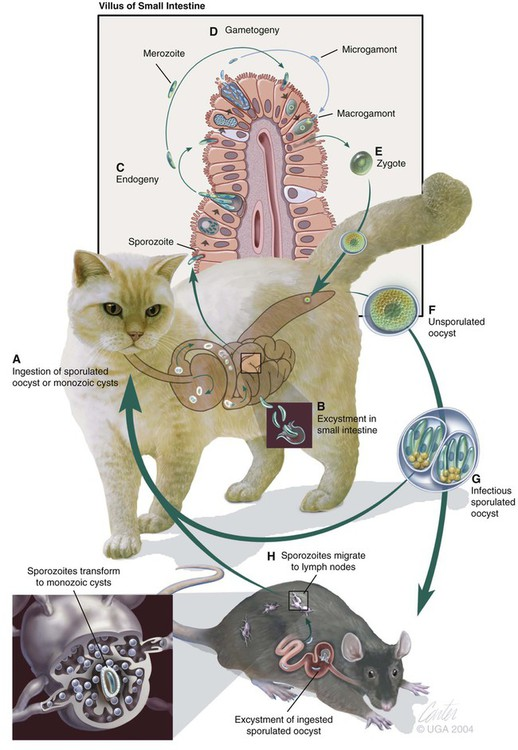
which parasite does this life cycle belong to?
Cystoisospora (genus), of family sarcocystidae of order Eimerida.
intermediate host: dormozoite/hypnozoite, DH: extraintestinal stage, Endogeny = schizogony/merogony. IP/PH: dormozoite/hypnozoite
Life cycle of Isospora felis, which is typical of the Isospora spp.
A) Ingestion: The host ingests sporulated oocysts or monozoic tissue cysts from contaminated food or water.
B) Excystation: In the gastrointestinal tract, the oocysts release sporozoites.
C) Sporozoite Stage: Sporozoites invade the epithelial cells of the intestine. Asexual reproduction happens (endodyogony - replicating once to make 2 daughter cells)
D) Merogony (Schizogony): Inside host cells, sporozoites replicate asexually in several rounds to produce multiple merozoites.
E) Gametogony: Some merozoites differentiate into male and female gametes, leading to sexual reproduction in the intestine. With the formation of a zygote.
F) Oocyst Formation: The fertilized gametes develop into unsporulated oocysts that are shed in feces.
G) Sporulation: The oocyst matures into an infectious sporulated oocyst, which can be ingested by the definitive or intermediate host.
H) Intermediate Host: excysted sporozoites migrate to tissues and form cysts.
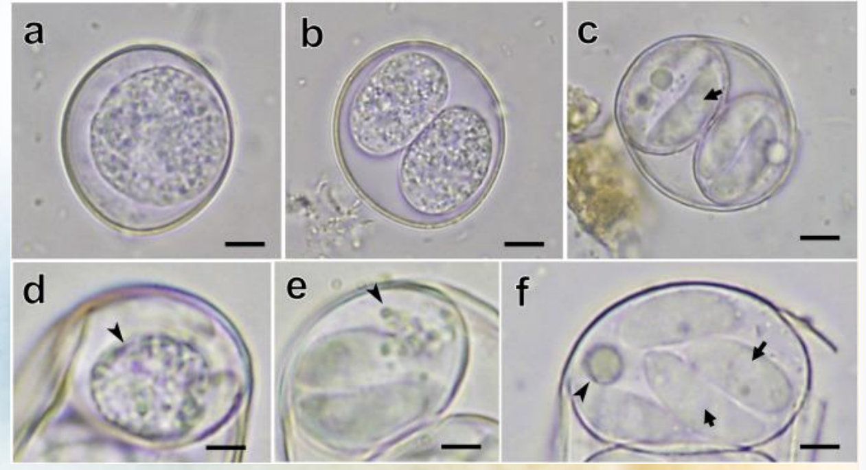
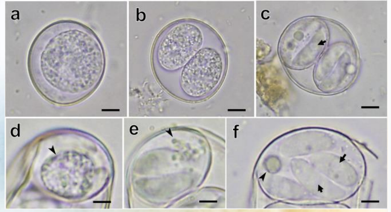
which species of parasite is this?
Photographs of Cystoisospora oocysts detected from Asian small-clawed otters
a) immature oocyst with 1 sporoblast
b) immature oocyst with 2 sporoblasts
c) Mature oocyst containing 2 sporocysts, each with 4 club-shaped sporozoites. Sporozoites contained rounded retractile vacuoles (arrow)
d-f) sporozoites containing sporozyst residuum, which was composed of numerous small granules (d and e) or a rounded granule (f)
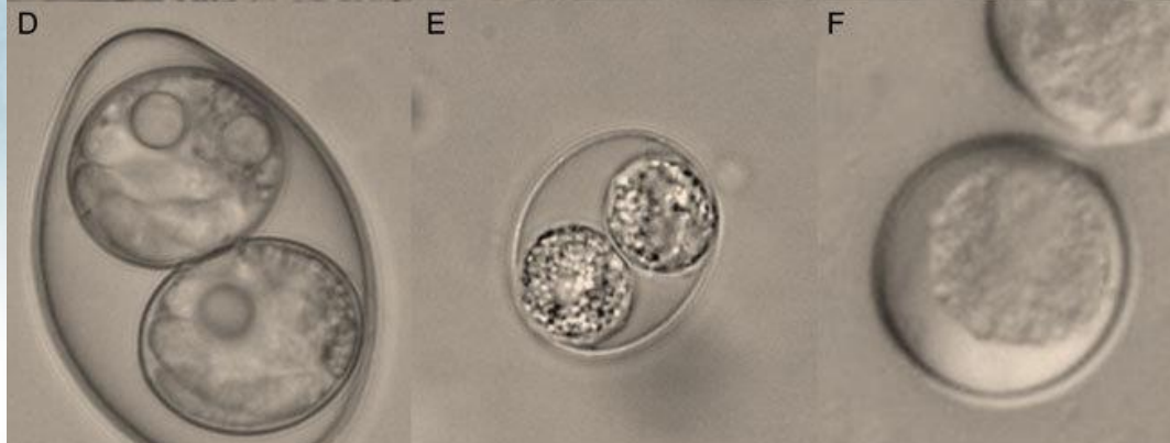
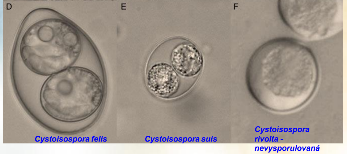
These pictures are of?
D. Cystoisospora felis
E. Cystoisospora suis
F. Cystoisospora rivolta
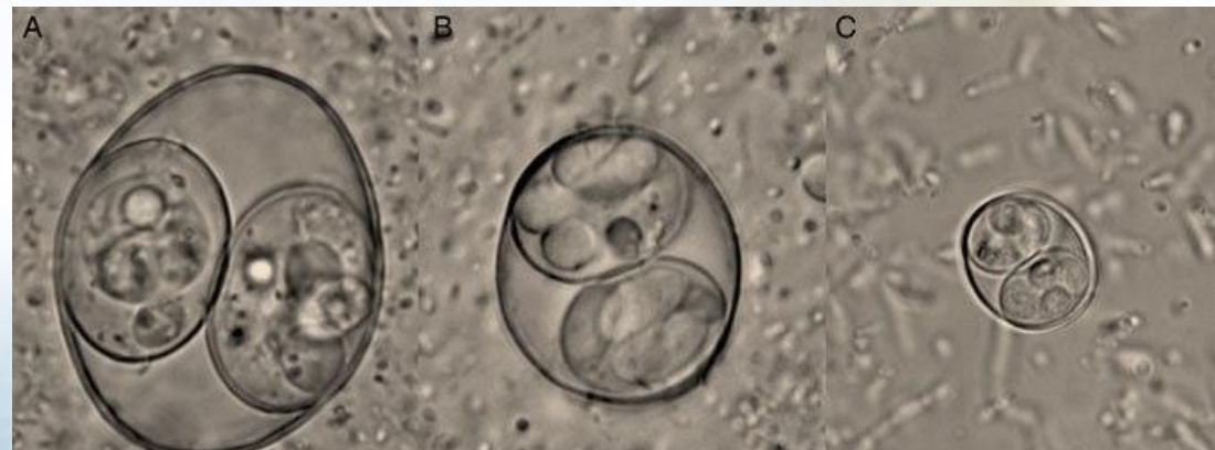
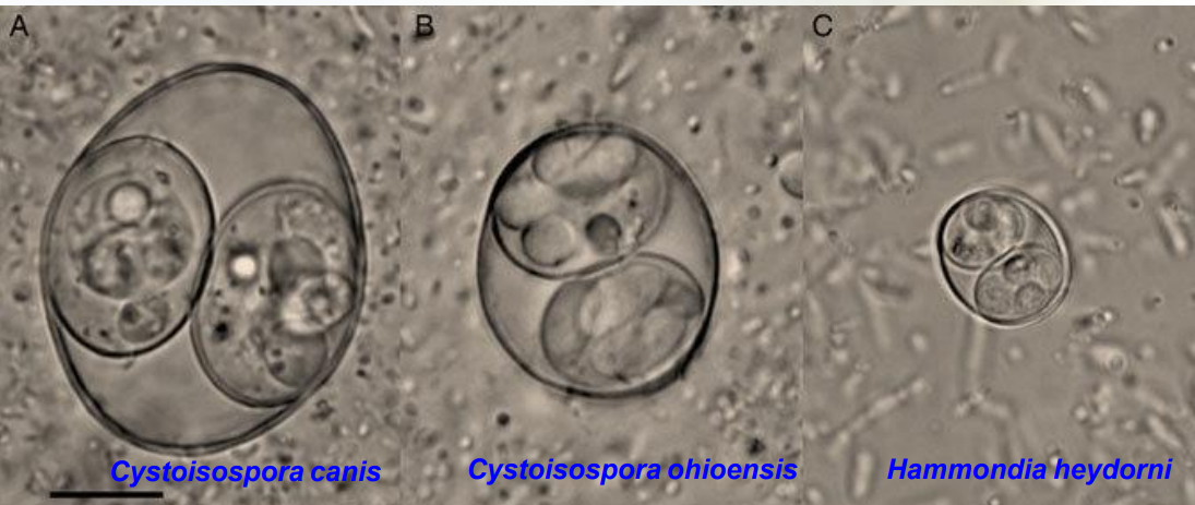
these are pictures of?
A. Cystoisospora canis (definitive host - dog, with 2 sporozyst and 4 sporozoits in each).
B. cystoisospora ohioensis
C. Hammondia Heydorni
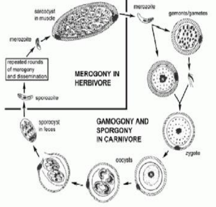
This is the life cycle of?
Genus: sarcocystis, of family sarcocystidae, of order Eimerida.
The life cycle of Sarcocystis species is heteroxenous, meaning it involves different hosts for its different life stages.
Definitive Host: such as carnivores including dogs and humans, harbors the sexual stages of the parasite, where gamogony (sexual reproduction) and sporogony (formation of oocysts) occur in the small intestine.
Intermediate Hosts: The intermediate hosts (which include herbivores, omnivores, and birds) play a crucial role in the life cycle as the parasite undergoes asexual reproduction, known as merogony.
In these hosts, the sporozoites that are ingested from oocysts excyst (are released) in the small intestine and invade the cells. They replicate asexually, generating meronts (by merogony) in endothelial of blood capillaries and after 3 rounds, subsequently forming cysts filled with bradyzoites, the slow-growing stage of the parasite → sarcocyst in muscle.
Transmission: When a carnivore consumes the infected tissues of the intermediate host, it can become infected with Sarcocystis again, thus continuing the life cycle.
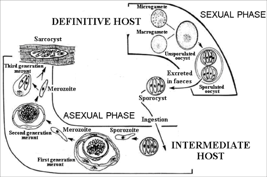
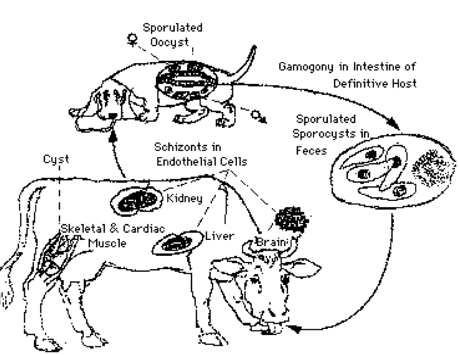
this is the life cycle of?
life cycle of S. Bovicans
Definitive Host (Dog)
Ingestion: The dog ingests tissue cysts containing bradyzoites from infected intermediate hosts (such as cattle).
Gametogony: In the dog's intestines, the bradyzoites invade epithelial cells and develop into gametes.
Sporulation: Oocysts are formed, which then sporulate within the dog's intestine.
Excretion: Sporulated oocysts are excreted in the feces.
Intermediate Host (Cow)
Ingestion: The cow ingests sporulated oocysts from contaminated food or water.
Sporozoite Release: The oocysts release sporozoites in the cow's intestines, which then invade endothelial cells.
Schizogony/merogony: Sporozoites undergo multiple rounds of nuclear division to form schizonts/meronts in the cow's tissues.
Cyst Formation: Schizonts/meronts release merozoites, which will develop into cysts containing bradyzoites within the muscles and other tissues of the cow.
The cycle continues when the definitive host ingests these cysts, and the process begins anew.
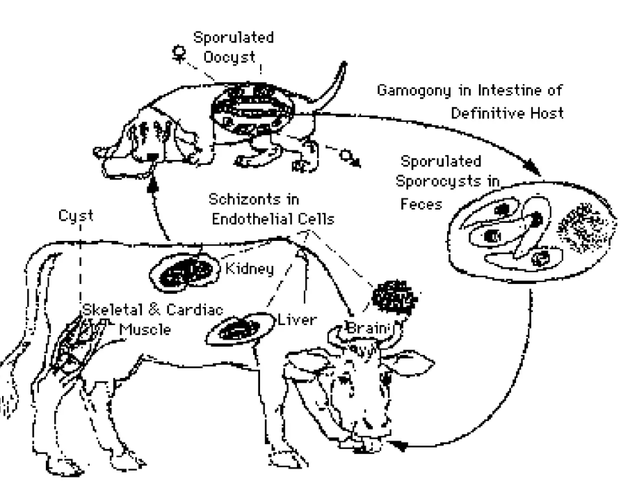
Endogenous sporulation/sporogony is the process of?
stage in the life cycle of some protozoan parasites, during this, sporozoites (infective forms) are made inside an oocyst within the host.
process: parasite infects the host, making oocysts within its tissues, sporogony happens where within these oocysts, the sporozoites form through multiple rounds of division → release of sporozoites.
host specificity of Eimeria spp., cryptosporidium spp., isospora spp., sarcocystis spp. and toxoplasma gondii?
categorization of parasites based on their host specificity:
Eimeria spp. parasites with sporogony, indicating they complete their life cycle within a single host.
Cryptosporidium spp. is similar to eimeria, completing life cycle within a single host. (sporogony endogenous)
isospora spp. requires a paratenic host to complete life cycle.
sarcosystis spp. involving multiple hosts. (sporogony endogenous)
toxoplasma gondii. involving multiple hosts.
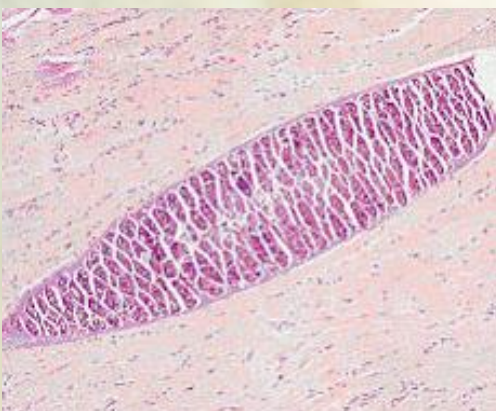
This is picture of?
sarcocystis in the muscle. (of intermediate hosts, like cattle)
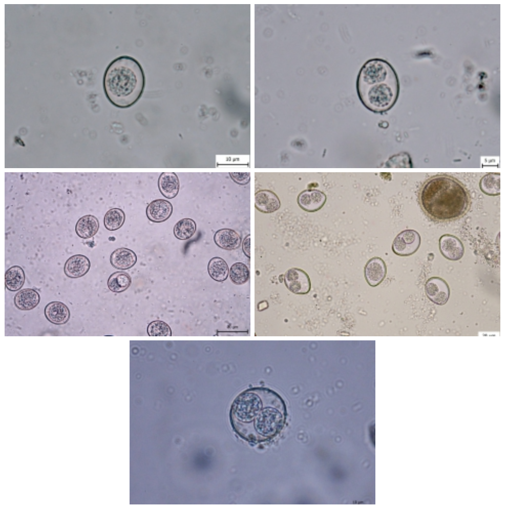
these are pictures of?
cystoisospora spp.
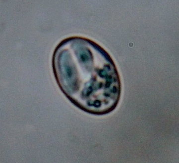
this is picture of?
sarcocystis spp.
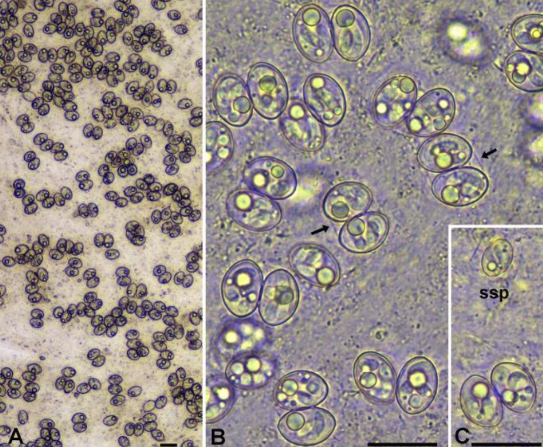
this is picture of?
Sporulated thin-walled oocysts of Sarcocystis halieti and Sarcocystis lari
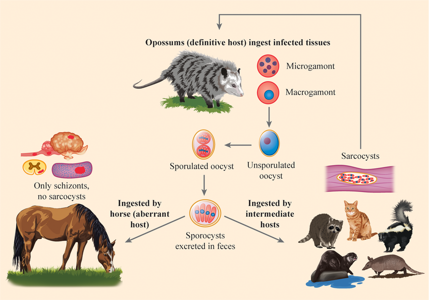
This is the life cycle of?
Life Cycle of Sarcocystis neurona.
Definitive Host (DH):
The opossum ingests tissue cysts containing bradyzoites from infected intermediate hosts.
nside the opossum, the parasite undergoes sexual reproduction, forming unsporulated oocysts.
Sporulation:
The unsporulated oocysts develop into sporulated oocysts within the opossum's intestines.
Excretion:
The sporulated oocysts are excreted in the feces of the opossum.
Intermediate Hosts (IH):
such as raccoons, armadillos, skunks, and felines ingest the sporulated oocysts from the environment.
Within the intermediate hosts, the parasite forms sarcocysts in the muscle tissues.
Aberrant Host (EQ - Horse):
Horses ingest sporocysts from the environment. In horses, only schizonts form, and no sarcocysts are produced.
Disease in Horses: The schizonts cause neurological disease in horses, known as Equine Protozoal Myeloencephalitis (EPM).
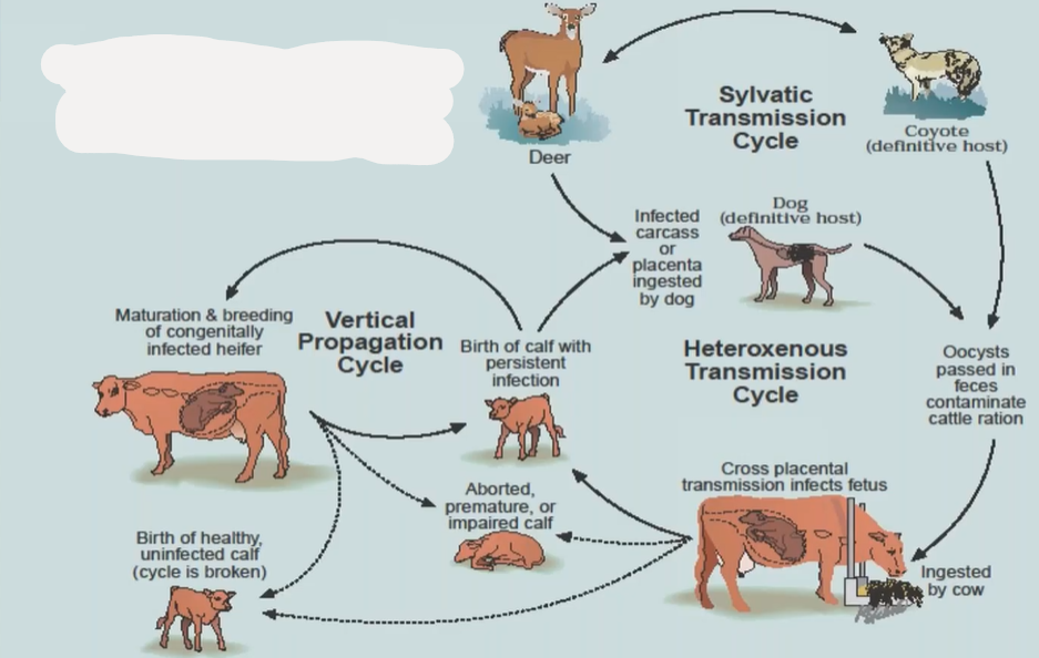
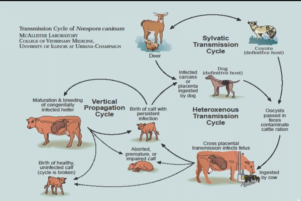
This is the life cycle of?
Lifecycle of Neospora caninum.
Definitive Host (Dog):
Dogs ingest tissue cysts from infected intermediate hosts (e.g., cattle).
intestine: sexual reproduction of parasite → oocyst → excreted in the feces → contaminates.
Intermediate Hosts (Cattle):
Cattle ingest sporulated oocysts from contaminated feed, water, or soil.
Sporozoites are released and invade tissues, leading to the formation of tachyzoites.
Tachyzoites → damage → converts later into tissue cysts with bradyzoites, in nervous tissue + muscle.
Vertical Transmission in Cattle:
Infected cows can transmit the parasite to their fetus via the placenta.
This vertical transmission can result in aborted, premature, or weak calves.
Wildlife Hosts (Sylvatic Cycle):
Deer and other wild animals can also act as intermediate hosts, maintaining the parasite's presence in the environment.
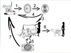
This is the life cycle of?
Toxoplasma gondii
Definitive Hosts (Cats and Wild Felids):
In the intestines of cats and wild felids → schizogony (asexual reproduction) and gamogony (sexual reproduction).
This results in the production of nonsporulated oocysts, which are shed in the feces into the environment.
Sporulation:
Once in the environment, these oocysts undergo a process called sporulation, becoming sporulated oocysts, which are infectious.
Intermediate Hosts (Mammals and Birds):
ingest sporulated oocysts, the oocysts release tachyzoites.
Tachyzoites rapidly invade various tissues, forming structures called pseudocysts.
Vertical (Transplacental) Transmission: (nr. 6 in picture)
Tachyzoites can also be transmitted from an infected mother to her fetus through the placenta, leading to congenital infection.
Tissue Cysts (Bradyzoites) (nr. 5 in the picture).
In the tissues of intermediate hosts, tachyzoites eventually transform into bradyzoites, which form tissue cysts.
These cysts can reside in muscles, the myocardium (heart muscle), and the central nervous system (CNS). → can be eaten by cat/wild felids → life cycle continues. (nr. 7 in picture).
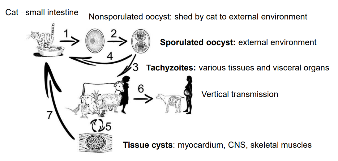
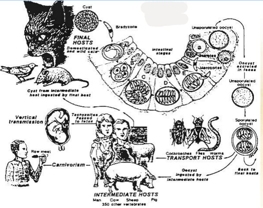
this is the life cycle of?
Toxoplasma.
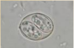
this is picture of?
Sporulated oocyst of Toxoplasma (T.gondii), with a thin oocyst wall, two sporocysts. Each sporocyst has four sporozoites.
sporulation in environment: up to 21 days (1-5 days).
cats are shedding oocysts only during the primoinfection up to 21 days.
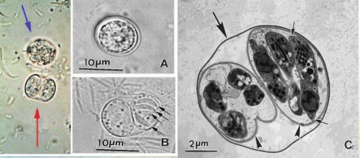
What are these pictures of?
Toxoplasma gondii oocysts.
blue arrow: unsporulated oocyst
red arrow: sporulated oocyst
A. unsporulated oocyst, note central mass (sporont) occupying most of the oocyst.
B. sporulated oocyst with 2 sporocysts, having 4 sporozoites (arrows) visible each.
C. sporulated oocyst, thin oocyst wall, two sporocysts and sporozoites (small arrows).
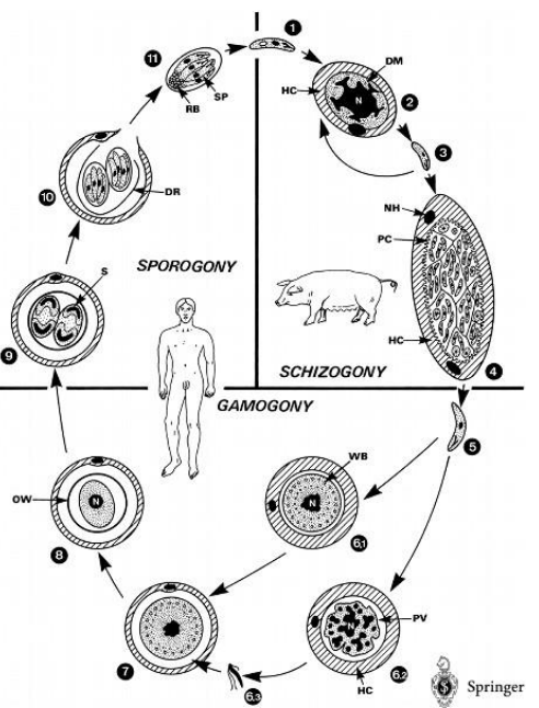
This is the life cycle of?
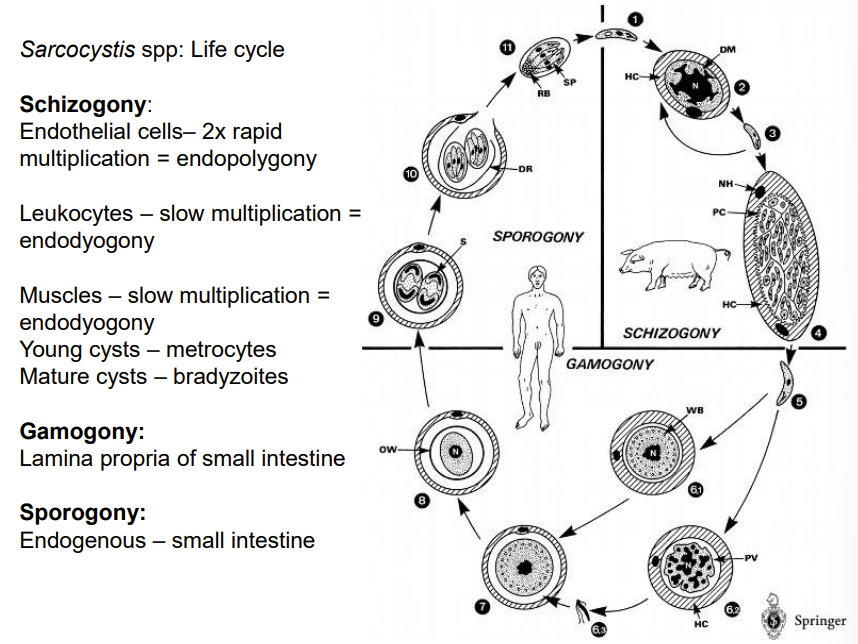
Sarcocystis spp.
1. Schizogony: Intermediate Host (e.g., pig):
Endothelial Cells: The parasite undergoes rapid multiplication (2x) via endopolygony, where multiple rounds of nuclear division occur within a single host cell.
Leukocytes and Muscle Cells: Here, the parasite multiplies slowly via endodyogony, where two daughter cells form inside the parent cell.
Young Cysts (Metrocytes): These develop within muscle tissues during the slow multiplication phase.
Mature Cysts: Over time, these young cysts mature and contain bradyzoites, which are the infective forms for the definitive host.
2. Gamogony: Definitive Host (e.g., dog, cat, human):
Lamina Propria of the Small Intestine: When the definitive host ingests tissue cysts from the intermediate host, the bradyzoites are released and invade the intestinal cells.
The parasite differentiates into male and female gametes. These gametes fuse to form zygotes, completing the sexual reproduction phase.
3. Sporogony: Definitive Host (e.g., dog, cat, human):
Small Intestine: The zygotes develop into oocysts within the small intestine. This process is known as endogenous sporulation.
Excretion: oocyst → sporulated with sporozoites → excreted in the feces → infect new intermediate host.
What is a cyst in protozoal parasites?
A cyst is the inactive stage of a protozoal parasite, formed when the active form (trophozoite) loses its motility and encloses itself in a tough wall. At this stage, the parasite can no longer grow or multiply.
Babesia species, B. canis and B. gibsoni are in which species? While B. motasi is in?
dog
small ru
Babesia species, B. bovis and B. divergens are in which species? B. caballi and B. equi are in which species?
catlle
horse
What is the location of babesia in vertebrates (IH)?
Intraerythrocytic (erythrocytes)
What is the definitive host and vector of babesia?
Ticks, definitive host too, as the sexual reproduction is done here.
vertebrates: intermediate hosts, where asexual reproduction is done.
vector name of b. canis and B. divergens?
B. canis: Dermacentor reticulatus (vector)
B. divergens: Ixodes ricinus (vector)
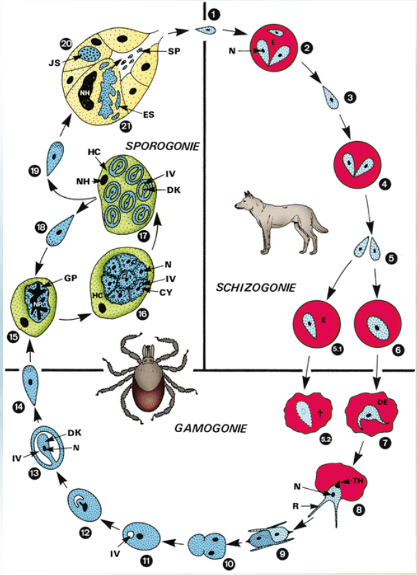
This is the life cycle of?
Life cycle of Babesia spp. - parasite causing babesiosis in mammals, involving a tick vector for transmission.
Sexual Cycle in Tick Host
tick eats gametocytes during blood meal on mammalian host.
gametocytes → male + female gametes in gut of tick.
zygote develops (gamogony) → meiosis of zygote, → kinete develops
2 options further:
Trans-ovarial transmission: in female tick, kinete will penetrate through ovaries and the eggs, thus when female lays eggs, larvae of tick hatch and are infected with babesia → continues in saliva.
trans-stage transmission: kinete penetrating several tissues, multiplicates, then invading salivary glands → sporogony happens → sporoblast is formed → sporozoites, ready to infect next vicitim. “larva taking blood from infected dog”.
Asexual Cycle in Mammalian Host
Sporozoites invade the red blood cells (RBCs) of host, when tick takes a blood meal.
Sporozoites develop into trophozoites.
Trophozoites divide by binary fission, producing merozoites.
Merozoites proliferate into new trophozoites and new merozoites.
Certain merozoites differentiate into male and female gametocytes within the RBCs.
Gametocytes are acquired by ticks during their feeding process.
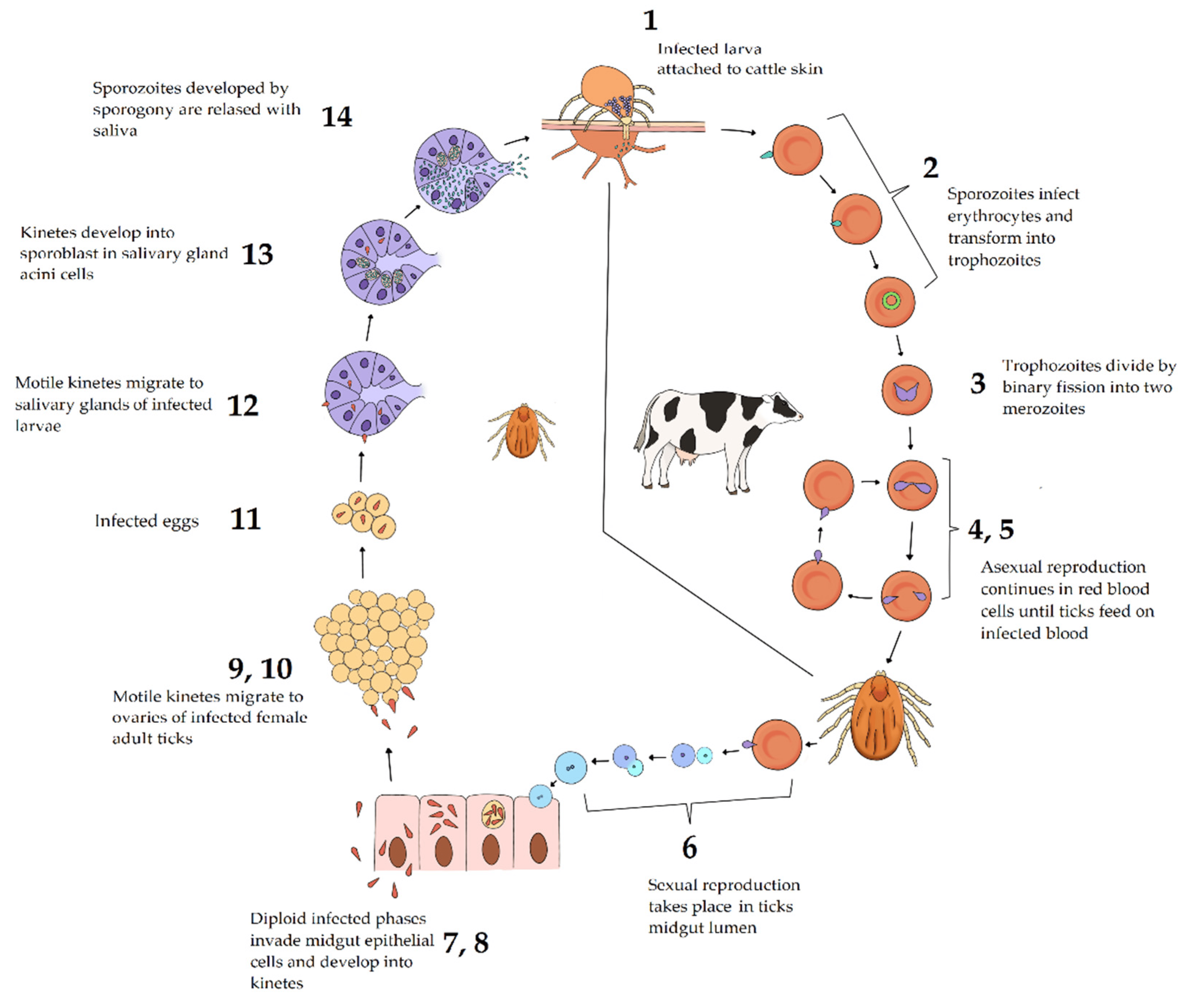
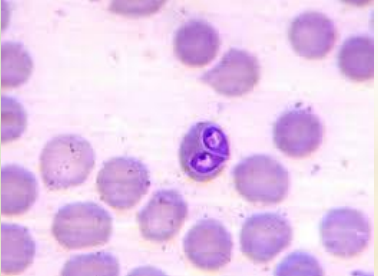
this is picture of? how to describe large vs. small?
Babesia canis.
large: 2-5 micrometer, 2 individuals forms acute angel, center of erythrocyte
small: 1-2 micrometer, 2 individuals forms obtuse angel, peripheral of erythrocyte.
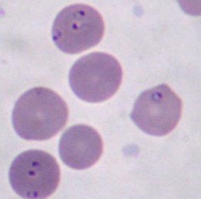
this is picture of?
Babesia gibsoni
where does these stages happen in babesia?
sporogony
gamogony
schizogony/merogony
Sporogony: tick`s salivary glands
Gamogony: Tick's gut. (intestine)
Schizogony/Merogony: Erythrocytes (red blood cells) of the mammalian host. (IH).
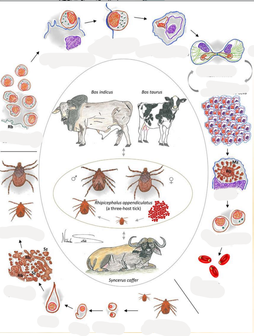
This is the life cycle of?
Theileria (genus) of the family Theileriidae.
Life Cycle of Theileria
Tick Feeding and Sporozoite Release: cycle starts when tick feeds on mammalian host ex. cattle. Tick releases sporozoites into the host bloodstream.
Sporozoite Attachment and Entry: sporozoites will attach to host lymphocytes, entering them through a zippering mechanism.
Transformation and Schizont/meront Formation: schizonts/meronts are formed inside the lymphoblast (mature vesion of lymphocyte), these mature later.
Proliferation of Schizont/meront-Infected Cells: the infected lymphoblasts proliferates.
Merozoite Release: Schizonts/meronts eventually differentiate into merozoites, which are then released when the host cell ruptures.
Piroplasms in Red Blood Cells: merozoites infect RBCs → forming piroplasms (intracellular parasites).
Tick Infection: when another tick feeds on the infected host → ingestion of the piroplasms
larvae and nymphs acquire the infection: will get infection through feeding or from infected mother (trans-stadial transmission).
gametes → zygote → kinete → sporogony → infected nymphal and adult ticks.
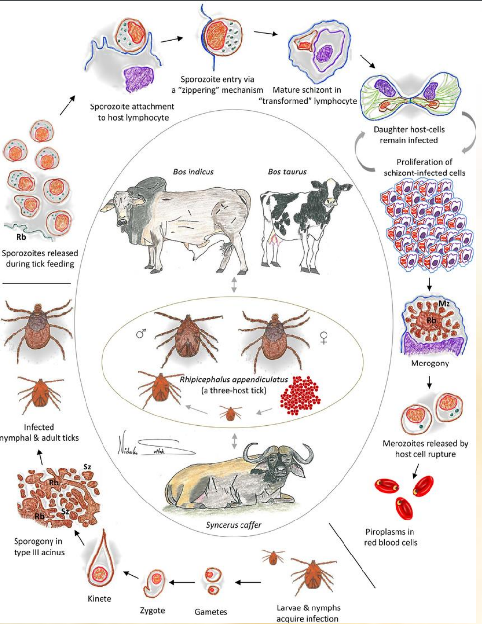
2 distinct differences of the life cycle of babesia vs. Theileria?
Cell Types: Theileria infects lymphocytes first, Babesia directly infects red blood cells.
Transmission: Theileria uses trans-stadial transmission; Babesia can use both trans-stadial and trans-ovarial transmission.
What does Theileria parva and Theileria annulata cause?
T. parva: East coast fever
T. annulata: Mediteranean coast fever
Both have ticks as vectors and as definitive host.
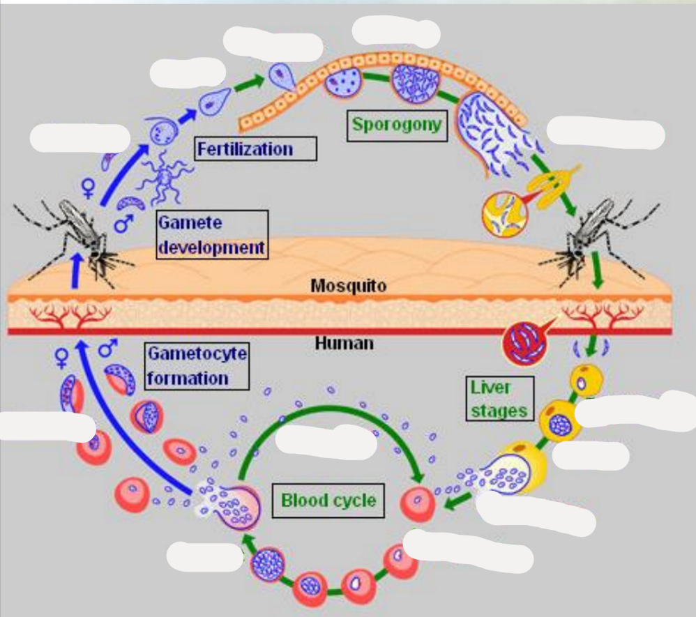
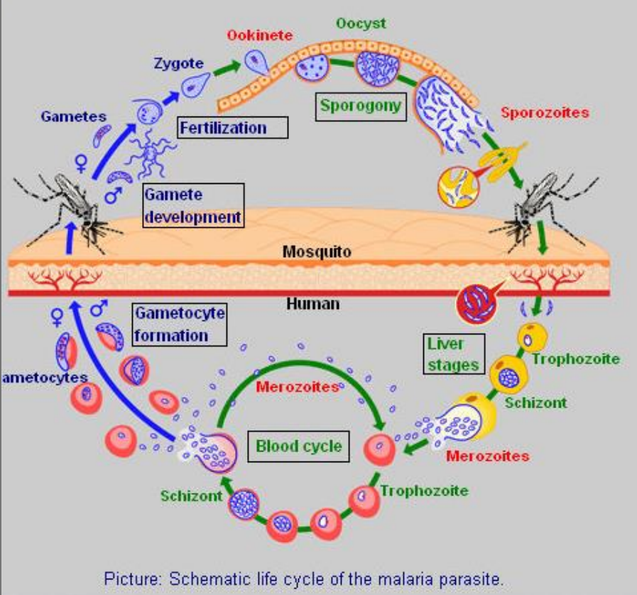
this is the life cycle of?
Cycle of Malaria parasite. (plasmodium malariae)
invasive forms: sporozoites and merozoites.
asexual stages: liver schizonts, blood schizonts.
sexual development: gametocytes and mosquito stages, where the gametocytes → gametes → zygote → ookinetes → oocysts.
Mosquito Bite: An infected mosquito injects sporozoites into a human.
Liver Stage: Sporozoites travel to the liver, becoming trophozoites, infect liver cells, and develop into schizonts, which release merozoites.
Blood Stage: Merozoites infect red blood cells, develop into trophozoites, mature into schizonts, and release more merozoites. Some merozoites develop into gametocytes.
Mosquito Uptake: Another mosquito bites the infected human, ingesting gametocytes.
Mosquito Development: Gametocytes develop into gametes, fertilize to form a zygote, which becomes an ookinete, then an oocyst. Oocysts produce sporozoites.
Completion: Sporozoites migrate to the mosquito's salivary glands, ready to infect another human, continuing the cycle.
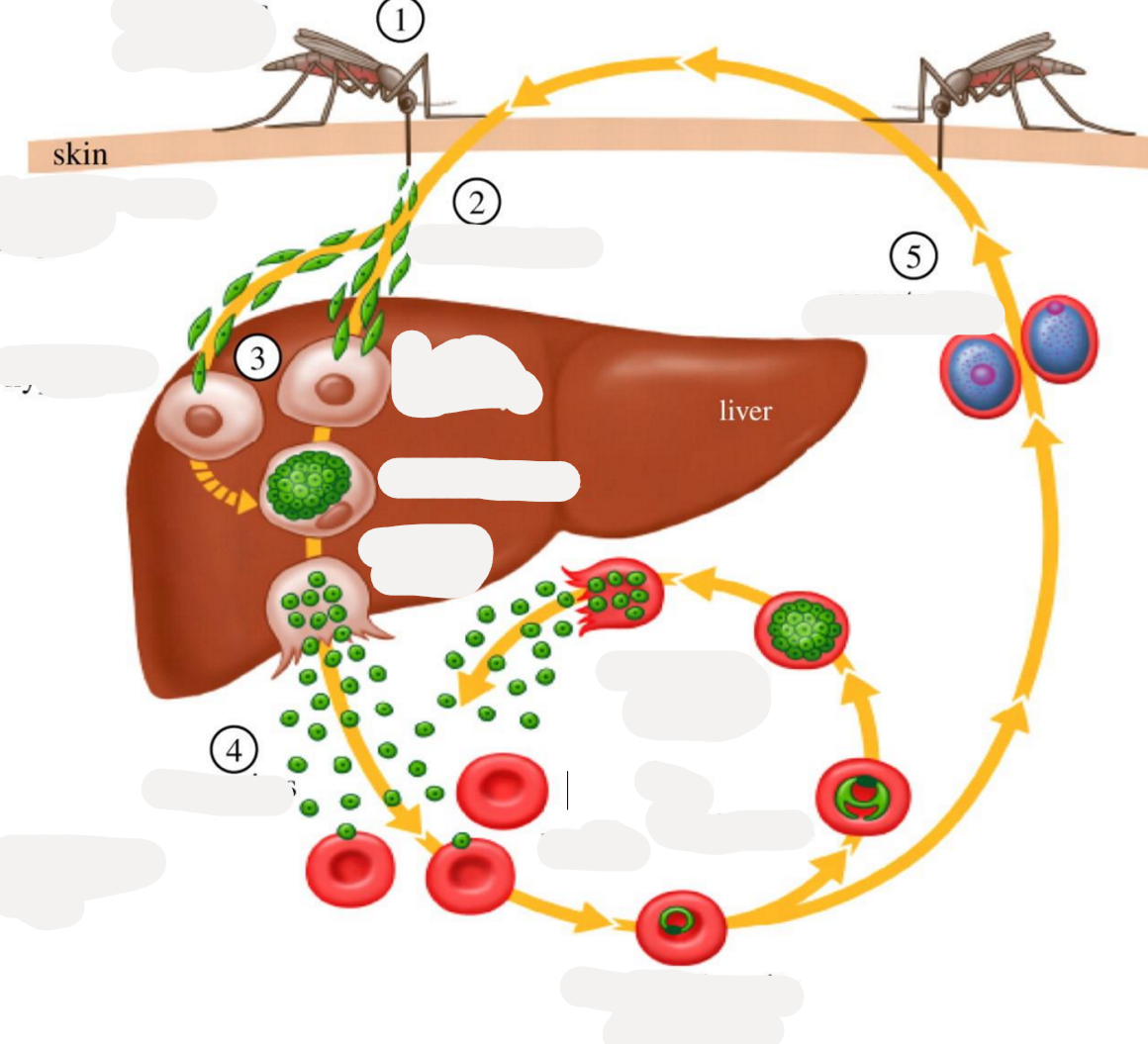
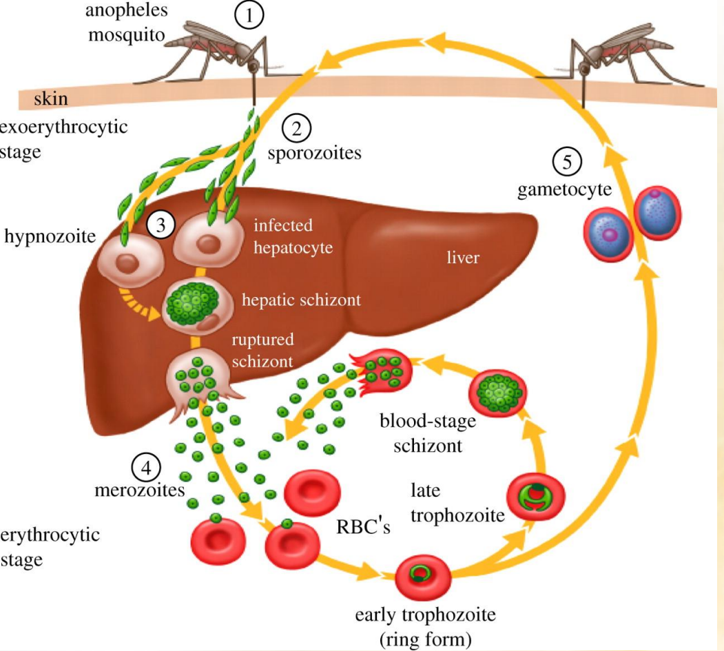
this is the life cycle of?
Plasmodium (genus), of family plasmodidae of order Haemosporida.
Mosquito Bite: An infected mosquito injects sporozoites into a human.
Liver Stage: Sporozoites travel to the liver, becoming trophozoites, infect liver cells, and develop into schizonts, which release merozoites.
Blood Stage: Merozoites infect red blood cells, develop into trophozoites, mature into schizonts, and release more merozoites. Some merozoites develop into gametocytes.
Mosquito Uptake: Another mosquito bites the infected human, ingesting gametocytes.
Mosquito Development: Gametocytes develop into gametes, fertilize to form a zygote, which becomes an ookinete, then an oocyst. Oocysts produce sporozoites.
Completion: Sporozoites migrate to the mosquito's salivary glands, ready to infect another human, continuing the cycle.
what is koch`s blue bodies?
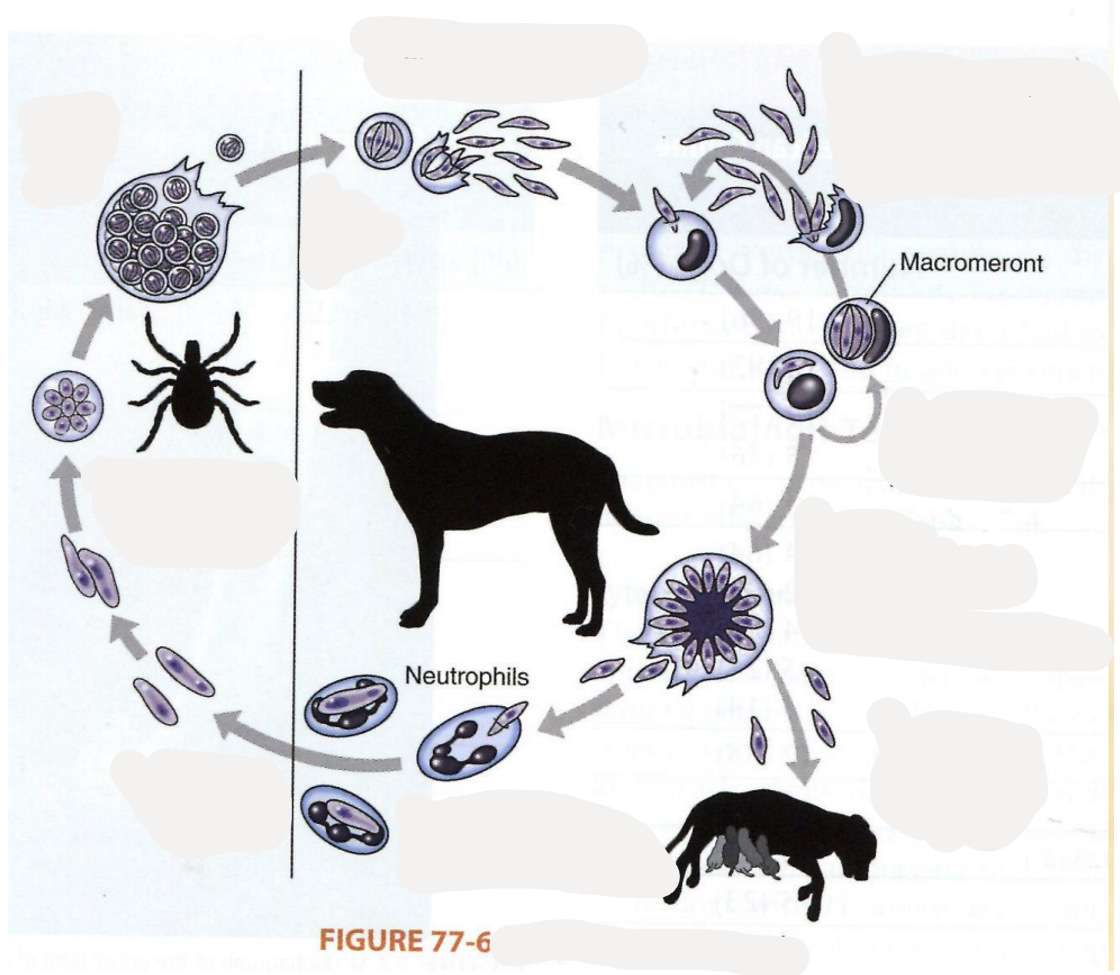
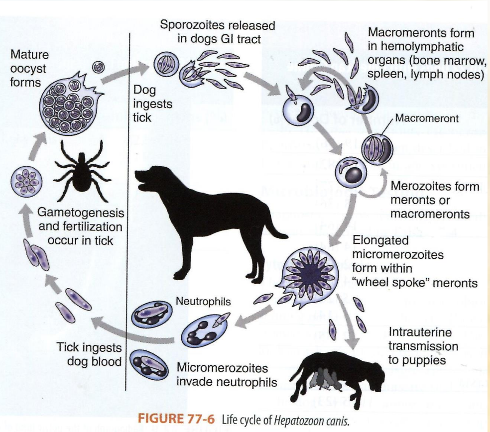
which parasite has this life cycle?
Hepatozoon canis (species of genus hepatozoon) of family hepatozoidae, order aleleida.
asexual reproduction in ca, fe (IH)
sexual reproduction: sporogony in tick (vector) DH.
Trans-stage transmission.
Dog ingests tick containing mature oocyst.
sporozoite release: oocyst release sporozoites in the dog`s GIT.
Macromeronts formation: sporozoites travel to the hemolymphatic organs like bone marrow, spleen, lymph nodes, forming macromeronts.
merozoite production from the macromeronts.
micromerozoites formation
neutrophil invasion: the micromerozoties invade the neutrophils in the dog`s blood.
tick feeding: a tick ingest the infected blood, taking up the micromerozoites.
gametogenesis + fertilization happen in the tick → mature oocyst formation.
cycle continuation - tick carries mature oocyst → infect another dog.
