Medsci 142 Exam MCQ Study
1/540
There's no tags or description
Looks like no tags are added yet.
Name | Mastery | Learn | Test | Matching | Spaced |
|---|
No study sessions yet.
541 Terms
Afferent nerves conduct nerve impulses from: a) one effector to another. b) receptors to the central nervous system. c) the central nervous system to receptors. d) effectors to the central nervous system.e) the central nervous system to effectors.
receptors to the central nervous system
The factor most affecting the rate of impulse conduction is the: a) length of the axon. b) rate of axonal transport. c) number of neuroglia associated with it.d) presence or absence of a myelin sheath.e) site of initial stimulation.
The factor most affecting the rate of impulse conduction is the presence or absence of a myelin sheath.
The posterior (dorsal) root ganglion is: a) the tapered end of the spinal cord. b) the roots of spinal nerves hanging inferiorly from the inferior end of the spinal cord in the vertebral column. c) an extension of the pia mater that anchors the spinal cord to the coccyx. d) an indentation on the dorsal side of the spinal cord. e) where the cell bodies of sensory neurons are located.
The posterior root ganglion is where the cell bodies of sensory neurons are located.
The part of a spinal nerve that contains only efferent fibres is the: a) dorsal root. b) ventral root. c) ventral ramus. d) dorsal ramus. e) plexus.
The part of a spinal nerve that contains only efferent fibres is the ventral root.
Cell bodies of motor neurons to skeletal muscles are located in the: a) posterior white columns. b) anterior white columns. c) central canal. d) anterior gray horns. e) lateral gray horns.
Cell bodies of motor neurons to skeletal muscles are located in the anterior gray horns.
Fine control of body motor coordination and balance is a function of the: a) pituitary gland. b) cerebellum. c) hypothalamus. d) thalamus. e) reticular activating system.
Fine control of body coordination and balance is a function of the cerebellum.
Paired masses of gray matter within the white matter of the cerebrum that are rich in dopamine and are involved in maintenance of muscle tone are the: a) substantia nigra. b) supraoptic nuclei. c) basal ganglia. d) pontine nuclei. e) mammillary bodies.
Paired masses of gray matter within the white matter of the cerebrum that are rich in dopamine and are involved in maintenance of muscle tone are the basal ganglia.
The main relay centre for conducting information between the spinal cord and the cerebrum is the: a) thalamus.b) corpus callosum. c) cerebellar peduncles.d) tentorium cerebelli.e) insula.
The main relay center for conducting information between the spinal cord and the cerebrum is the thalamus.
The brain stem is made up of the: a) medulla oblongata, thalamus, and midbrain. b) midbrain, hypothalamus, and thalamus. c) medulla oblongata, hypothalamus, and pons. d) medulla oblongata, pons, and midbrain.e) cerebellum, pons, and hypothalamus.
The brain stem is made up of the medulla oblongata, pons, and midbrain.
Damage to the cerebellum would result in: a) altered pituitary function. b) inability to dream. c) loss of memory. d) uncontrollable body temperature. e) uncoordinated movement.
Damage to the cerebellum would result in uncoordinated movement.
Question 11: The cerebral hemispheres are connected internally by the: a) corpus callosum. b) basal ganglia.c) intermediate mass.d) arachnoid villi.e) pons.
The cerebral hemispheres are connected internally by the corpus callosum.
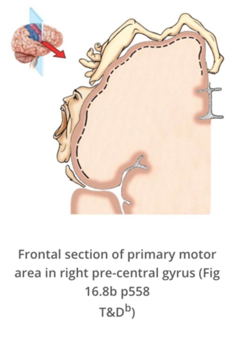
The primary motor area of the cerebral cortex is located in the: a) postcentral gyrus. b) precentral gyrus. c) occipital lobe. d) temporal lobe. e) insula.
The primary motor area of the cerebral cortex is located in the precentral gyrus.
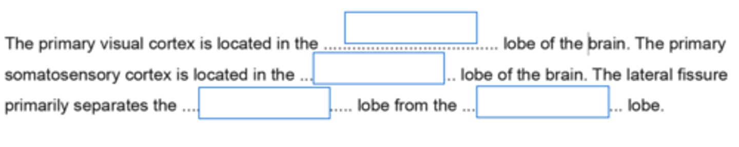
The primary visual area and visual association area of the cerebral cortex are both located in the: a) temporal lobe. b) parietal lobe. c) occipital lobe. d) insula. e) frontal lobe.
The primary visual area and visual association area of the cerebral cortex are both located in the occipital lobe.
Precise location and identification of specific sensations occurs in the: a) cerebral cortex. b) brain stem. c) thalamus. d) spinal cord. e) all of these options.
Precise location and identification of specific sensations occurs in the cerebral cortex.
Receptors for pressure that are widely distributed in the subcutaneous tissue and the submucosal tissues are the: a) Ruffini corpuscles. b) Merkel discs. c) Meissner corpuscles.d) intrafusal muscle fibres. e)
Receptors for pressure that are widely distributed in the subcutaneous tissue and the submucosal tissues are the Pacinian corpuscles.
Injury to lower motor neurons results in: a) flaccid paralysis. b) long-term potentiation. c) coma. d) kinesthesia. e) spastic paralysis.
Injury to lower motor neurons results in flaccid paralysis.
The cell bodies of first-order neurons in the posterior column-medial lemniscus pathway to the cortex are located in the: a) medial lemniscus. b) posterior root ganglia of spinal nerves. c) medulla. d) precentral gyrus. e) anterior gray horns of the spinal cord.
The cell bodies of first-order neurons in the posterior column-medial lemniscus pathway to the cortex are located in the posterior root ganglia of spinal nerves.
The medial lemniscus is a projection tract of second-order neurons extending from the: a) upper motor neurons to the lower motor neurons. b) proprioceptors to the anterior gray horns of the spinal cord. c) posterior root ganglia to the medulla. d) thalamus to the postcentral gyrus of the cerebral cortex.e) medulla to the thalamus.
The medial lemniscus is a projection tract of second-order neurons extending from the medulla to the thalamus.
First-order sensory neurons conduct impulses from: a) the spinal cord to the brainstem.b) a receptor to the CNS.c) one part of the spinal cord to another. d) the thalamus to the somatosensory cortex. e) the CNS to an effector.
First-order sensory neurons conduct impulses from a receptor to the CNS.
Cell bodies for upper motor neurons are located in the: a) cerebellum. b) thalamus. c) anterior gray horns of the spinal cord. d) connective tissues surrounding skeletal muscles. e) precentral gyri of the cerebral cortex.
Cell bodies for upper motor neurons are located in the precentral gyri of the cerebral cortex.
Third-order sensory neurons in the posterior column-medial lemniscus pathway extend from the: a) medulla oblongata to the thalamus. b) spinal cord to the medulla oblongata. c) skin to posterior root ganglia. d) thalamus to the somatosensory area of the cerebral cortex. e) posterior root ganglia to the posterior gray horn of the spinal cord.
Third-order sensory neurons in the posterior column-medial lemniscus pathway extend from the thalamus to the somatosensory area of the cerebral cortex.
The parietal lobe of the brain contains: a) Wernicke's area b) Primary visual cortex c) Primary somatosensory cortex d) Primary motor cortex e) Broca's area
The parietal lobe of the brain contains the primary somatosensory cortex.
Lower motor neurons are: a) located in the dorsal root ganglia b) located in the ventral horn of the spinal cord c) responsible for conveying touch and pressure sensations to the spinal cord d) located in the motor cortexe) sensory neurons in the spinal cord
Lower motor neurons are located in the ventral horn of the spinal cord.
The lateral spinothalamic tract conveys: a) sensory impulses regarding discriminative touch b) sensory impulses regarding two point discrimination c) sensory impulses regarding vibrationd) sensory impulses regarding proprioception e) sensory impulses regarding pain
The lateral spinothalamic tract conveys sensory impulses regarding pain.
The medial lemniscus: a) contains mainly unmyelinated fibres b) conveys sensory information from the thalamus to the sensory cortex c) conveys sensory information on pain and temperature only d) is located in the brainsteme) is located in the spinal cord
The medial lemniscus is located in the brainstem.
The corticospinal (pyramidal) tract has all of the following characteristics, EXCEPT: a) where affected by a disease results in a flaccid paralysis b) passes down the spinal cord as the lateral corticospinal tract c) is involved in the voluntary control of muscles on the opposite side of the body d) arises from upper motor neurons in the precentral gyrus e) contains mainly fast conducting myelinated fibres
The corticospinal (pyramidal) tract has all of the following characteristics, EXCEPT: where affected by a disease results in a flaccid paralysis.
The basal ganglia consists of all of the following structures, EXCEPT: a) Putamen b) Substantia nigra c) Caudate nucleus d) Globus pallidus e) Cerebellum
The basal ganglia consists of all of the following structures, EXCEPT: cerebellum.
The cells in the peripheral nerves which produce myelin are the: a) sensory neurons b) lower motor neurons c) astrocytes d) Schwann cells e) oligodendrocytes
The cells in the peripheral nerves which produce myelin are the Schwann cells.
The fibres responsible for conveying discriminative touch and pressure sensations from the skin to the spinal cord: a) are heavily myelinated fibres b) are slow conducting fibres c) enter the spinal cord via the ventral root d) are associated with non-encapsulated receptors e) have their cell bodies in the dorsal horn
The fibres responsible for conveying discriminative touch and pressure sensations from the skin to the spinal cord are heavily myelinated fibres.
The temporal lobe of the brain contains: a) Primary visual cortex b) Wernicke's area c) Broca's aread) Primary motor cortex e) Primary somatosensory cortex
The temporal lobe of the brain contains Wernicke's area.
The cell bodies of upper motor neurons are: a) located in the ventral horn of the spinal cord b) located in the dorsal root ganglia c) located in the basal ganglia d) sensory neurons in the spinal cord e) located in the motor cortex
The cell bodies of upper motor neurons are located in the motor cortex.
The fibres responsible for conveying pain and temperature sensations from the skin to the spinal cord are: a) heavily myelinated fibres b) unmyelinated fibres c) associated with encapsulated receptors d) fast conducting fibres e) enter the spinal cord via the ventral root
The fibres responsible for conveying pain and temperature sensations from the skin to the spinal cord are unmyelinated fibres.
The frontal lobe of the brain contains: a) Wernicke's area b) Primary sensory cortex c) Primary auditory cortexd) Primary visual cortex e) Broca's area
The frontal lobe of the brain contains Broca's area.
Which of the following symptoms results from a lesion of the primary motor cortex on the right side of the brain? a) a flaccid paralysis on the right side of the body b) a spastic paralysis on the left side of the body c) a flaccid paralysis on the left side of the body d) muscle rigidity on the right side of the body e) a spastic paralysis on the right side of the body
A lesion of the primary motor cortex on the right side of the brain is likely to result in spastic paralysis on the left side of the body.
If a stroke caused damage to the posterior third of the inferior frontal gyrus in the left hemisphere, what would the expected result be? a) Sensory aphasia b) Motor/Expressive aphasia c) Conduction aphasiad) Paralysis of the right side of the face e) Loss of sensation to the left side of the face
A stroke causing damage to the posterior third of the inferior frontal gyrus in the left hemisphere would expect to result in motor / expressive aphasia.
The primary somatosensory cortex is located in the:a) Frontal lobe b) Temporal lobe c) Parietal Lobe d) Occipital Lobe e) Brainstem
The primary somatosensory cortex is located in the parietal lobe.
The spinothalamic tracts convey: a) sensory impulses regarding temperature. b) sensory impulses regarding vibration. c) sensory impulses regarding discriminative touch. d) sensory impulses regarding proprioception. e) motor impulses to skeletal muscles.
The spinothalamic tracts convey sensory impulses regarding temperature.
The posterior column tracts convey ALL of the following, EXCEPT: a) sensory impulses regarding vibration. b) sensory impulses regarding two-point discrimination. c) sensory impulses regarding pain. d) sensory impulses regarding discriminative touch. e) sensory impulses regarding proprioception.
The posterior column tracts convey ALL of the following, EXCEPT sensory impulses regarding pain.
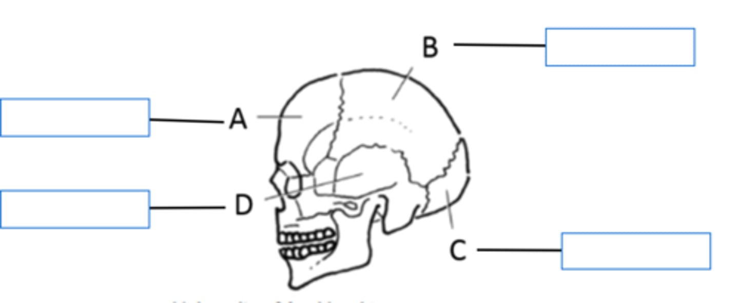
Identify the lobes of the human brain.
A - Frontal Lobe
B - Parietal Lobe
C - Occipital Lobe
D - Temporal Lobe
Fill in the blanks.
1. Occipital
2. Parietal
3. Frontal
4. Temporal
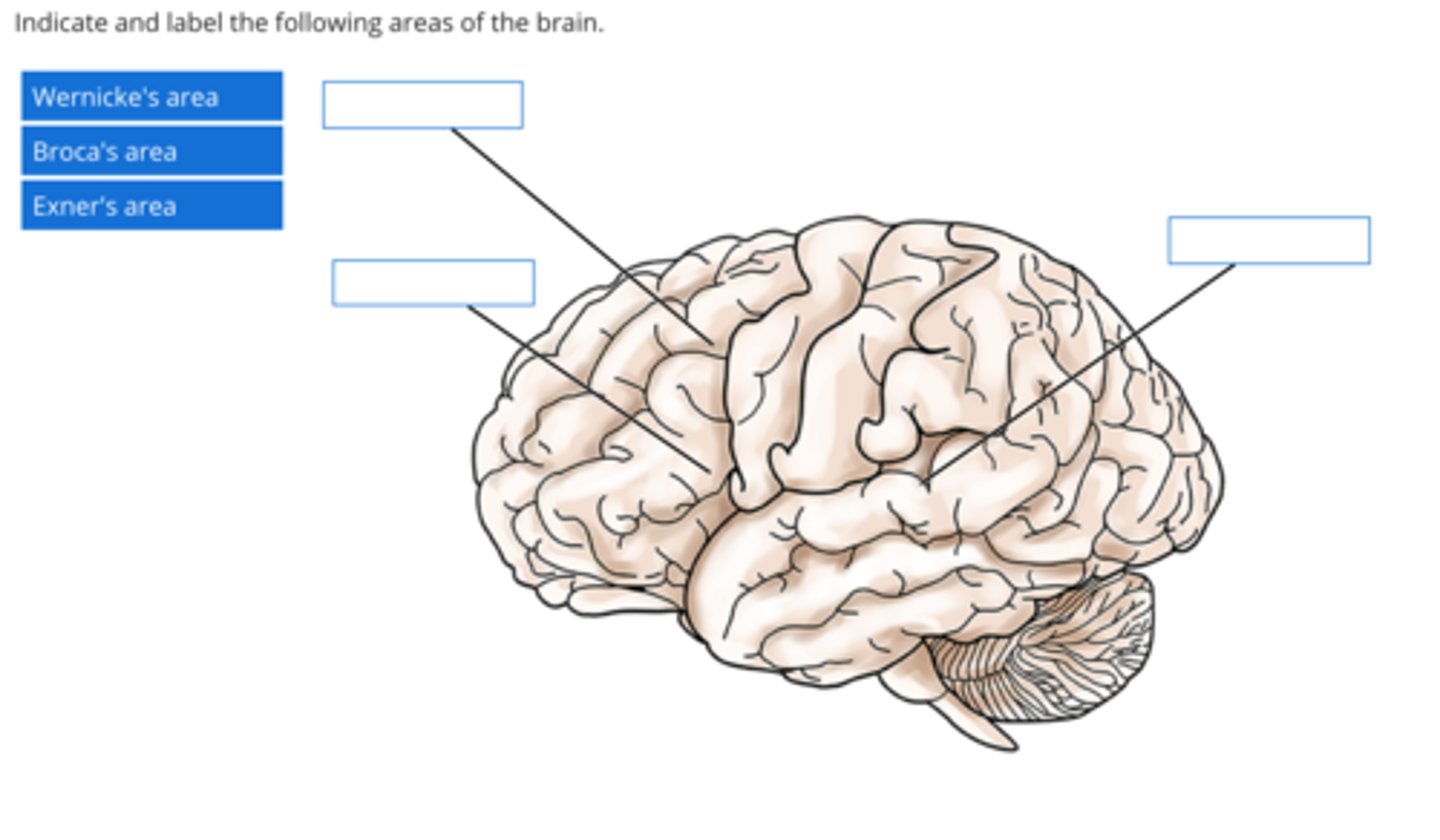
State the locations of the following areas of the brain using the appropriate letters from the diagram below.
Top Left: Exner's Area
Bottom Left: Broca's Area
Right: Wernicke's Area
Describe the key functions of the temporal association cortex
Memory, mood, aggression, intelligence
Describe the key functions of the frontal association cortex
Intelligence, personality, behaviour, cognitive function, some aspects of mood
Describe the key functions of the parietal association cortex
Spatial skills - e.g. determining whether a couch can fit inside the door frame3D recognition: Shapes, faces, concepts, abstract perceptionHelps us to understand ambiguity and nuances in language - e.g. "a curry with a kick in it" or "a loud shirt"
Describe the function of the arcuate fasciculus
An arching bundle of white matter fibres that connect Wernicke and Broca's Areas. It functions to align speech recognition/comprehension with speech production
Describe the major symptom of a person with a non-fluent aphasia
Results from damage to Broca's speech area. The major symptom is the inability to coordinate muscular movements required to physically produce/articulate speech/form of words / what they mean to say despite knowing what they want to say
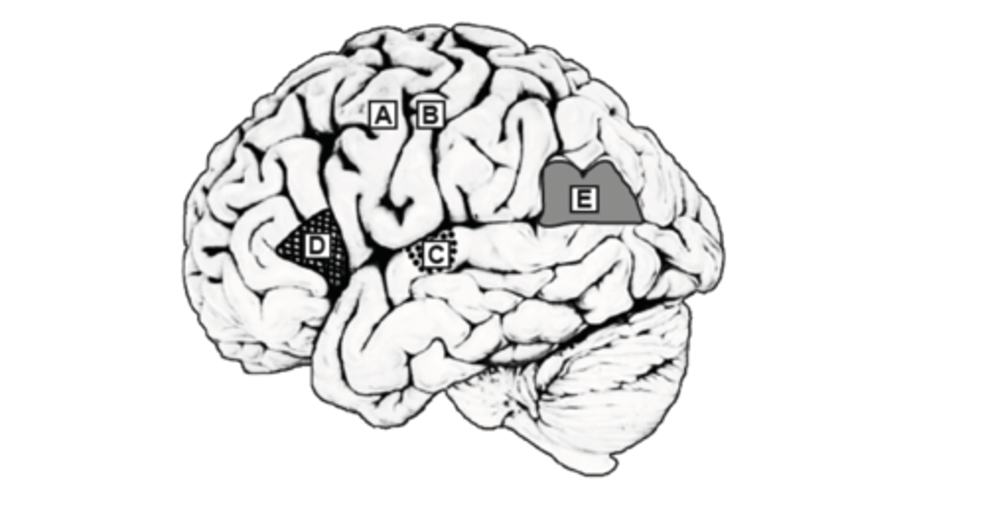
explain the functional effects of lesions (e.g. strokes) of the cerebral cortex in each of the areas indicated
A - Difficulty with movement of the right arm - specifically, spastic paralysis
B - No sensation from the right arm region
C - Deafness (loss of hearing)
D - Motor (non-fluent) aphasia - inability to coordinate muscular movements for generating speech
E - Inability to understand reading and writing
List the protective structures of the spinal cord
dura mater, pia mater and arachnoid mater. Also Meninges and Vertebrae
How is the grey and white matter arranged in the spinal cord?
In the spinal cord, there is a butterfly-shaped core of grey matter in the centre, and the exterior is made up of white matter tracts.
How is the grey and white matter arranged in the brain
In the brain, grey matter forms the outer cortex and white matter is found more internally / deeply
the pathway of discriminative sensation from the toe to the dorsal columns
the pathway for pain and temperature at this level of the spinal cord
The pathway conveying non-discriminative pain and temperature sensation from the skin to the cerebral cortex has the following characteristics (be specific):
- The first order neuron terminates in the (a) ........ .
- The second order neuron terminates in the (b) .......... of the (c) ........ .
- Has fibres that are relatively (d) [ fast / slow ] conducting and that decussate via the (be specific) (e) ......... in the spinal cord.
A - Dorsal Horn of the spinal chord
B - Vetro Posterior nucleus
C - thalamus
D - slow
E - anterior white commissure
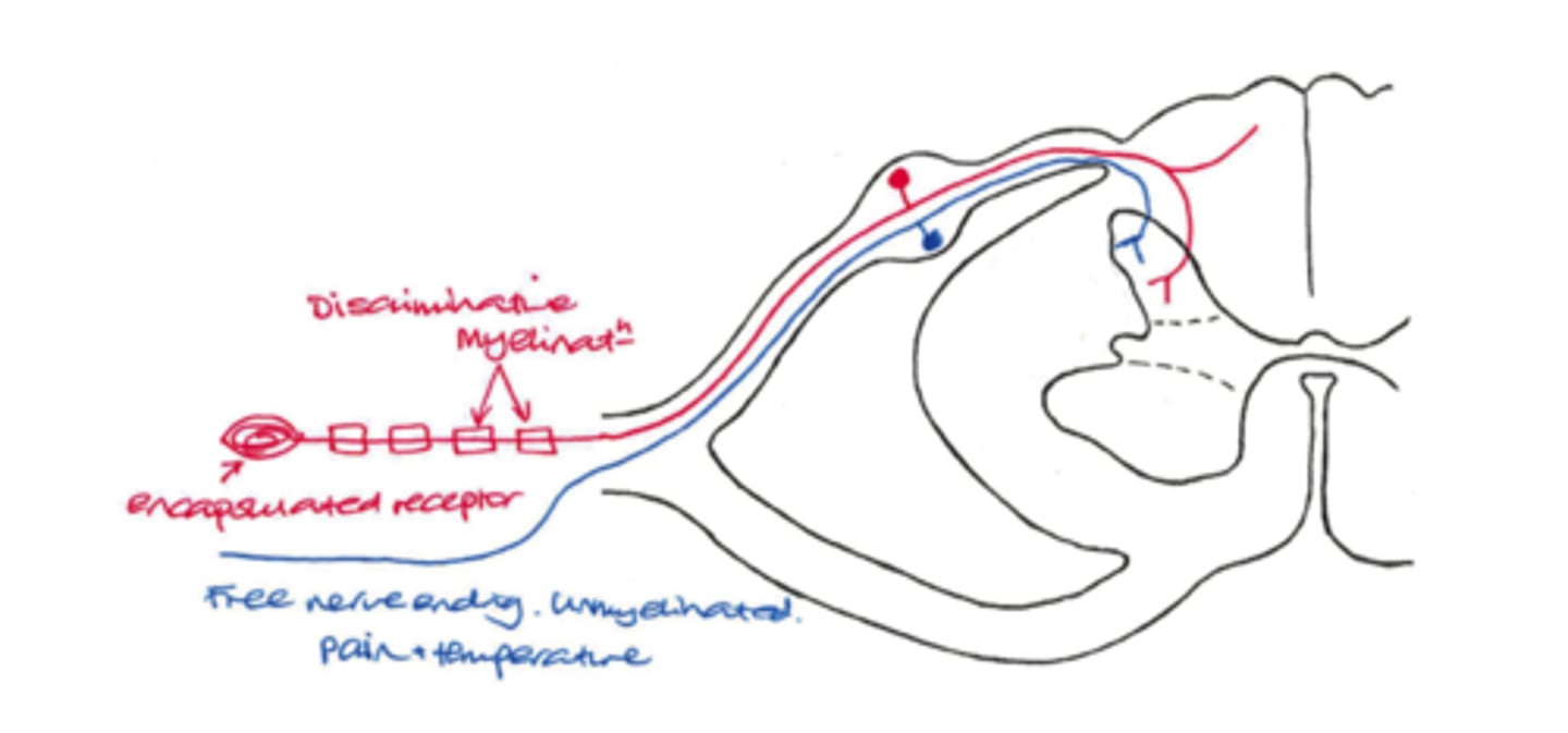
Fill in the blanks to complete the sentences below.
The pathway in the brainstem which conveys discriminative touch and pressure sensation is known as the (a) .......... , whereas the pathway in the brainstem for non-discriminative pain and temperature sensation is the (b) ........ . Both pathways terminate in the (c) ........ nucleus in the thalamus.
A - Medial Leminiscus
B - Lateral spinothalmic tract
C- Ventro-posterior
Fill in the blanks to complete the sentence below:
Spinal nerves contain a mixture of myelinated and unmyelinated nerve fibres. Many of the myelinated fibres convey information on (a) .......... sensations from (b) .......... receptors in the skin. The unmyelinated fibres convey information on (c) .......... sensation from (d) .......... receptors in the skin.
Enter your answer here
Check Answer
A - Discriminatory
B - Encapsulated
C - Non-discriminitory
D - Free nerve ending
The foot has a ..... representation that the hand
A) smaller
B) larger
Smaller
The neurons representing the face are located more ..... than those from the elbow
A) Lateral
B) medial
lateral
A larger area of skin is ...... represented as a larger region of sensory neurons
A) always
B) not always
not always
A larger representation in the sensory homunculus represents ..... sensitive areas of the body
A) more
B) less
More
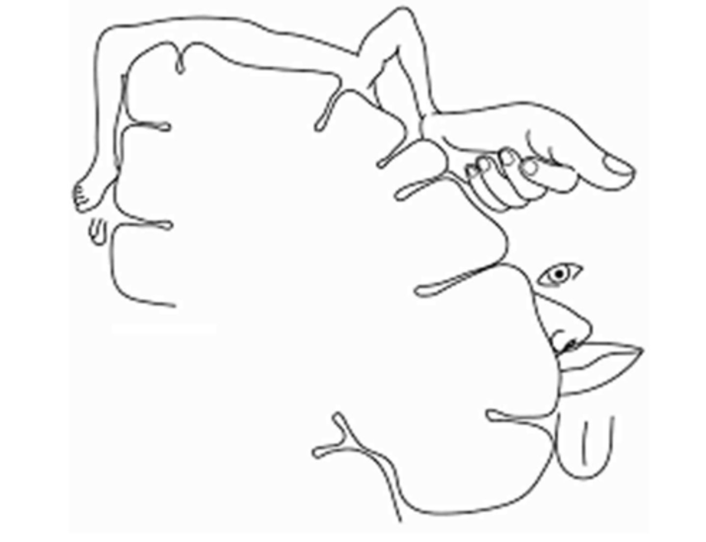
an approximate sketch of the sensory homunculus on the cortex schematic
see image
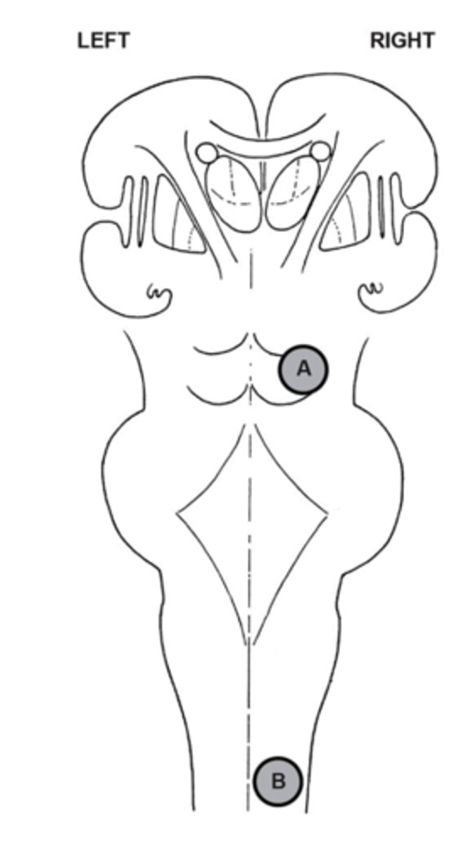
describe the sensory symptoms that would result from lesions at location A
This is known as ....... sensory loss
Pain and Temperature: Left side
Touch and Pressure: Left side
Associative
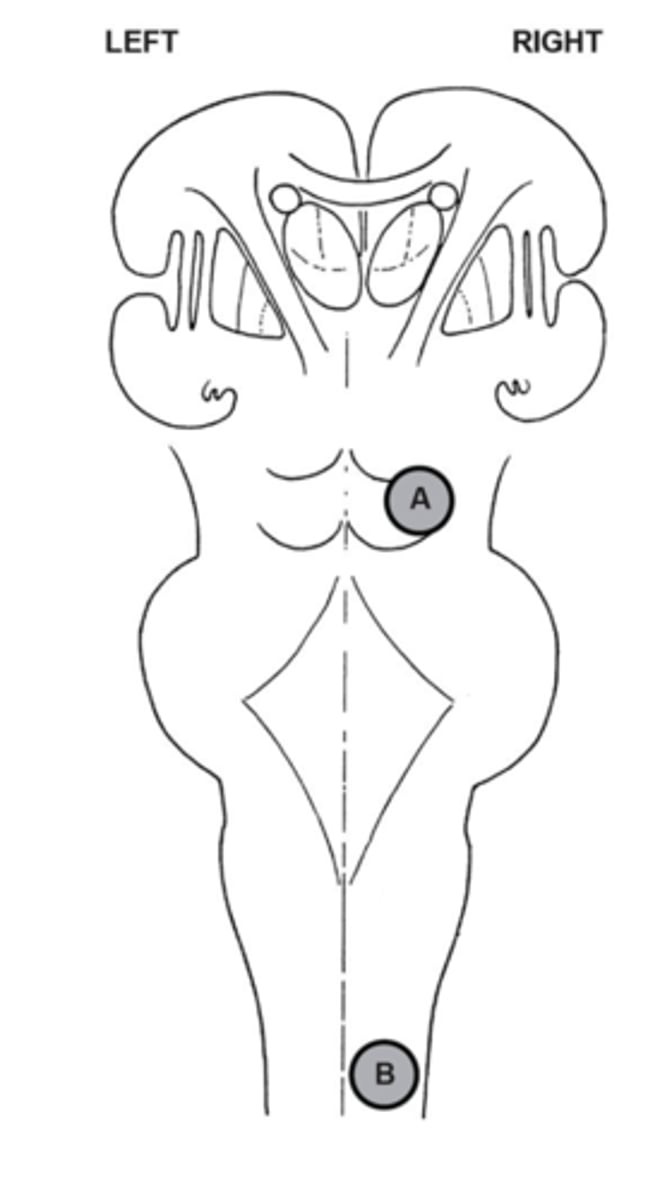
describe the sensory symptoms that would result from lesions at location B
This is known as ....... sensory loss
Pain and Temperature: Left side
Touch and Pressure: Right side
Dissociative
When the pain and temperature pathway and the discriminative touch and pressure pathway are both affected by a lesion on the right side of the spinal cord, we refer to the pattern of sensory loss as being (a/an) (a) ....... sensory loss, because touch and pressure are lost on the (b) ....... side of the body and pain and temperature are lost on the (c) ....... side of the body. When both these pathways are affected on the right side of the brain stem, touch and pressure are lost on the (d) ....... side of the body and pain and temperature are lost on the (e) ....... side of the body, resulting in (a/an) (f) ....... sensory loss.
A) dissociative
B) right
C) left
D) left
E) left
F) associative
Why do some parts of the body have a larger representation in the primary motor cortex than others?
Some areas of the body are specialised for making fine / complicated movements. each of these motor units needs to be innervated by a motor neuron and so there is proportionally more representation on the primary motor cortex
show how the basal ganglia contributes to movement control
Initiation of movement
Conveying mood through movement
Modifies movement after practice to be smoother, more controlled and precise
No direct input / output to spinal cord
show how the basal cerebellum contributes to movement control
Coordinates, maps, terminates movement, and works with unconscious movement e.g. swinging of arms while walking
Maintains balance
Adjusts movements to account for discrepancy between planned and actual movements
Direct output to spinal cord
Diseases involving the left side of the cerebellum result in (a) ...... movements and the loss of balance on the (b) ...... side of the body, and lesions of the basal ganglia on the left side of the brain result in (c) ...... movements on the (d) ...... side of the body.
A) uncoordinated
B) left
C) unrefined / imprecise
D) right
A patient presents with a sudden stroke, which causes a spastic paralysis of the muscles on the left side of the face and left upper limb, a loss of sensation on the left hand, a loss of hearing in the left ear, and problems with nonverbal communication. Specifically list the regions of the cerebral cortex which would be involved in this stroke.
Spastic paralysis would have been due to damage in the primary motor cortex in the right hemisphere.
Loss of sensation in the left hand would have been due to damage in the primary somatosensory cortex in the right hemisphere
Loss of hearing in the left ear would be due to damage to the primary auditory cortex in the right hemisphere
Problems with non-verbal communication would be due to damage in the non-dominant hemisphere - which for most people is the right.
Symptoms of a lesion on the right side of the Basal Ganglia
Left side of the body may be affected in the following ways:
- Difficulty initiating movements
- Voluntary movements will be coarse/unrefined due to the loss of smoothing of muscle movement for fluid motion
- Handwriting, expressing mood, and other aspects of fine motor control become difficult because of a loss of this smoothing system
Symptoms of a lesion on the right side of the Arm are of the Motor Cortex
The Left side of the body will be affected.
The left arm will exhibit spastic paralysis, in which muscle tone in heightened and the overexcited lower motor neurons cause rigid, exaggerated muscle movement, especially in regards to flexion and extension. There will be a loss of voluntary control of the limb
Symptoms of a lesion on the right side of the Cerebellum
The right side of the body may be affected in the following ways:
- Difficulty stopping movement
- Loss of normalisation of actual movement to align with planned movement (i.e. there may be a discrepancy between the actual and planned movement)
- Difficulty mapping and coordinating movement
- Loss of balance
- Loss of unconscious movement, such as holding postural muscles in appropriate tone in relation to gravity, depending on the desired function e.g. standing, resting horizontally.
Symptoms of a lesion on the right side of the Lower motor neurons of the spinal chord
The right side of the body will be affected, resulting in flaccid paralysis in which affected muscles are unable to be controlled either voluntarily or involuntarily.
The characteristic motor symptoms of Parkinson's disease consist of: (a) ...... , (b) ...... , and (c) ...... . The most recently developed surgical treatment of Parkinson's disease is (d) ...... of the region in the basal ganglia known as the (e) ...... .
A) tremor at rest
B) bradykinesia / Hypokinesia
C) rigidity
D) deep brain stimulation
E) subthalamic nucleus or globus pallidus internus
A patient presents with Parkinson's disease symptoms on the left side of the body. Describe the major motor symptoms that the patient would show.
Bradykinesia / Hypokinesia
Tremor at rest
Muscle rigidity
Which neurotransmitter is used by pre-ganglionic nerve axons of both the sympathetic and parasympathetic divisions of the autonomic nervous system? a) Acetylcholine b) Epinephrine c) Norepinephrine d) Aldosterone e) Cortisol
The neurotransmitter used by pre-ganglionic nerve axons of both the sympathetic and parasympathetic divisions of the autonomic nervous system is acetylcholine.
A person with a chronically activated sympathetic autonomic nervous system pathway is likely to have all of the following EXCEPT: a) elevated concentration of epinephrine in the blood b) dilated pupils c) elevated heart rate d) elevated rate of salivation e) elevated blood pressure
A person with a chronically activated sympathetic autonomic nervous system pathway is likely to have all of the following EXCEPT elevated rate of salivation.
After a satisfying lunch, you are likely to experience the results of: a) A stress response b) Upregulated sympathetic system activation c) Upregulated parasympathetic system activation d) Increased epinephrine production e) Increased acetylcholine production
After a satisfying lunch, you are likely to experience the results of upregulated parasympathetic system activation.
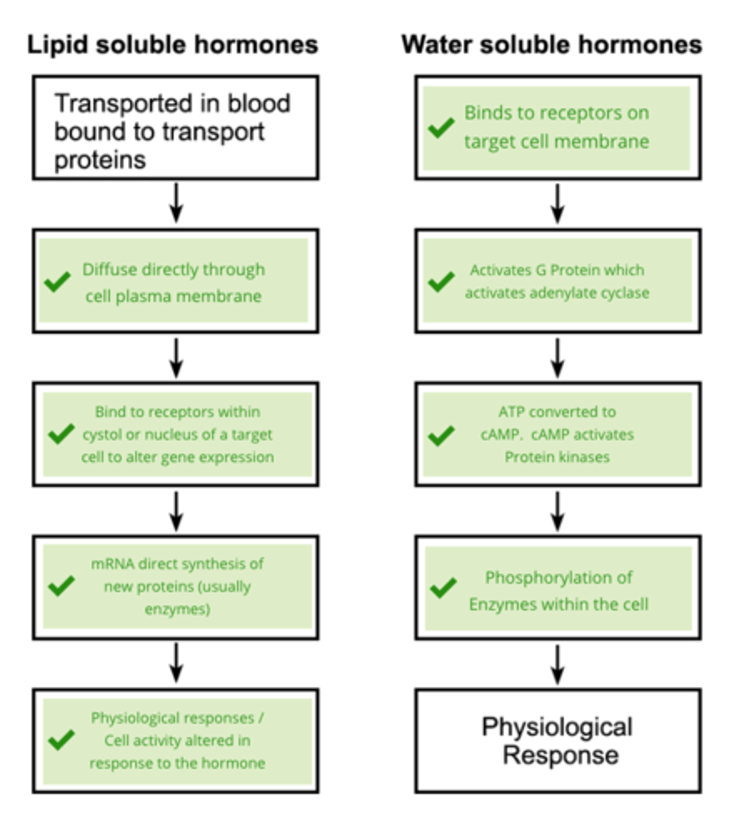
When a hormone that uses a second messenger binds to a target cell, the next thing that happens is that: a) adenylate cyclase is activated by a G protein b) voltage-regulated ion channels open in the plasma membrane c) a gene is activated in the nucleus d) phosphodiesterase is activated e) a protein kinase is activated
When a hormone that uses a second messenger binds to a target cell, the next thing that happens is that adenylate cyclase is activated by a G protein.
The compound that most often acts as a second messenger is: a) CRH b) phosphodiesterase c) COMT d) cholesterol e) cyclic AMP
The compound that most often acts as a second messenger is cyclic AMP.
When a steroid hormone binds to its target cell receptor, it: a) turns specific genes of the nuclear DNA on or off. b) is converted into cholesterol, which acts as a second messenger. c) alters the membrane's permeability to G proteins. d) causes the formation of cyclic AMP. e) causes the formation of releasing hormones.
When a steroid hormone binds to its target cell receptor, it turns specific genes of the nuclear DNA on or off.
Releasing and inhibiting hormones are produced by the: a) hypothalamus to control the posterior pituitary b) hypothalamus to control the anterior pituitary c) posterior pituitary to control the anterior pituitary d) pineal gland to control the hypothalamus e) anterior pituitary to control the posterior pituitary
Releasing and inhibiting hormones are produced by the hypothalamus to control the anterior pituitary.
Paracrines are: a) local hormones that act on the cells that produced them b) inactive forms of circulating hormones c) local hormones that act on neighboring cells d) the receptors for steroid hormonese) circulating hormones that are never broken down
Paracrines are local hormones that act on neighboring cells.
The specific effect of a water-soluble hormone on a target cell depends on the: a) particular gene that is activated. b) specific protein kinase activated. c) extent to which the hormone can diffuse into the target cell. d) source of the phosphate for the phosphorylation reaction. e) type of adenylate cyclase present.
The specific effect of a water-soluble hormone on a target cell depends on the specific protein kinase activated.
The hypothalamus: a) provides the major link between the target organs and the pituitary. b) forms the walls of the ventral half of the lateral ventricles. c) is connected to the pituitary via the infundibulum. d) is connected to the neural hypophysis of the anterior pituitary via blood vessels, and the posterior pituitary via axons. e) is part of the cerebral hemispheres of the hindbrain.
The hypothalamus is connected to the pituitary via the infundibulum.
Lipid soluble hormones: a) activate a second messenger signalling pathway b) bind to receptors embedded in the cell's plasma membrane c) travel in the blood in a "free" form d) must dissociate from a transporter protein in order to leave the blood stream e) have no effect on gene expression
Lipid soluble hormones must dissociate from a transporter protein in order to leave the blood stream.
The anterior pituitary gland: a) is not regulated by the hypothalamus b) secretes all of the releasing hormones c) is situated well anterior to the optic chiasm d) produces the hormone hGH which causes pituitary dwarfism when hypersecreted e) synthesises at least 7 different hormones critical in the regulation of all aspects of homeostasis
The anterior pituitary gland synthesises at least 7 different hormones critical in the regulation of all aspects of homeostasis.
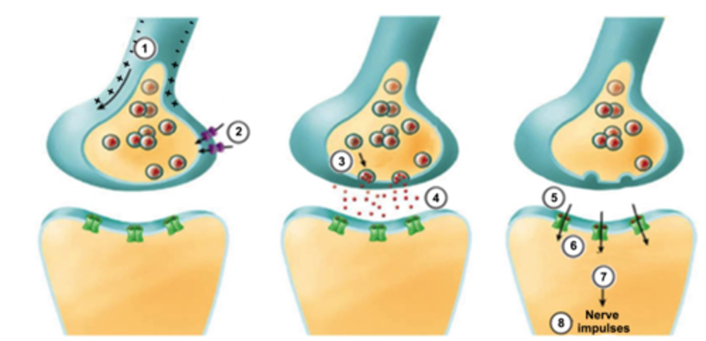
Describe in order (briefly) the events happening between 1 (action potential reaching the pre-synaptic axon terminal) and 8 (action potential / nerve impulses propagating in the post synaptic cell)
1) Action potential arrives at pre-synaptic axon terminal
2) Voltage gated Ca++ channels open on the pre-synaptic terminal
3) Ca++ causes synaptic vesicles to fuse to the cell membrane release NT via exocytosis
4) Neurotransmitter diffuses across synaptic cleft
5) Neurotransmitter binds to receptors on the post-synaptic cell membrane
6) Ligand-gated ion channels open on the post synaptic membrane and Na++ flows into post-synaptic cell
7) Postsynaptic potential reaches threshold and fires an action potential
8) Nerve impulses continue to propagate
Chemical synapses are characterised by all of the following, except:
a) the release of neurotransmitter by the pre-synaptic membranes.
b)receptors on the post-synaptic membranes that bind neurotransmitter.
c) ions flowing directly from the pre-synaptic to the post-synaptic neuron through protein channels.
d) a gap filled with fluid separating the pre- and post- synaptic neurons.
c) ions flowing directly from the pre-synaptic to the post-synaptic neuron through protein channels.
With respect to size and myelination of cell axons, the velocity of nerve impulse conduction is greatest in
a) small-diameter myelinated fibers
b) large-diameter nonmyelinated fibers
c) large-diameter heavily myelinated fibers
d) small-diameter nonmyelinated fibers
c) large-diameter heavily myelinated fibers
Clinical application: Multiple sclerosis (MS) is a disease that causes the progressive destruction of myelin sheaths / demyelination of nerves in the CNS and PNS.
a. What effect would this have on impulse conduction in the affected fibres?
b. What are some of the possible symptoms this might cause (explain which nerves would be affected)
a) The destruction of myelin sheaths slows the propagation of nerve impulses
b)
- Numbness or weakness in one or more limbs that typically occurs on one side of your body at a time, or the legs and trunk
- electric-shock sensations that occur with certain neck movements, especially bending the neck forward (Lhermitte sign)
- Tremor, lack of co-ordination or unsteady gait
- Effect on vision: partial or complete loss, double or blurry vision
-Other symptoms may include slurred speech, fatigue, dizziness, tingling or pain in parts of the body
Explain the features of the alarm response
1. Pupils dilate to help take in more visual information
2. Responses associated with exercise, emotion, excitement
3. Heart: increased rate and force of contraction
4. fight or flight
5. blood is channeled toward core / vital organs away from the periphery
6. Decreased gut functions (loss of apetite, decreased salivation
Explain the features of the relaxation response
1. pupils contract
2. responses associated with rest, repletion, relaxation
3. increased gut functions
4. Heart rate: Decreased rate and force of contraction
5. dilation of peripheral blood vessels
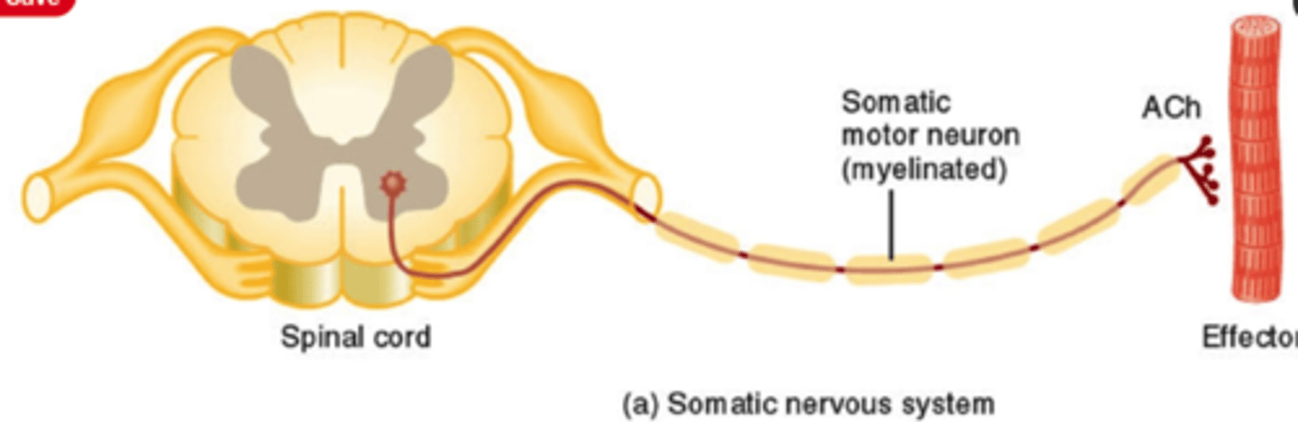
Central Nervous system to the effector for the Somatic nervous system
See Image
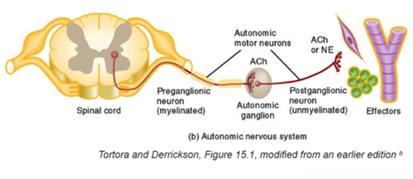
Central Nervous system to the effector for the Autonomic nervous system,
See image
Match the name of the endocrine gland to its location on the image below.
A) Hypothalamus
B) pituitary gland
C) Thyroid
D) Thymus
E) Pancreas
F) Ovaries
G) Testies
H) Adrenals
I) Pineal Gland
Fill in the table below by dragging and dropping the actions of each class of hormone in order of it actions.
See image
describe the relationship between the hypothalamus and Anterior pituitary gland.
Connects to the Hypothalamus via: Hypophyseal portal system (vascular connection)
Response to (hormones or neural connections) : releasing and inhibiting hormones from ventral hypothalamus: GHRH, GHIH, TRH, CRH, GnRH, PIH
Releases (hormones):GH, TSH, ACTH, FSH, LH, PRL
describe the relationship between the hypothalamus and Posterior pituitary gland.
Connects to the Hypothalamus via: Hypothalamic-hypophyseal tract (neural connection)
Response to (hormones or neural connections) :Respond directly to hypothalamic neurons
Releases (hormones):Oxytocin + ADH
The physiological 'stress response' has three components: an alarm response, an extended alarm response and a resistance reaction. The alarm response is mediated by activation of the (a) ...... nervous system.The extended alarm response is mediated by the hormones (b) ...... and (c) ....... secreted by the (d) ..... gland.The resistance reaction is mediated by the hormone (e) ....... secreted by the (f) ...... gland
(a)
sympathetic
(b)
epinephrine / norepinephrine (order independent)
(c)
epinephrine / norepinephrine (order independent)
(d)
adrenal medulla
(e)
cortisol
(f)
adrenal cortex
Complete the diagram below with the most appropriate terms for, or descriptions of, what happens in your brain and body when you perceive yourself to be in a threatening situation.
See Image
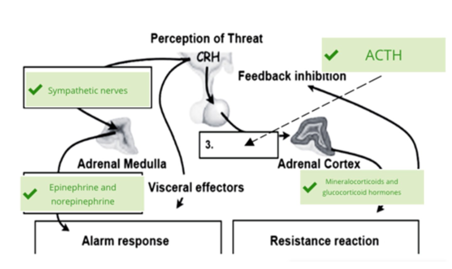
List SIX changes that occur in your body very rapidly as a result of sympathetic autonomic activation when you sense that you are in immediate danger
Heart: Increased rate and force of contraction
Skin: contraction of erector pilli muscles, increased secretion from sweat glands
Pupils: Dilation
Peripheral capillaries: Constriction of blood vessels
Lungs: Dilation of bronchi
Adrenal gland: release of epinephrine / norepinephrine to supplement and prolong the alarm response