A&P Unit 4 - Muscles: Part 1
1/38
There's no tags or description
Looks like no tags are added yet.
Name | Mastery | Learn | Test | Matching | Spaced |
|---|
No study sessions yet.
39 Terms
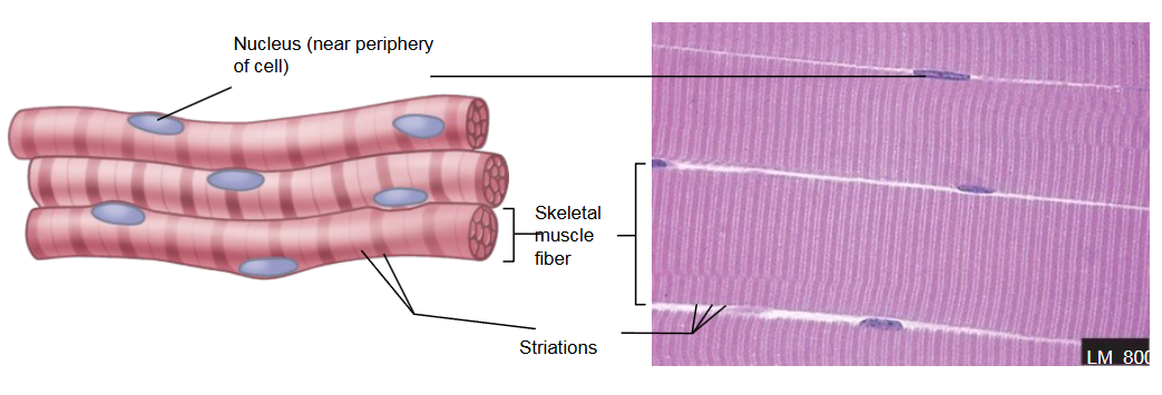
Skeletal muscle
Location: attached to skeleton or other connective tissue
Cell appearance: long and cylindrical
Nucleus: multinucleated, located on periphery
Function: move the body
STRIATED
VOLUNTARY
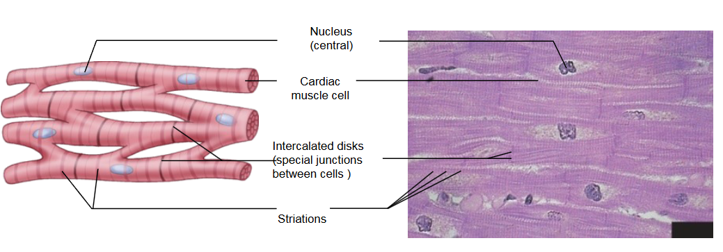
Cardiac muscle
Location: heart
Cell appearance: cylindrical, branch
Nucleus: single nucleus, centrally located
Function: contract the heart; force for moving blood through vessels
Features: branching fibers; intercalated discs with gap junctions
STRIATED
INVOLUNTARY
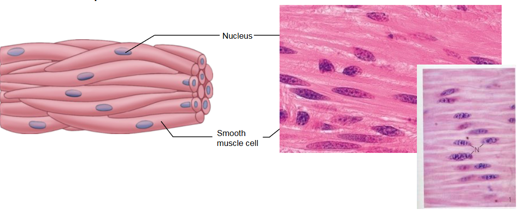
Smooth muscle
Location: walls of organs, blood vessels, eyes, glands, skin
Cell appearance: spindle-shaped
Nucleus: single, centrally located
Function: muscular function of organs; multiple functions
Features: gap junctions
NON-STRIATED
INVOLUNTARY
Functions of skeletal muscle tissue
Produce skeletal movement
Maintain posture and body position
Support soft tissues
Guard body entrances and exits
Maintain body temperature
Provide nutrient reserves
Properties of skeletal muscle tissue
Excitability: react to stimuli
Conductivity: spread electrical impulse through muscle cell
Contractility: shorten when stimulated
Extensibility: can stretch without harm
Elasticity: can recoil from stretch
Skeletal muscles are organs made of
Skeletal muscle tissue
Connective tissue
Nerves
Blood vessels
Fascia
Wraps muscle group
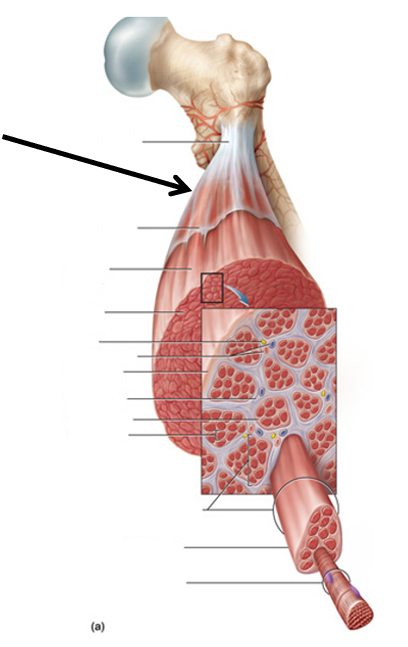
Epimysium
Wraps muscle
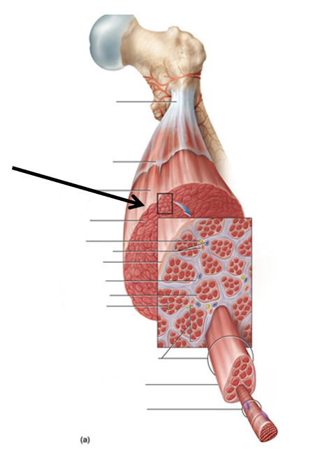
Perimysium
Wraps fascicle
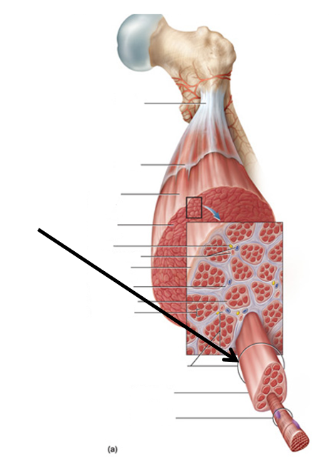
Endomysium
Wraps cell
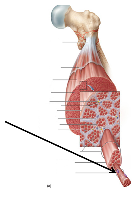
Muscle cell (myocyte) =
Muscle fiber
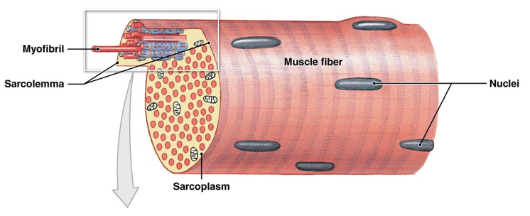
Muscle words start with
Myo- or sarco-
Sarcoplasm
Muscle cytoplasm
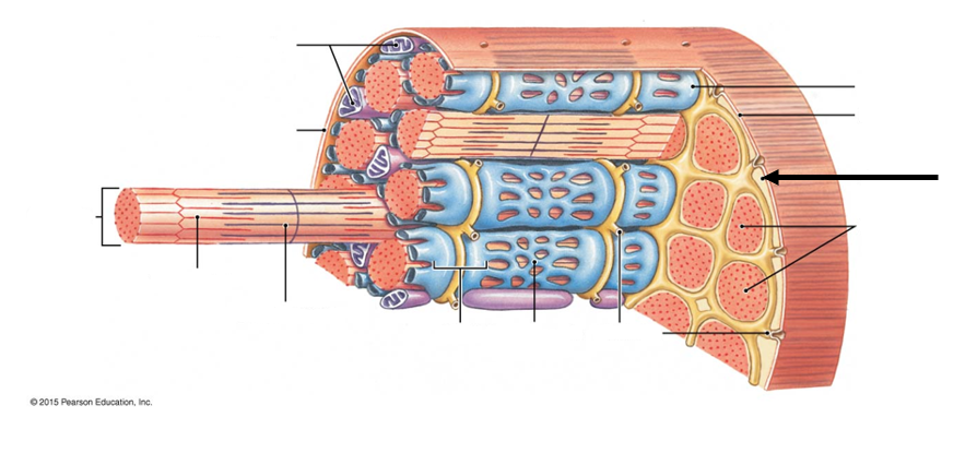
Sarcolemma
Muscle cell plasma membrane
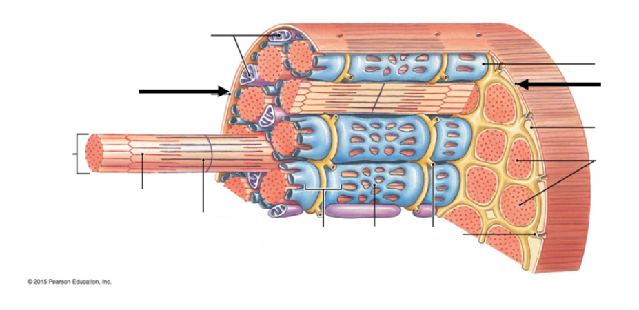
Myofibril
Inside the muscle cell
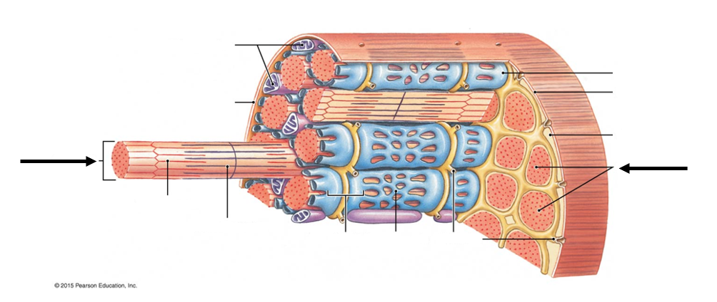
Myofilament
Make up the myofibrils
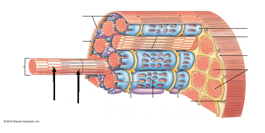
Transverse (T)-Tubules
Tubular infoldings of sarcolemma, go through cell
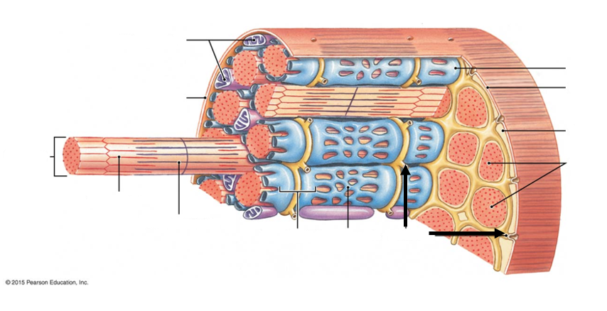
Sarcoplasmic Reticulum
Muscle cell ER
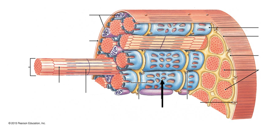
Terminal Cisternae
Enlarged portions of sarcoplasmic reticulum
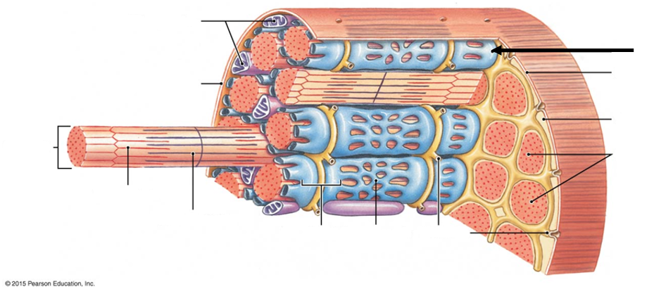
Myofibrils features
Repeating units of sarcomeres that run the length of the muscle fiber
Has stripes called striations
Made of thick and thin myofilaments
Sarcomere
Muscle cell contractile unit
During contraction, sarcomeres shorten
Runs from one Z line to the next Z line
Z line
In the middle of I band, hold sarcomere together, actin connected here
A band
Runs the length of the thick filament
H band
Composed of thick filament only, in the middle of A band
M line
In the middle of H band
I band
Composed of thin filament only
Thick filaments
Composed of myosin
Thin filaments
Composed of mostly actin
Contains troponin and tropomyosin
Troponin
Switch - binds Ca2+, actin, tropomyosin
Tropomyosin
Blocks binding sites for myosin
Sliding Filament Theory of Contraction
When a skeletal muscle fiber contracts, the thin filaments slide past the thick filaments
When muscle contracts
I band - decreases
H band - decreases
A band - no change
Zone of overlap - increases
Distance between Z-lines - decreases
Neuromuscular Junction (NMJ)
Composed of the axon terminal, motor end plate, and the synaptic cleft
Special type of synapse
Events at the NMJ
Electrical signal called an action potential travels along a nerve fiber
An action potential is a sudden change in membrane potential
The action potential causes the opening of voltage-gated Ca2+ channels
Calcium (Ca2+) ions enter into the axon terminal
This causes the exocytosis of vesicles filled with Acetylcholine
Acetylcholine is a neurotransmitter
Acetylcholine is released into the synaptic cleft and binds to Acetylcholine (Ach) Receptors
Ach-Receptors are ligand-gated Na+ channels
Bind of Ach to Ach-Receptors causes sodium (Na+) to enter the cell and change the membrane permeability
This generates an action potential along the sarcolemma of the muscle fiber
Excitation-Contraction Coupling
Neural control - A skeletal muscle fiber contracts when stimulated by a motor neuron at a neuromuscular junction. The stimulus arrives in the form of an action potential at the axon terminal
Excitation - The action potential causes the release of Ach into the synaptic cleft, which leads to excitation - the production of an action potential in the sarcolemma
Release of calcium ions - This action potential travels along the sarcolemma and down T tubules to the triads. This triggers the release of Calcium ions (Ca2+) from the terminal cisternae of the sarcoplasmic reticulum
Contraction cycle begins - The contraction cycle begins when the calcium ions (Ca2+) bind to troponin, resulting in the exposure of the active sites on the thin filaments. This allows cross-bridge formation and will continue as long as ATP is available
Sarcomere shortening - As the thick and thin filaments interact, the sarcomeres shorten, pulling the ends of the muscle fiber closer together
Generation of muscle tension - During the contraction, the entire skeletal muscle shortens and produces a pull, or tension, on the tendons at either end
Cross-bridge formation
Contraction cycle begins - Arrival of calcium ions (Ca2+) within the zone of overlap in a sarcomere
Active-site exposure - Calcium ions bind to troponin, weakening the bond between actin and the troponin-tropomyosin complex. The troponin molecule then changes position, rolling the tropomyosin molecule away from the active sites on actin and allowing interaction with the energized myosin heads
Cross-bridge formation - once the active sites are exposed, the energized myosin heads bind to them, forming cross-bridges
Myosin head pivoting - After cross-bridge formation, the energy that was stored in the resting state is released as the myosin head pivots toward the M line. This is called a power stroke. When it occurs, the bound ADP and phosphate groups are released
Cross-bridge detachment - When another ATP binds to the myosin head, the link between the myosin head and the active site on the actin molecule is broken. The active site is now exposed and able to form another cross-bridge
Myosin reactivation - Myosin reactivation occurs when the free myosin head splits ATP into ADP and P. The energy released is used to recock the myosin head
Steps that initiate a muscle contraction
Ach released - Ach is released at the NMJ and binds to Ach receptors on the sarcolemma
Action potential reaches T Tubule - An action potential is generated and spreads across the membrane surface of the muscle fiber and along the T tubules
Sarcoplasmic reticulum releases Ca2+ - The sarcoplasmic reticulum releases stored calcium ions
Active site exposure and cross bridge formation - Calcium ions bind to troponin, exposing the active sties on the thin filaments. Cross-bridges form when myosin heads bind to those active sites
Contraction cycle begins - The contraction cycle begins as repeated cycles of cross-bridge binding, pivoting, and detachment occur - all powered by ATP
Steps that end a muscle contraction
Ach is broken down - Ach is broken down by acetylcholinesterase (AchE), ending action potential generation
Sarcoplasmic reticulum reabsorbs Ca2+ - As the calcium ions are reabsorbed, their concentration in the cytosol decreases
Active sites covered, and cross-bridge formation ends - Without calcium ions, the tropomyosin returns to its normal position and the active sites are covered again
Contraction ends - Without cross-bridge formation, contraction ends
Muscle relaxation occurs - The muscle returns passively to its resting length
Important to know about contraction cycle
Starts when calcium ions arrive from the sarcoplasmic reticulum
Calcium ions bind to a protein called troponin, causing the exposure of the active (binding) site on actin
Activated myosin heads form cross-bridge with the actin binding site
When myosin heads pivot, they move the thin filament, this is the “power stroke”
Myosin needs another ATP to repeat the cycle
The cycle continues as long as calcium and ATP are present
Can repeat several times each second