Human Physiology
1/149
There's no tags or description
Looks like no tags are added yet.
Name | Mastery | Learn | Test | Matching | Spaced | Call with Kai |
|---|
No analytics yet
Send a link to your students to track their progress
150 Terms
1st Step of Scientific Method
Make observations
2nd step of Scientific Method
Form a hypothesis, the hypothesis must be testable
3rd step in scientific method
Design a conductible experiment
Homeostasis
Dynamic Constancy in body’s Internal Environment

External Environment
The space that surrounds the body
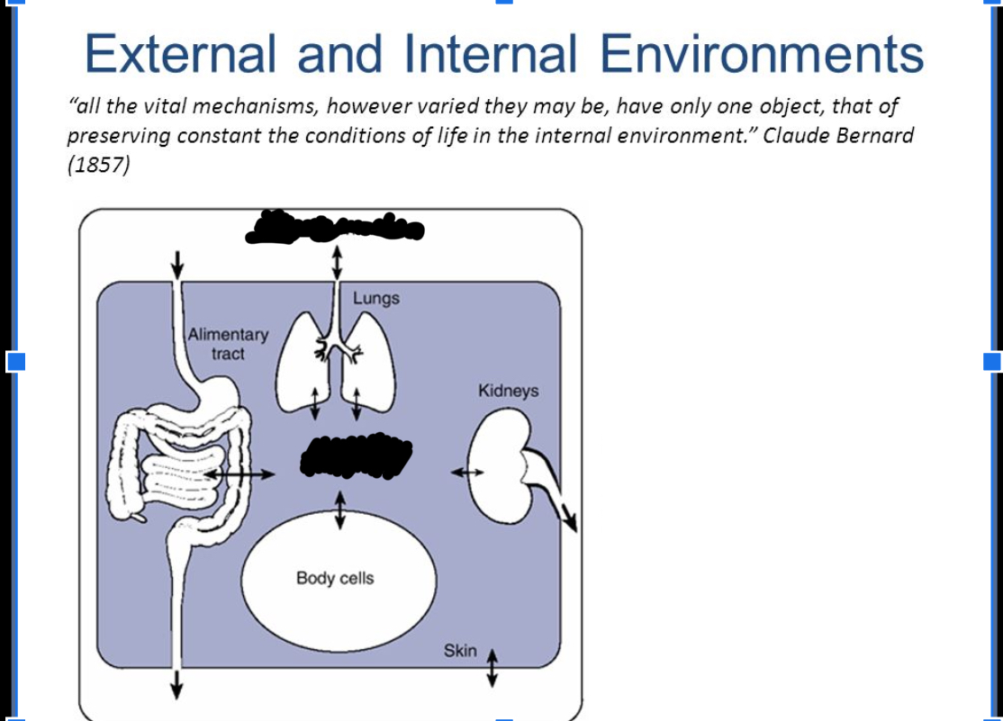
Internal Environment
The extracellular fluid surrounds the cells of the body
What is Homeostasis controls
Body Temperature
Blood Volume
Blood Sugar
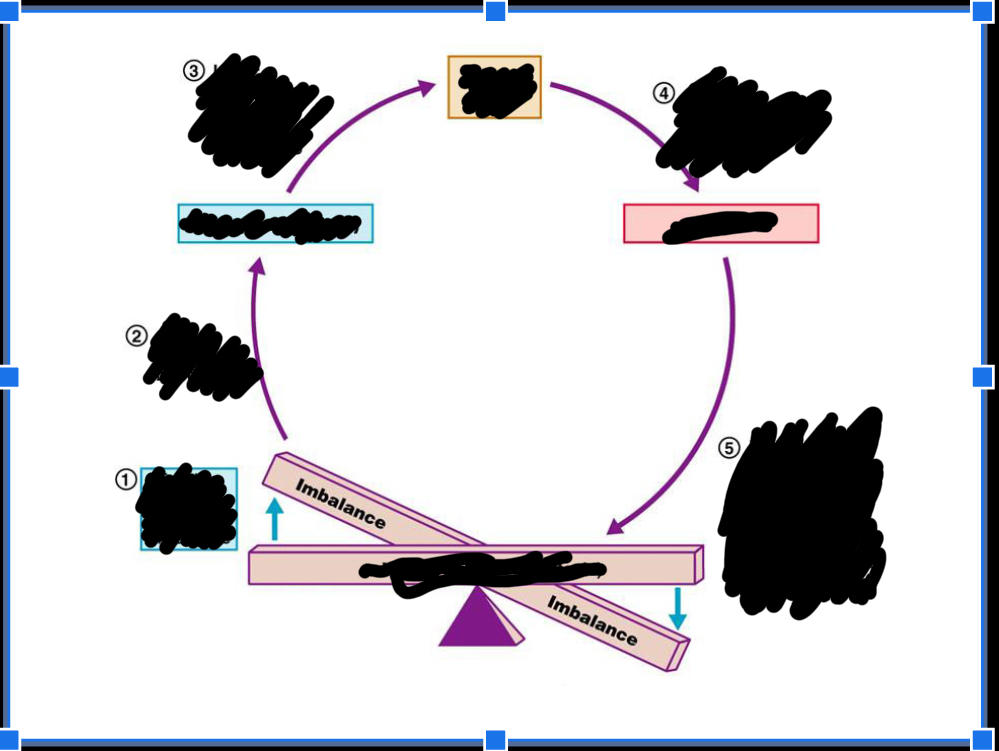
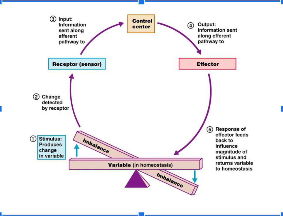
How is Homeostasis Maintained in the Body (Feedback System)
Controlled Variables
Receptor
Control Center
Effector
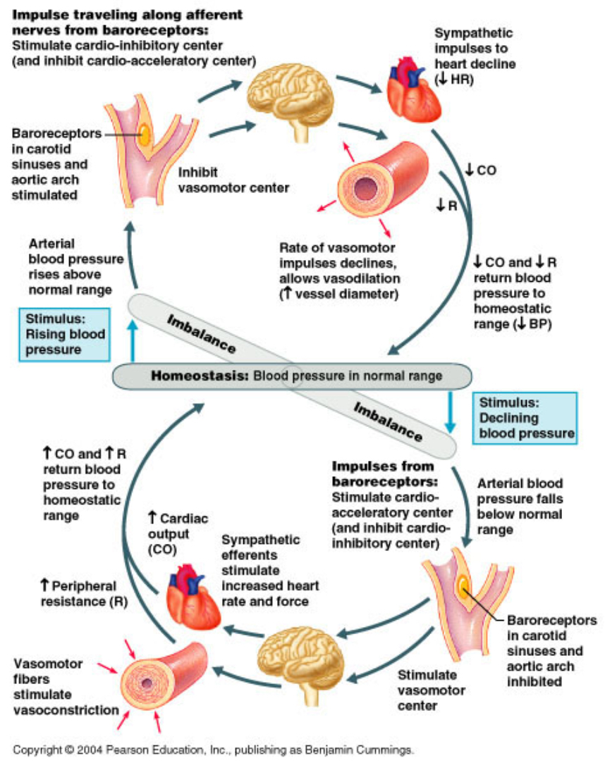
Negative Feedback System
Blood Loss → Baroreceptor → Brain → Increased HR, Contractility, and cardiac output; Vasoconstriction
Sympathetic Nervous System Activated
Endocrine Regulation of Homeostasis
Decreased Blood Sugar → (Consume sugars) → Increased Blood Sugar → Pancreatic Cells → Elevated Insulin Protection → Repeat
Positive Feedback System
Strengthens or reinforces a change in a controlled variable
Can only be stopped by some events outside the system
Not Common
Negative feedback regulates homeostatic conditions over long periods
4th step of Scientific Method
Analyze the data
5th Step of Scientific Method
Duplicate results to accept conclusion
6th Step of the Scientific Method
Several verified Hypotheses become a theory
Good Physiological Research requires
1.) Quantifiable measurements
2.) An Experimental and Control group
3.) Statistical Analysis
4.) Review and publication by peer-reviewed Journal
1st Step of Developing Pharmaceuticals
Basic research is conducted for years before drug given to person
2nd Step of Developing Pharmaceuticals
Research begins by studying the effects of a chemical on cells in vitro (in culture dish)
3rd Step of Developing Pharmaceuticals
Next, studies are done in animals (usually rats and mice) to see if the same effect occurs in vivid and if there’s any toxic side effects.
Phase 1 Clinical Trials
Test drug on healthy human volunteers to test for side effects, rates of passage and dosage
Phase II Clinical Trials
Tests Effectiveness on people with a particular disease
Phase III Clincal Trials
Conducted on large # of people, different races, sexes, and complex health conditions. Many drugs fail here
Phase IV Clinical Trials
Tests other applications of the drugs
1st Level of Organization
Molecule - Microscopic Level
2nd Level of Organization
Cell- Basic unit of structure and function of living things
3rd Level of Organization
Tissue- Group of similar cells that perform a similar function
4th Level of Organization
Organ - Group of two or more tissues into structure and functional units
5th Level of Organization
System- Group of organs that work together to perform related functions
6th Level of Organization
Organism- Systems working together in coordination
4 Major Primary Tissues
Muscle Tissue
Nervous Tissue
Epithelial Tissue
Connective Tissue
Muscle Tissue
Specialized for Contraction
Three Types
Skeletal Muscle (Voluntary Muscles)
Cardiac Muscle (Involuntary Muscles)
Smooth Muscle (Hollow Organs; Bladder, blood vessels, bronchioles)
Nerve Tissue
Found In Brain, spinal cord, and nerves
Composed of Nerves and neuroglia
Function in communication
Epithelial Tissue
Forms Membrane that cover surfaces, line inside of hollow organs
Epithelial membranes are classified by # of layers
Simple; one layer and for transport
Stratified; multiple layers, provides protection
Connective Tissue
Characterized by a matrix made up of; protein fibers, extracellular materials, and specialized cells
Function of connecting and supporting
4 Major categories:
Connective Tissue proper
Cartilage
Bone
Blood
Organ Systems
Integumentary
Organs
Skin, Hair, Nails
Primary Functions
Protection, Thermoregulation
Organ Systems
Nervous
Organs:
Brain, Spinal Cord, Nerves
Primary Functions:
Regulation of other body systems
Organ Systems
Endocrine
Organs:
Hormone-secreting glands, pituitary, thyroid, and adrenal glands
Functions
Secretion of regulatory molecules called hormones
Organ Systems
Skeletal
Organs:
Bones, Cartilages
Functions:
Movements and support
Organ Systems
Muscular
Organs:
Skeletal Muscles
Functions:
Movements of the skeleton
Organ Systems
Circulatory
Organs:
Heart, Blood Vessels, Lymphatic vessels
Functions:
Movement of blood and lymph
Organ System
Immune
Organs:
Red Bone Marrow, Lymphoid Organs
Functions
Defense of the body against invading pathogens
Organs Systems
Respiratory
Organs:
Lungs, Airway
Functions:
Gas Exchange
Organ Systems
Urinary
Organs:
Kidneys, Ureters, Urethra
Functions
Regulation of blood volume and composition
Organ Systems
Alimentary
Organs:
Mouth, Stomach, Intestine, Liver, Gallbladder, Pancreas
Function:
Breakdown of food into molecules that enter the body
Organ Systems
Reproductive
Organs:
Gonads, External genitalia, Associated glands and ducts
Functions:
Continuation of the Human Species
Principle parts of cells
Plasma Membrane
Flexible, separates from the external environment of the cell, selectively permeable, communication.
Principal parts of cells
Cytoplasm
Fluid part of the cells (Cytosol) and little organelles that do the functions
Principal parts of cells
Nucleus
Contains DNA and directs cell activities
What passes through Plasma Membrane
Small, hydrophobic substances, such as Oxygen, CO2 and small lipid soluble molecules
Functions of Membrane Proteins
Ion Channels (integral)
Forms a pore, which specific ion can flow to cross membrane, include specific channels for specific ions
Functions of Membrane Proteins
Carrier (Integral)
Transports a specific substance across membrane by undergoing a change in shape. Example amino acids needed to, synthesis new proteins enter via ____ Proteins. Also known as ______
Functions of Membrane Proteins
Receptor (Integral)
Recognizes specific ligands and alters cell function in some ways. Example antidiuretic hormone binds to ______ in the kidneys and changes the water permeability of certain plasma membranes
Functions of Membrane Proteins
Enzyme (Integral and Peripheral)
Catalysts reactions occur both ______ and _____, the cell. Example Lactase protruding from epithelial cells lining small intestine splits the disaccharide lactose in the milk you drink
Functions of Membrane Proteins
Linker (Integral and Peripheral)
Anchors filaments ____ and _____ the plasma membrane giving shape and structural stability. May also link 2 cells together
Functions of Membrane Proteins
Cell Identity Marker (Glycoprotein)
Distinguishes cells, from anyone else’s. Important class of markers are major hisocompatibility proteins.
Other Plasma Membrane Components
Carbohydrates
Attached to lipids (glycolipids) and to proteins (glycoproteins); serve as antigens and interactions with regulatory molecules
Other Plasma Membrane structures
Cholesterol
Gives flexibility to membrane
Phagocytosis
Large extracellular substances into the cell
Some cells, like neutrophils and macrophages , can perform amoebiod movement by extending psuedopods to pull the cell forward.
Pseudopods engulf bacteria, dead cells, r other organic materials and then fuse together to form a food vauole
The food vacuole fuses with a lysosome, and the material is digested
Very important for body defense, inflammation, and apoptosis
Endocytosis
Process for bringing large materials into the cells
Plasma membrane furrows inward rather than extending outward. A Small part of the membrane surrounding the substance pinches off and is brought in as a vesicle
Encdocytosis (Pinocytosis)
Nonspecific
Endocystosis (Receptor-mediated endocytosis)
Specific, has receptor proteins in the membrane that will bind to the substance to be brought in
Exocytosis
Large cellular products (proteins) are moved out of the cell
The golgi apparatus packages proteins into vesicles that fuse to the plasma membrane, and the contents spill out of the cell
Used to release digestive enzymes, hormones, and neurotransmitters from certain cells
Cytoplasm
Materials in a cell
Includes organelles, a fluid called cytosol, the cytoskeleton, inclusions
Inclusions- Stored chemical aggregates such as glycogen granules, melanin granules, and triglycerides.
Network of protein filaments that extend through cytosol
Provides structural framework
Aids in movement
Three Types
Microfilaments
Intermediate filaments
Microtubules
Microfilaments (Actin Filaments)
Thinnest elements, made up of actin
Most prevalent at the edge of the cell
Functions
Help generate movements
Provide mechanical Support
Form microvilli
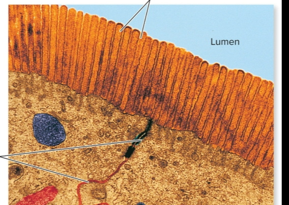
Microvilli
Folds in the plasma membrane that increase the surface area for chemical reacations and rapid diffusion
(Intestines)
Intermediate Filaments
Intermediate in size
Smaller than microtubules. Larger than microfilaments
Functions
Found in places where cells encounter stress
Help position organelles
Attach cells to one another
Microtubules
Largest of the cytoskeletal components
Hollow tubes composed mainly of tubulin
Functions
Determine cell shape
Movements of organelles
Found in cilia and flagella
Cilia
Tiny hairlike Structures composed of microtubules that project from the plasma membrane
Primary Cilium
Most cells have this nonmotile with “9+0” structure; may have a sensory functions in some cells
Motile Cilia
Propel fluid across surface of cells
Found in respiratory tract and uterine tubes
Has a “9+2” arrangement
Flagellum
A single whip-like structure that can propel a cell forward
Composed of microtubules with a “9+2” arrangement— similar to cilia but typically much longer
The sperm is the only cell in the human body with it
Organelles
Tiny specialized structures within the cell that have characteristic shapes
Perform specific functions in cellular growth, maintenance and reproduction
types
Ribosomes
ER
Golgi
Mitochondria
Lysosomes, peroxisomes, and proteasomes
Mitochondria
Site of energy production (ATP)
Structure
Inner membrane and an external membrane seperated by a intermemcrabous space
Inner membrane is folded into cristae to increase surface area for reactions
Central area is fluid and called the matrix
Ribosomes - Protein factory
Messanger RNA takes genetic information to the ribosome so a protein can be assembled
Very small; made of 2 subunits
Found free in the cytoplasm or associated with the granular endoplasmic reticulum
Serves as enzymes called ribozymes that are needed for protein synthesis
Endoplasmic Reticulum (ER)
The is a system of membranous passageways from the nuclear membrane to the plasma membrane
Granular (rough )
Has ribosomes embedded on the outer surface
Functions in protein synthesis and secreation
Agranular (Smooth ) has many functions, depending on the cell. Fatty acid and steroid synthesis
Golgi Complex (apparatus)
Transport vesicles made in the rough ER
Transport vesicles move to ____ _____
Proteins enter lumen of ____
Proteins modified as they travel through cristernae
Modified proteins are packaged and sorted
Secretory/new transport vesicles are formed, bring the proteins to the final destination
Lysosomes - Digestive Enzymes
Membrane-enclosed vesicles containing digestive enzyme that digest a wide variety of substrates
Peroxisomes
Smaller than lysosomes
Contain oxidases that help metabolize amino acids and fatty acids
Proteasomes
Degrades unwanted or damaged proteins that are in the cytosol
Cell Nucleus
Most cells have one ___
Muscle cells have hundreds; mature RBC have none
The ___ is enclosed by the nuclear envelope made of two membranes
Outer membrane continuous with rough ER
Inner membrane often fused to outer by nuclear pore complexes, which allow small molecules and RNA to move in and out
Cluster of DNA,RNA and proteins
Gene expression
Genetic Transcription- The gene on the DNA is transcribed as messenger RNA, which can leave the nucleus
Genetic translation- The messenger RNA is then translated at the ribosomes to assemble the proper amino acid sequence
Genome
Is all the genes in a particular individual or all the genes of a particular species
Proteome
All the proteins that are produced from the genome
How can a gene code fore more than one protein
Posttransalational modification by
Methylation
Phosphorylation
Cutting into small units
Extracellular Environment
Includes everything located outside the cells
Cells receive nourishment from and release wastes into the ______ ______
Cells communicate with each other by secreting chemical regulators into the _____ _______
Body Fluids
67% of our water is within cells in the intracellular compartments
33% is in the extracellular compartment. Of this:
a. 20% is in blood plasma
b. 80% makes up what is called tissue fluid, or interstitial fluid; connects the intracellular compartment with the blood plasma
Transport of Bulk Molecules
Take in
Phagocytosis
Endocytosis (pintocytosis, receptor-mediated)
Spit out
Exocytosis
Plasma membrane permeability
Is selectively permeable
Passive Transport
Molecules move from higher to lower concentration without using metabolic energy
Active Transport
Molecules move from lower to higher concentration using ATP and specific carrier pumps
Noncarrier-mediated (passive)
Simple diffusion of lipid-soluble molecules
Simple diffusion of ions through channels
Simple diffusion of water = Osmosis
Carrier-mediated
Facilitated diffusion (Passive)
Active Transport (Active)
Diffusion Rate
Measured by the # of diffusing particles per unit of time
Steepness of concentration gradients — Driving force
Permeability of the membrane
Temperature of the solution
Surface area of the membrane — microvilli
Simple diffusion
Nonpolar molecules (oxygen, Carbon Dioxide, Steroid)
Ions and water molecules through specific channels
Facilitated Diffusion: Carrier proteins
What can pass through the plasma membrane
Charged Ions can pass through an ion channel, that maybe gated. K+,Na+,Ca+2,Cl-
Osmotic Pressure
Osmotic pressure is the force surrounding a cell required to stop Osmosis
A higher solute concentration would require a higher osmotic pressure
Osmolarity
Unit of concentration that gives the total molarity of all soutes
2M glucose solution = 1M glucose + 1M fructose
Water always goes from lower to higher
Normal Blood Plasma Osmolarity
300mOsM
Isotonic
Same osmolarity in the 2 environments (No net movement)
Hypotonic
Lower than plasma osmolarity, Will push water into the cell; cell will swell and could lyse
Hypertonic
Higher than plasma osmolarity; Will pull water out of the cell, cell will shrivel up and could crenate
Facilitated Diffusion —
Transport carriers for glucose are designated GLUT followed by the number of the isoform
GLUT 1
GLUT 2
GLUT 3
GLUT 4