PNS: Neuro Reflexes
1/26
Earn XP
Description and Tags
Write an appropriate answer to each definition.
Name | Mastery | Learn | Test | Matching | Spaced | Call with Kai |
|---|
No analytics yet
Send a link to your students to track their progress
27 Terms
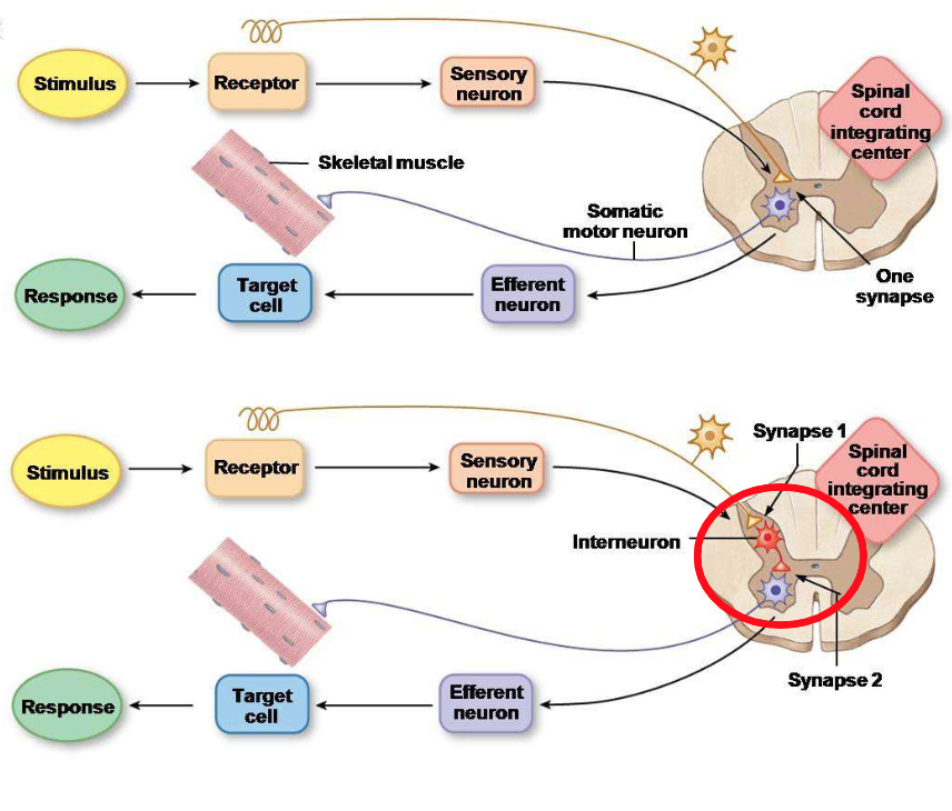
Specify the difference between a monosynaptic reflex and a polysynaptic reflex.
A monosynaptic reflex involves only one synapse between the sensory neuron and the motor neuron, allowing for a quick response, while a polysynaptic reflex involves two or more synapses and includes interneurons, resulting in a longer response time.
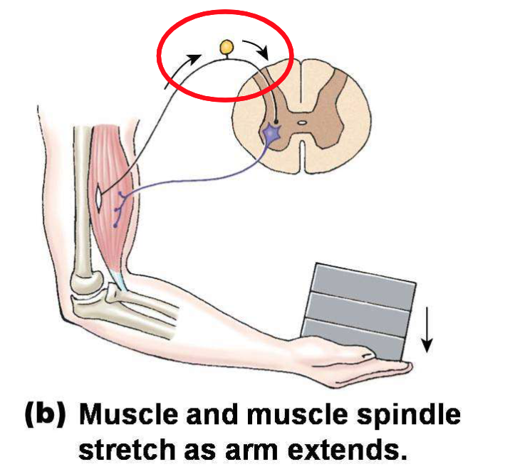
Explain the concept of the Golgi tendon reflex happening in this photo.
Once the load is placed on the hand, the muscle and muscle spindle stretch as the arm extends. Muscle spindle afferents fire more frequently.
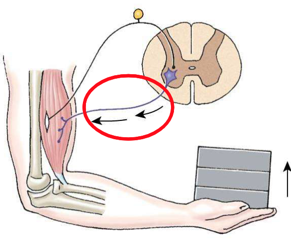
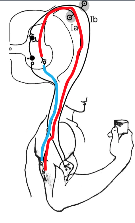
Explain the release of the alpha motor neurons happening during this stretch reflex.
During a stretch reflex, the activation of muscle spindle afferents leads to the stimulation of alpha motor neurons, causing the muscle to contract in response to the stretch.
To stabilize the joint posture, how would the inhibitory interneuron come into play?
The inhibitory interneuron inhibits motor neurons, allowing opposing muscles to relax and helping maintain joint stability.
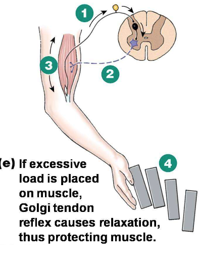
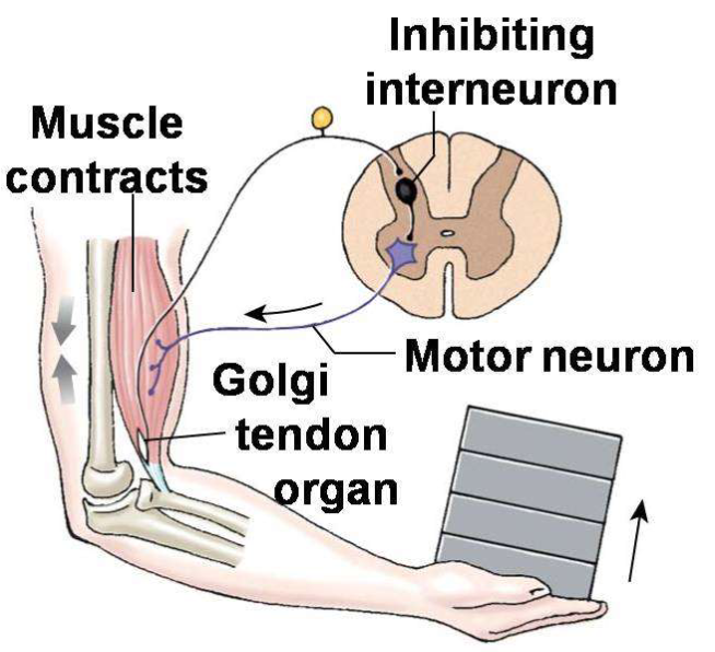
When you are holding more than enough load for your body to handle, how would the muscle relax?
Neurons from the GTO fire, allowing the inhibitory interneuron to inhibit the motor neuron; the muscle relaxes and the load drops. This prevents excessive contraction of antagonistic muscles, allowing for smoother and more controlled movement.
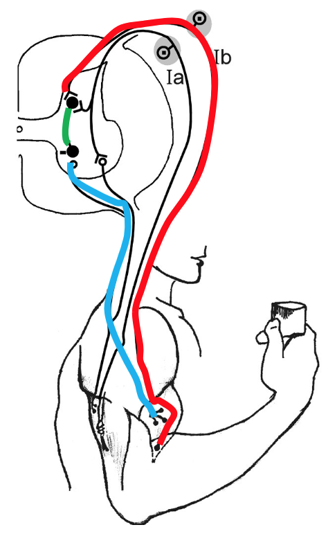
When the biceps are overactivated, what reduces their activity?
The GTR responds by inhibiting alpha motor neurons, leading to decreased biceps activity. The GTR sends Ib afferent fibres to the inhibitory interneuron to allow this process.
How could the triceps counteract the overactivation of the biceps?
The triceps activate a stretch reflex that facilitates contraction of the triceps while the biceps are inhibited, promoting balance in muscle activation.

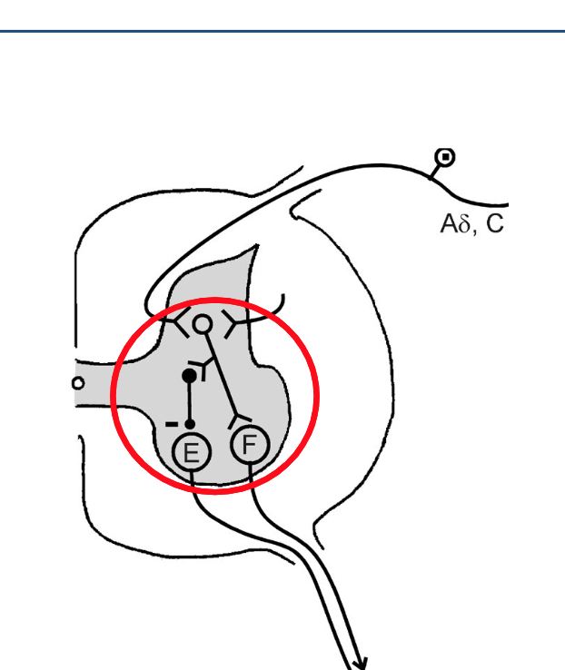
The person was holding a drink in their hand until a candle was lit under their arm. Explain step one of the “Flexion Withdrawal Reflex” in this image.
Step one involves the activation of nociceptors in the skin, which detect the painful stimulus (heat from the candle). This generates an action potential that travels along sensory neurons to the spinal cord.
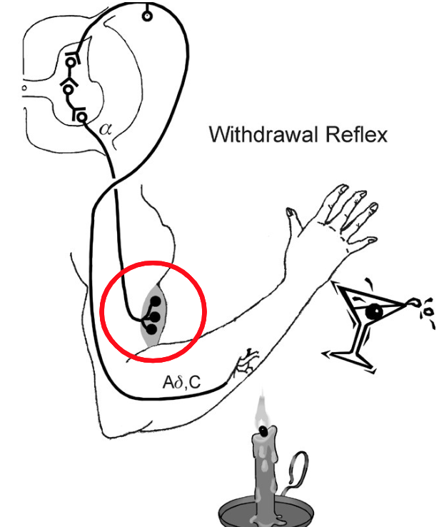
Explain step two of the “Flexion Withdrawal Reflex” in this image.
In step two, the sensory neurons synapse with excitatory interneurons in the spinal cord, which then stimulate alpha motor neurons to activate flexor muscles, flexing their arm away from the flame.
When you want to just activate your biceps, how does reciprocal inhibition come into play?
Activation of flexor motoneurons inhibits extensor motoneurons. That way, triceps muscles are relaxed while biceps contract, allowing smooth movement.
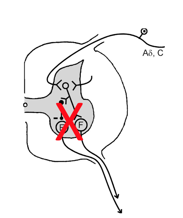
When is co-contraction of antagonists used (in terms of reciprocal inhibition)?
Co-contraction occurs when both flexors and extensors are activated simultaneously to stabilize a joint during movement, allowing for fine motor control and balance.
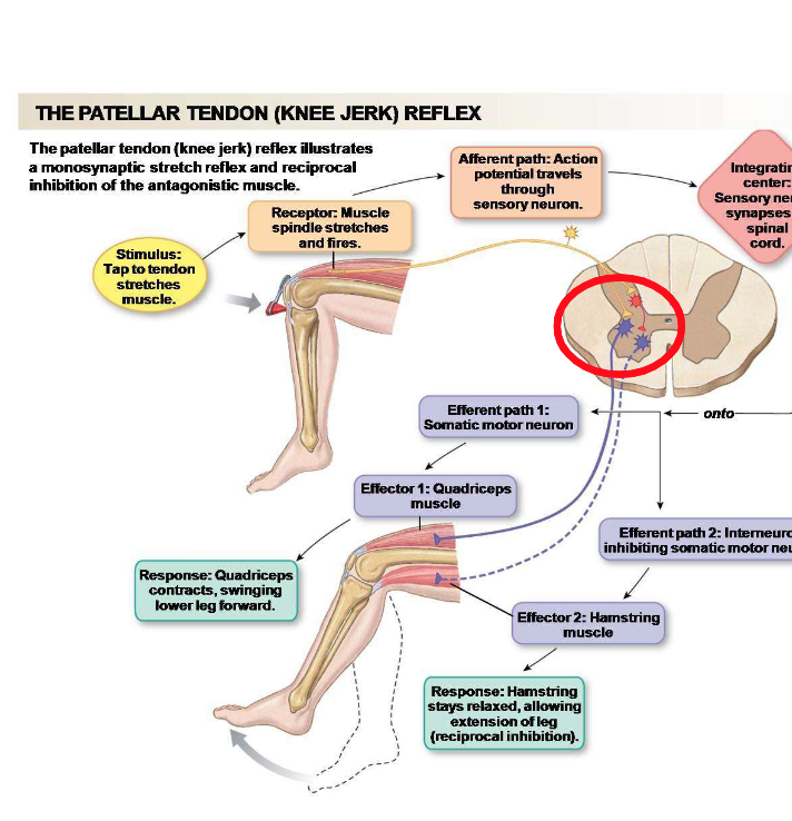
Describe the first step of the patellar tendon reflex.
Stimulus: Tap the tendon to stretch the quadriceps muscle.

Describe the second step of the patellar tendon reflex.
Stretching activates the muscle spindle receptors, which allow afferent fibres to fire action potentials.

Describe the third step of the patellar tendon reflex.
The afferent fibres make a direct excitatory synapse on the alpha motor neurons.

Describe the fourth step of the patellar tendon reflex.
Alpha motor neurons send signals back to the quadriceps, jerking the knee. This contraction results in the knee extending.

Describe the fifth step of the patellar tendon reflex.
The inhibitory neurons inhibit the alpha motor neurons to prevent excessive contraction of the hamstring muscles. This allows for fine motor control and prevents injury.
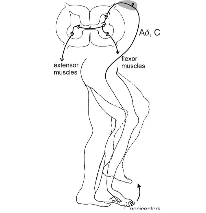
Explain the first step of the cross extension reflex based on this image.
When a painful stimulus is detected, sensory neurons transmit the signal to the spinal cord.

Explain the second step of the cross extension reflex based on this image.
The extensors of the leg that felt the pain are inhibited, while flexors contract. Interneurons in the spinal cord cross over to activate alpha motor neurons on the opposite side.
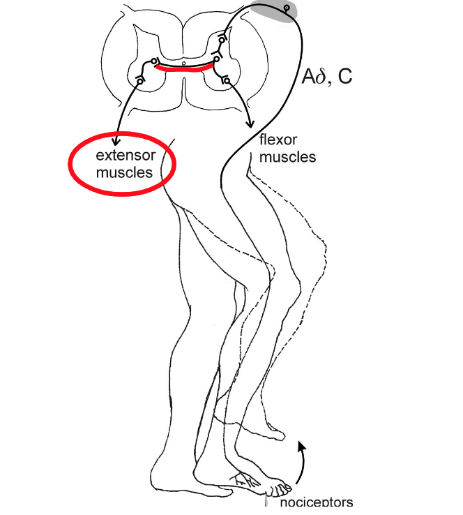
Explain the third step of the cross extension reflex based on this image.
The opposite leg's extensors contract to support the body, while the flexors are inhibited. This helps maintain balance during withdrawal from the painful stimulus.
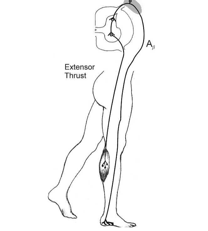
Explain the act of the extensor thrust reflex.
If you feel pressure on the bottom of your feet, you’ll push and stand up by using your extensor muscle contractions to extend the legs.
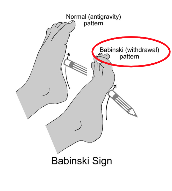
Provide an example of a Babinski sign. Explain the first step.
The first step of the Babinski sign occurs when the sole of the foot is stroked; this stimulation causes the big toe to extend upward while the other toes fan out.

Provide an example of a Babinski sign. Explain the second step.
Nociceptors detect stimulus. Afferent neurons travel to the spinal cord through the dorsal root.

Provide an example of a Babinski sign. Explain the third step.
Alpha motor neurons activate the muscles to respond; this typically results in the flexor withdrawal response in infants.

Provide an example of a Babinski sign. Explain the response in healthy adults or if the pathway is damaged/immature.
In healthy adults, the response is typically a downward flexion of the toes (plantar response). In cases of pathway damage or immaturity, an upward extension of the big toe may occur, indicating neurological dysfunction.

Explain the first step of the vestibulospinal reflex based on this image.
The woman tilts her head to the right, activating the otolith organ on the right side.

Explain the second step of the vestibulospinal reflex based on this image.
The tilt activates the vestibulospinal nuclei on the right side, which sends signals to the spinal cord.
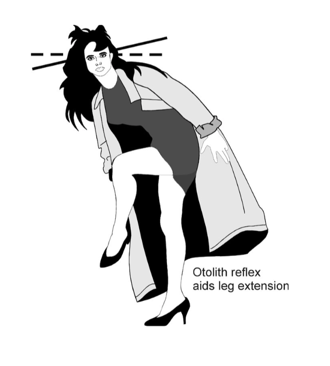
Explain the third step of the vestibulospinal reflex based on this image.
Extensor activity is reinforced on the right side, while flexor activity is inhibited on the left side, maintaining balance. The body is supporting weight on the right leg.