Health Q2
1/33
There's no tags or description
Looks like no tags are added yet.
Name | Mastery | Learn | Test | Matching | Spaced |
|---|
No study sessions yet.
34 Terms
MCL
prevents the leg from moving from side to side, works together with the LCL

PCL
Attaches to posterior tibia
Prevents backward sliding of tibia and forward sliding of femur. thicker compared to ACL.

Sacrum
connects the two hip bones, stabilization from behind.

ischium
the curved bone forming the base of each half of the pelvis. seat down bone.

Pelvic Gridle
ilium, ischium, pubis (hip bone)

patella
kneecap, prevent the knees from hyper extension

malleolus
the rounded bony protuberance on each side of the ankle

shin splint
a painful condition caused by the muscle tearing away from the tibia. The pain usually disappears after warm up
shin bone hairline fracture
pain either increases or stay the same after warming up.
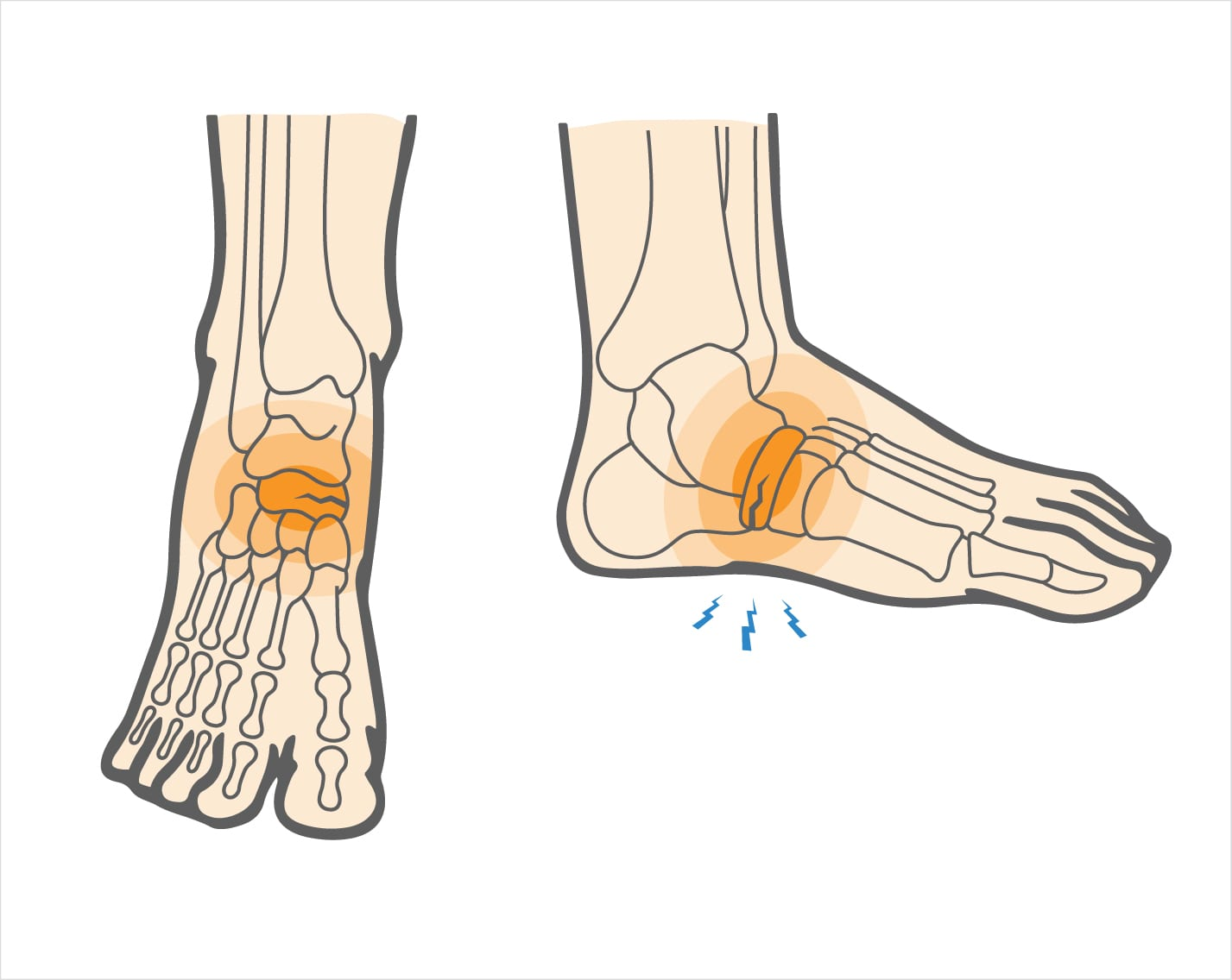
Tarsals
ankle bones. Includes
- talus
- navicular bone
- calcaneus

calcaneus
heel bone, where the AST (achilles tendon) is attached

metatarsals
foot bones

Phalanges
toes

ACL
holds sides and top and bottom of knees together
can be injured by twisting or tearing. X shaped. Thinner compared to PCL. Prevents the leg from moving forward and backward.

Meniscus
provides cushion in between the knees

lateral meniscus
cartilage in the knee

Meniscus tear
Impact form outside of the knee or not knowing how to properly decelerate or land a jump.
iliac crest
very prominant
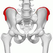
sacroiliac joint
responsible to hold the sacrum and ilic crest together
pubic symphysis
Provide stability for the hip bone in the front

Acetabulum
large socket in the pelvic bone for the head of the femur

neck of femur
usually where fracture occur

shaft of femur
body of femur

Fibula
The lateral and smaller bone of the lower leg, calf bone

tibia
the medial and larger bone of the lower leg (shin bone)

tibiotalar joint
joint between the tibia and the talus

Talus
ankle bone. the only bone in contact with the lower leg.

LCL
prevents the leg from moving from side to side, works together with the MCL

medial meniscus
C-shaped cartilage in the knee. the outer 1/3 has more blood supply, therefore faster at recovery. the inner 2/3 of the medial meniscus has little to none blood supply therefore hard to recover form injuries.

knee joint
includes
patella
meniscus
ligaments
labrum
cartilage between femur head and acetabulum
lateral malleolus
the longer malleolus
medial malleolus
the shorter malleolus
navicular bone
sentisve with lots of nerves in the tarsals