chp. 14: circulatory system
1/125
There's no tags or description
Looks like no tags are added yet.
Name | Mastery | Learn | Test | Matching | Spaced |
|---|
No study sessions yet.
126 Terms
circulatory system consists of
blood, blood vessels, and a muscular, pumping heart.
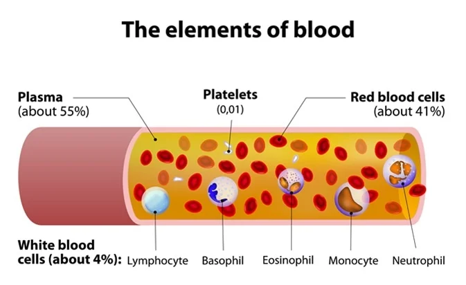
blood
a connective tissue made of:
Formed elements (cellular part)
erythrocytes, leucocytes, and thrombocytes
Plasma (extracellular part)
plasma proteins (albumen, globulins, fibrinogen)
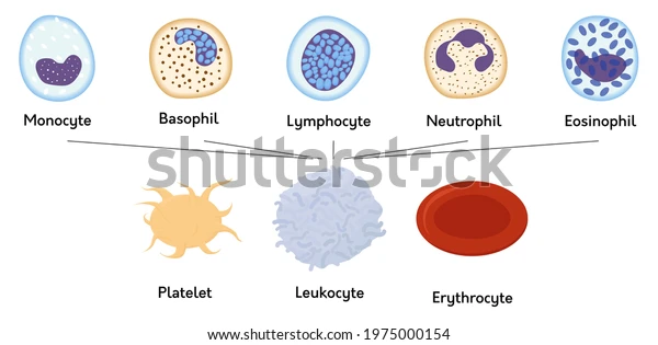
formed element of blood
the cellular component of blood
3 types:
Erythrocytes (Red Blood Cells)
Most numerous blood cell.
Contain hemoglobin, which carries oxygen.
Leukocytes (White Blood Cells)
Protect the body from infection.
Two categories:
Granular leukocytes → neutrophils, eosinophils, basophils
Agranular leukocytes → lymphocytes, monocytes
Thrombocytes (Platelets)
Cell fragments involved in blood clotting.
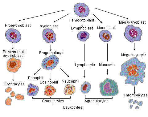
formed by Hemopoiesis

erythrocytes
a subclass of the formed element of blood
RED BLOOD CELLS
they are the most numerous type of blood cell
contain hemoglobin to carry oxygen

leucocytes
a subclass of the formed element of blood
WHITE BLOOD CELLS
Protect the body from infection.
Two categories:Granular leukocytes → neutrophils, eosinophils, basophils
Agranular leukocytes → lymphocytes, monocytes
agranular leukocytes
a subclass of leukocyte in the formed element of blood
includes
lymphocytes (T and B cells)
monocytes

granular leukocytes
a subclass of leukocyte in formed element of blood
Includes:
neutrophils (most abundant + can tell gender)
eosinophils
basophils
thrombocytes
a subclass of the formed element of blood
THE PLATELETS
involved in the clotting response

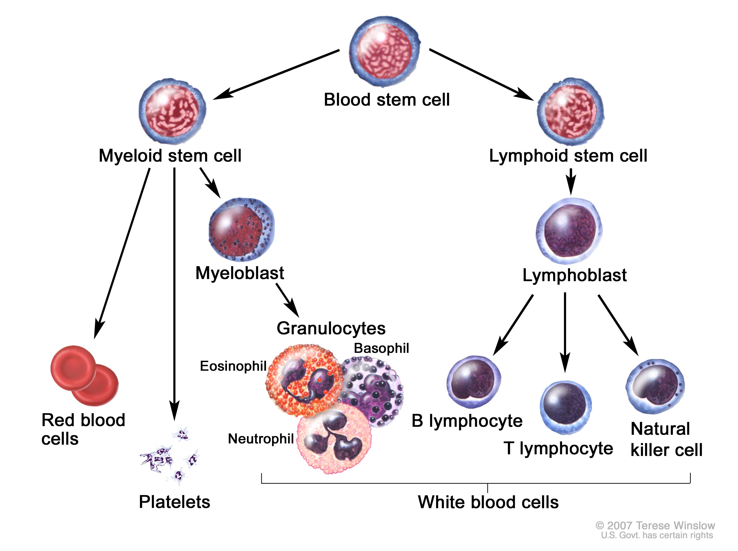
Hemopoieses/Hematopoiesis
formation of all formed elements.
During embryonic/fetal development, hemopoiesis occurs in:
yolk sac, liver, spleen, thymus, lymph nodes, bone marrow
In adults, hemopoiesis occurs mainly in red bone marrow.
Found in: sternum, vertebrae, ribs, pelvis.
Agranulocytes leave the red marrow and finish maturing in lymphoid tissues (thymus, lymph nodes, tonsils).
Because red marrow produces formed elements, it is called myeloid tissue

agrunolocytes
a type of white blood cell that will migrate out of red marrow and mature elsewhere in lymphoid tissues (thymus, lymph nodes, tonsils).
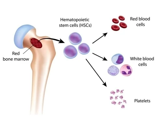
myeloid tissue
red bone marrow!!
called this cuz red marrow produces all the formed elements in adults
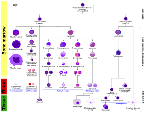
hemocytoblasts
undifferentiated stem cells of red bone marrow that become hemocytoblasts.
are multipotent and form five stem cell lines that produce all formed elements.
plasma
extracellular component of the blood
liquid component of blood.
Mostly water, plus:
nutrients, gases, hormones, electrolytes, wastes and
plasma proteins, which include:
Albumen → contributes to blood viscosity
Globulins → include immunoglobulins (antibodies)
Fibrinogen → inactive form of fibrin, required for clotting
plasma proteins
the proteins of the blood
in plasma (extracellular)
Include:
Albumen → contributes to blood viscosity
Globulins → include immunoglobulins (antibodies)
Fibrinogen → inactive form of fibrin, required for clotting
albumen
a type of plasma protein in plasma (liquid extracellular component of blood)
contributes to blood viscosity
globulins
a type of plasma protein in plasma (liquid extracellular component of blood)
include immunoglobulins (antibodies)
fibrinogen
a type of plasma protein in plasma (liquid extracellular component of blood)
inactive form of fibrin, the protein required for clotting
fibrin
protein req. for blood clotting
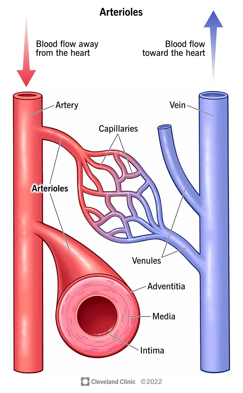
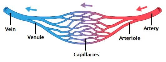
blood vessels
hollow organs that carry blood throughout the body.
3 types:
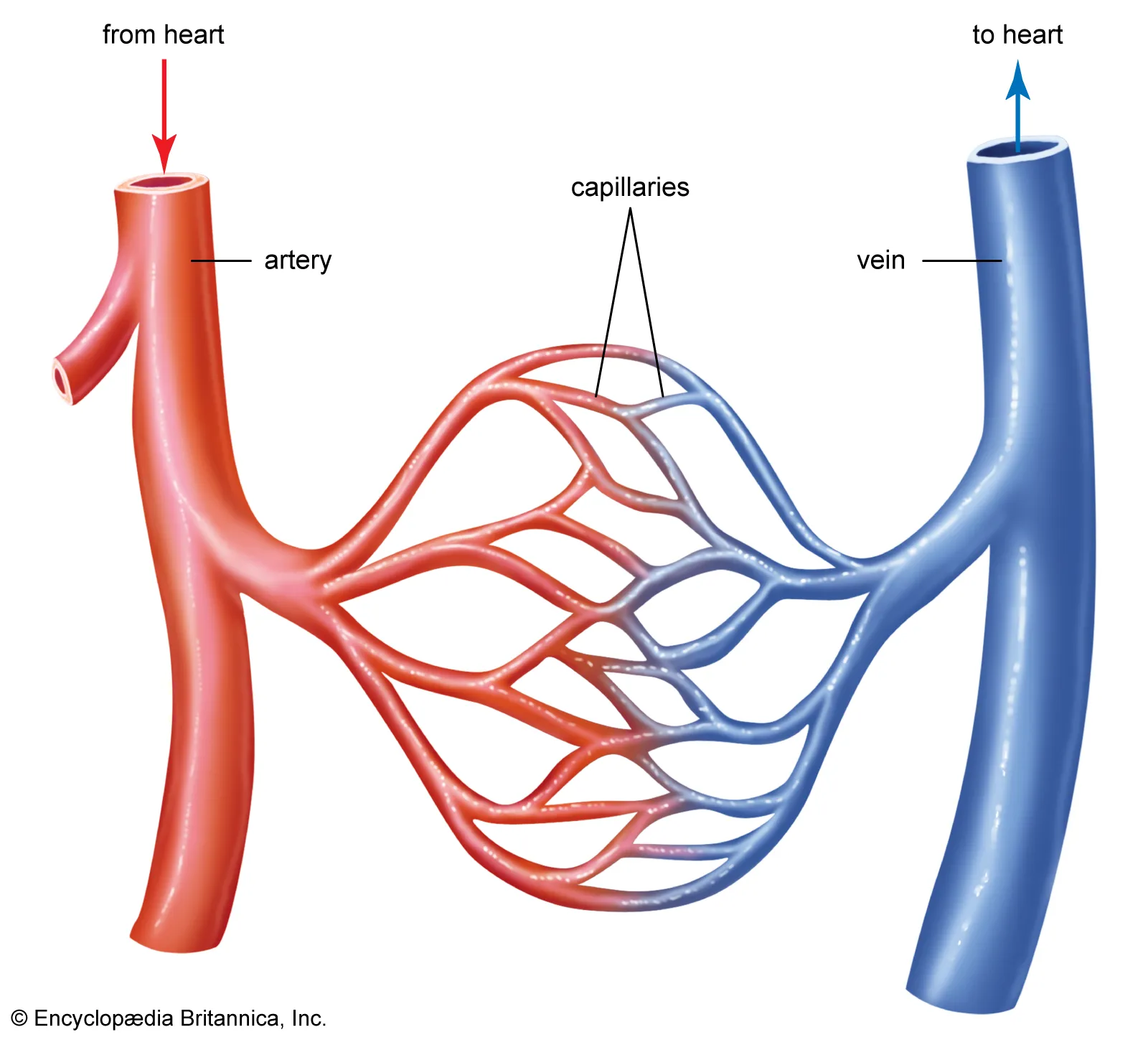
arteries ( carry blood from heart to tissues of bod)
veins (carry blood back to heart from tissues
capillaries

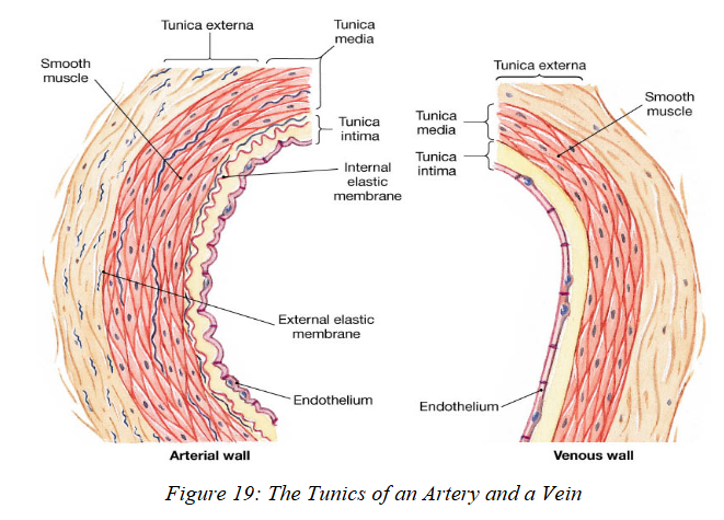
blood vessel tunics
have 3 layers (tunics):
Tunica Intima (Tunica Interna)
Innermost layer; touches the blood.
Made of endothelium on top of areolar c.t
Tunica Media
Middle layer; contains smooth muscle.
Thickest layer in arteries.
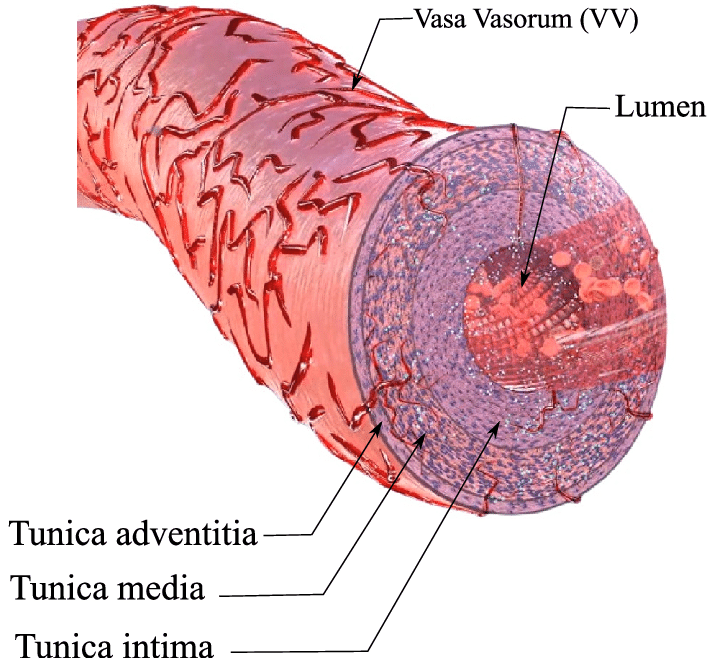
Tunica Externa (Tunica Adventitia)
Outermost layer; mostly loose connective tissue.
Contains small blood vessels called vasa vasorum, which supply nutrients to large arteries and veins.

tunica intima
innermost layer of blood vessels
in direct contact w/ blood.
Made of endothelium on top of areolar connective tissue.
tunica media
middle layer of blood vessels
contains smooth muscle.
Thickest layer in arteries!!!!

tunica externa/tunica adventitia
outermost layer of blood vessels
composed of loose connective tissue.
Contains small blood vessels called vasa vasorum, which supply nutrients to large arteries and veins.
These vessels enter the adventitia, the media, and sometimes partly the intima.

vasa vasorum
these r on the tunica advintitia/ externa
are v small blood vessels that supply nutrients to large arteries and veins.
These vessels enter the adventitia, the media, and sometimes partly the intima.
“blood vessel of the blood vessel”
arteries
a blood vessel that carry blood from the heart to body tissues.
Carry blood at high pressure, so they have the thickest walls.
Tunica media(muscular layer) is thickest layer, filled with elastic fibers and/or muscle fibers.
3 subclasses (largest → smallest):
Elastic arteries
Muscular arteries
Arterioles
Smallest arteries.
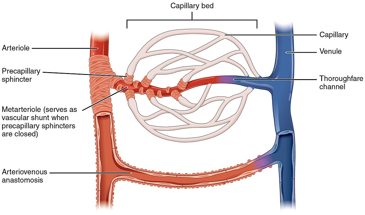
Deliver blood into capillary beds

arterioles
the smallest arteries and deliver blood into capillary beds
Entrance to each capillary bed is guarded by a precapillary sphincter (a circular muscle at the end of an arteriole).
Controls which organs receive blood based on metabolic needs.
When closed, blood bypasses the capillary bed through an arteriovenous shunt.
precapillary sphincter
guards the entrance into the capillary bed
a circular cuff of muscle located on terminal arteriole
Controls blood flow and which organs receive blood based on metabolic needs.
When closed, blood bypasses the capillary bed through an arteriovenous shunt.
an abnormal or surgically created connection between an artery and a vein
capillaries
microscopic blood vessels where exchange occurs between blood and tissues.
Very thin-walled for diffusion.
ONLY made of endothelium LACK THE 3 TUNICS!!!!!
types of capillaries:
Continuous Capillaries
Most common.
Endothelial cells form uninterrupted rings.
Fenestrated (Discontinuous) Capillaries
Have pores (fenestrae) where cytoplasm is absent.
Allow easier diffusion.
Sinusoids
Similar to capillaries but:
Wider lumen
Fenestrae guarded by macrophages
Still microscopic, still used for exchange.
continuous capillaries
the most common and numerous subclass of capillaries
Endothelial cells form uninterrupted rings.
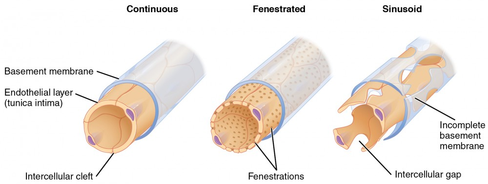
fenestrated/discontinuous capillaries
a type of capillary
Have pores (fenestrae) in endothelial walls where cytoplasm is absent.
Allow easier diffusion.

sinusoids
microscopic blood vessels found in certain regions that may or may not be true capillaries
like capillaries:
microscopic
involved in exchange of materials
have wall of endothelium
unlike capillaries:
have wider lumen
their fenestrae (pores) are guarded by macrophages

Veins
Veins carry blood back to the heart from the tissues
Thinner walls than arteries because they carry low-pressure blood.
Subclasses:
Venules
First vessels to receive blood from capillary beds.
Very thin walls; small venules may lack all three tunics.
True Veins (small → medium → large)
Wall thickness increases as blood approaches the heart.
Tunica externa is the thickest tunic, rich in collagen.
Blood pressure remains much lower than in arteries.
Important feature:
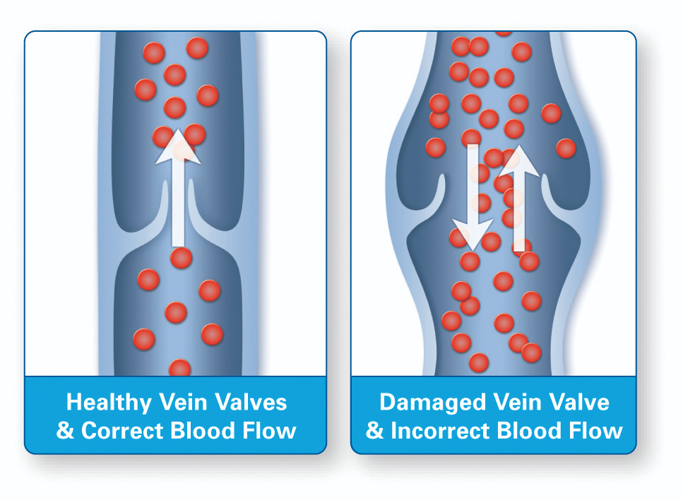
Veins contain numerous valves to prevent backflow due to low pressure.

venules
smallest subclass of vein
First vessels to receive blood from capillary beds.
Very thin walls; small venules may lack all three tunics.
this is when blood is at its LOWEST PRESSSURE
venules drain into “true veins”
vein bloodflow
blood drains from capillaries to very small venules where its at its lowest pressure
venules then drain intro “true veins” which can be small, medium, and large
Wall thickness and blood pressure gradually increases as blood approaches the heart.
Tunica externa is the thickest tunic, rich in collagen.
Blood pressure in veins remains much lower than in arteries.
veins have valves to prevent backflow of blood

valves
Veins contain numerous valves projecting into lumen to prevent backflow due to low pressure.
only in veins NOT arteries!
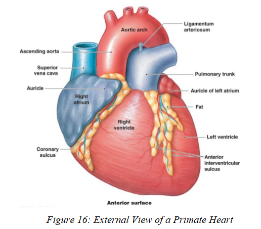
heart
a muscular pump
It sits in the Pericardial Cavity and is surrounded by the Pericardium, a serous membrane.
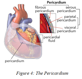
pericardium
serous membrane covering heart

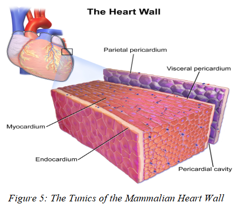
heart wall layers
Endocardium – inner lining; includes endothelium.
Myocardium – middle, thickest layer; rich in cardiac muscle, provides pumping force.
Epicardium – outer layer made of connective tissue and a serous membrane.
The heart receives its own blood through coronary vessels.

endocardium
inner lining of heart wall
contains endothelium

myocardium
the middle and THICKEST layer of the heart wall
composed of cardiac muscle to pump blod

epicardium
outer layer of the heart wall
composed of c.t. and a serous membrane
single circuit hearts (fish)
Blood travels heart → gills → body → heart.
Gills oxygenate the blood; tissues use the oxygen; deoxygenated blood returns to heart.
This forms one continuous loop (single circuit).
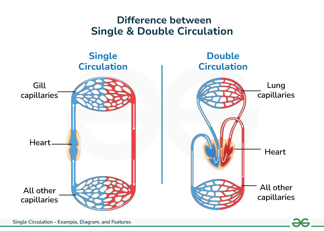
double circuit hearts (amniotes)
2 separate pathways:
Pulmonary Circuit
Carries deoxygenated blood → lungs
Returns oxygenated blood → heart
Systemic Circuit
Carries oxygenated blood → body tissues
Returns deoxygenated blood → heart
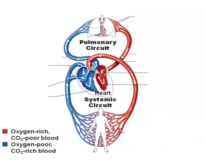
pulmonary circuit
one of the circuits in the double circuit heart
Carries deoxygenated blood → lungs to pu oxygen
Returns oxygenated blood → heart

systematic circuit
one of the circuits in the double circuit heart
Carries oxygenated blood → body tissues
Returns deoxygenated blood → heart

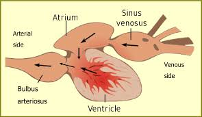
Fish Hearts (Gill-Breathers, except Dipnoans)
fish have 2-chambered hearts: one atrium + one ventricle
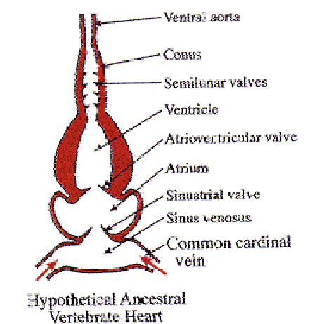
Fish have 4 sequential structures:
Sinus Venosus – receives deoxygenated blood
Very little contraction; blood enters due to ventricular pressure
Blood passes through sinoatrial aperture (2 unidirectional valves)
Atrium – thin-walled muscular chamber
receives blood from sinus venosus to push to ventricle
Ventricle – thick-walled major pump
Main force for fish circulation
Sends blood to the conus arteriosus
Conus Arteriosus – leads blood to the gills
Has cardiac muscle + elastic CT
Contains semilunar valves to prevent backflow
Large in cartilaginous fishes; short in teleosts
sinus venosus
receives deoxygenated blood
Thin-walled w/ little muscle, mainly fibrous CT
Very little contraction; blood enters due to ventricular pressure
Blood passes through sinoatrial aperture (2 unidirectional valves)
NOT IN MAMMALS OR BIRDS!
ancestral location of sinus venosus in mammals is marked by location of the sinoatrial node

sinoatrial aperture
blood travels from the sinus venosus through the sinoatrial aperture into the atrium
is guarded by a pair of unidirectional valves
blood moves to atrium when atrium relaxes after emptying

atrium
thin-walled muscular chamber
receives blood from the sinus venosus and pushes it into ventricle
Pushes blood through the atrioventricular aperture (also 2 valves) into the ventricle

atrioventricular aperture
inbtwn atrium and ventricle
guarded by pair of unidirectional valves
ventricle
muscular thick-walled major pump
Main pumping chamber of the heart!
Sends blood to the conus arteriosus
generates main force for fish

conus arteriosus
receives blood from the ventricle and conducts it to the gills
Has cardiac muscle + elastic CT
Contains semilunar valves to prevent backflow of blood back into ventricle
Maintains steady blood pressure to ventral aorta
Large in cartilaginous fishes; short in teleosts
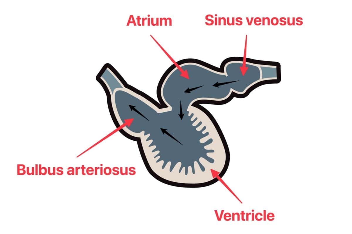
Teleosts compensate with a muscular Bulbus Arteriosus
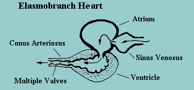
semilunar valves (in fish)
prevent backflow of blood going back to ventricle in the conus arteriosus
bulbus arteriosus
since bony fishes have a shorter conus arteriosus, they compensate with a muscular Bulbus Arteriosus
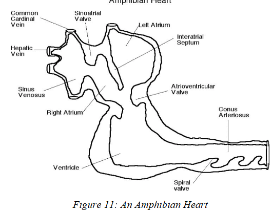
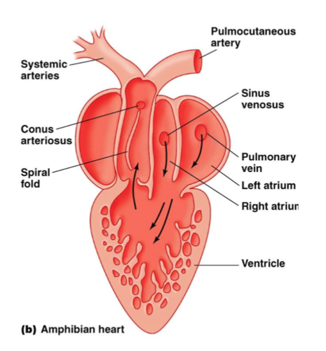
Dipnoan and Amphibian Hearts
Modified for air-breathing, allowing separation of oxygenated and deoxygenated blood.
Four Key Modifications:
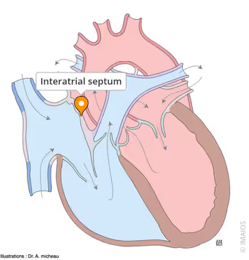
Interatrial Septum (partial or complete)
Separates right and left atria
Left atrium receives oxygenated blood from lungs/swim bladder
Right atrium receives deoxygenated blood from sinus venosus
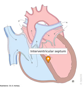
Interventricular Septum (partial) or Ventricular Trabeculae
Both reduce mixing of oxygenated/deoxygenated blood
Spiral Valve in Conus Arteriosus
Separates oxygen/deoxblood
Routes oxygenated blood → systemic arches
Routes deoxygenated blood → lungs or gills
Shortened Ventral Aorta
Blood goes straight from conus arteriosus to the correct vessel
Urodeles are the exception—they keep a prominent ventral aorta

interartial septum
modification of amphibian heart from fish heart (adapted to land)
separates right and left atriums
Complete in anurans, some urodeles
Absent in lungless urodeles
Left atrium receives oxygenated blood from lungs/swim bladder
Right atrium receives deoxygenated blood from sinus venosus
Interventricular Septum (partial)/ Ventricular Trabeculae
modification of amphibian heart from fish heart (adapted to land)
Both reduce mixing of oxygenated/deoxygenated blood. separates right and left ventricles
Partial septum: dipnoans, sirens
Trabeculae: most amphibians (ridges inside ventricle)
spiral valve
modification of amphibian heart from fish heart (adapted to land) ONLY IN AMPHIBIANS
established spiral valve in cornus arteriosus to separate deoxygen/oxygen blood
Routes oxygenated blood →aortic archesthat go to tissues
Routes deoxygenated blood → lungs or gills
Found in dipnoans and anurans
Shortened Ventral Aorta
modification of amphibian heart from fish heart (adapted to land)
Blood goes straight from conus arteriosus to the correct vessel
Urodeles are the exception—they keep a prominent ventral aorta

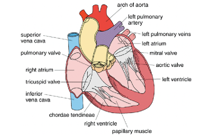
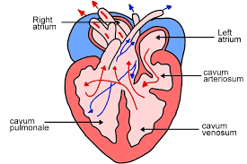
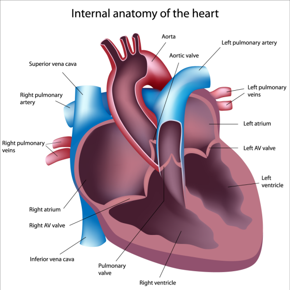
heart in amniotes
4-chambered hearts: 2 atria + 2 ventricles
Turtles and squamates have a third ventricle (Cavum venosus)
Birds and mammals lack a sinus venosus
Their veins dump directly into right atrium
The Sinoatrial node marks the ancestral sinus venosus position
Crocodilians partially incorporate sinus venosus into right atrium

atria (atriums) in amniotes
Separated by a complete interatrial septum in all amniotes
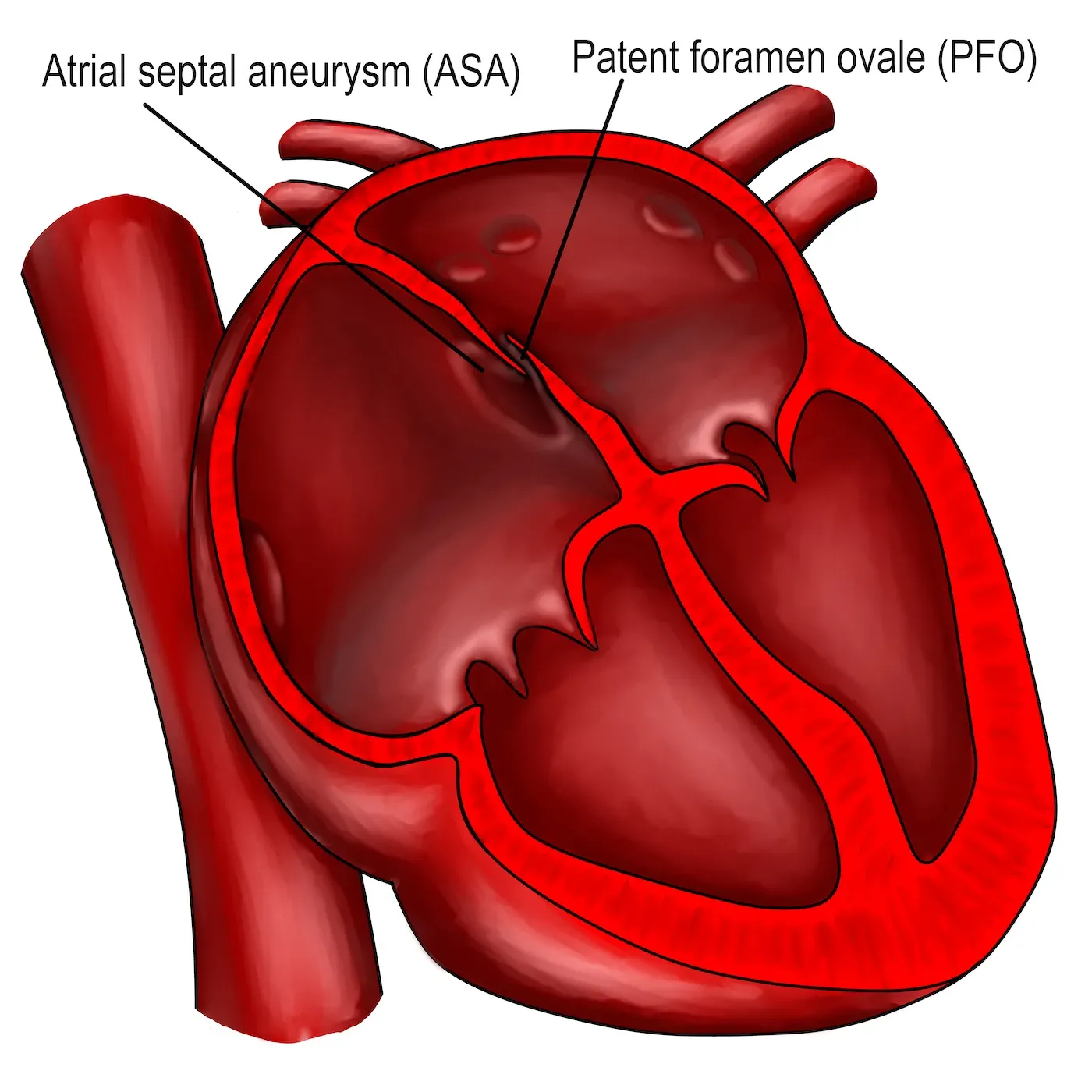
Embryos have a temporary opening: Foramen Ovale
Closes before birth/hatching
Adult remnant in mammals = Fossa Ovalis
Right atrium receives deoxygenated blood
From sinus venosus (reptiles) or
From superior/inferior vena cava (birds & mammals)
Left atrium receives oxygenated blood via pulmonary veins
Mammals have atrial outpocketings called auricles
foramen ovale/foramen ovalis
mbryos have a temporary opening: Foramen Ovale
Closes before birth/hatching
Adult remnant in mammals = Fossa Ovalis
right atrium
receives deoxygenated blood
From sinus venosus (reptiles) or
From superior/inferior vena cava (birds & mammals)

left atrium
Left atrium receives oxygenated blood via pulmonary veins

superior/inferior vena cava
the two largest veins in the body that collect deoxygenated blood from body and return it to the heart's right atrium

pulmonary veins
a vein carrying oxygenated blood from the lungs to the left atrium of the heart

auricles
the atriums in mammals have outpocketings

ventricles in amniotes
Crocodilians, birds, mammals: complete interventricular septum → true 4-chambered heart
Most reptiles: incomplete septum → 3-chambered heart
Turtles + squamates have extra chamber: Cavum Venosus
Helps separate oxygenated vs. deoxygenated blood
IN MAMMALS: As the heart develops, the conus arteriosus is absorbed into the right ventricle
cavum venosus
3rd ventricle in hearts of turtles and scaled reptiles
helpes separating deoxygen/oxygen blood
cardiac muscle in amniotes
All chambers use cardiac muscle
Ventricles have more muscle (pump farther than atria)
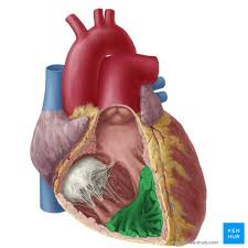
Ventricular walls reinforced by Trabeculae Carnae (interlacing ridges)
trabeculae carnae
muscular ridges and columns on the inner surfaces of the heart's ventricles.
These structures provide support
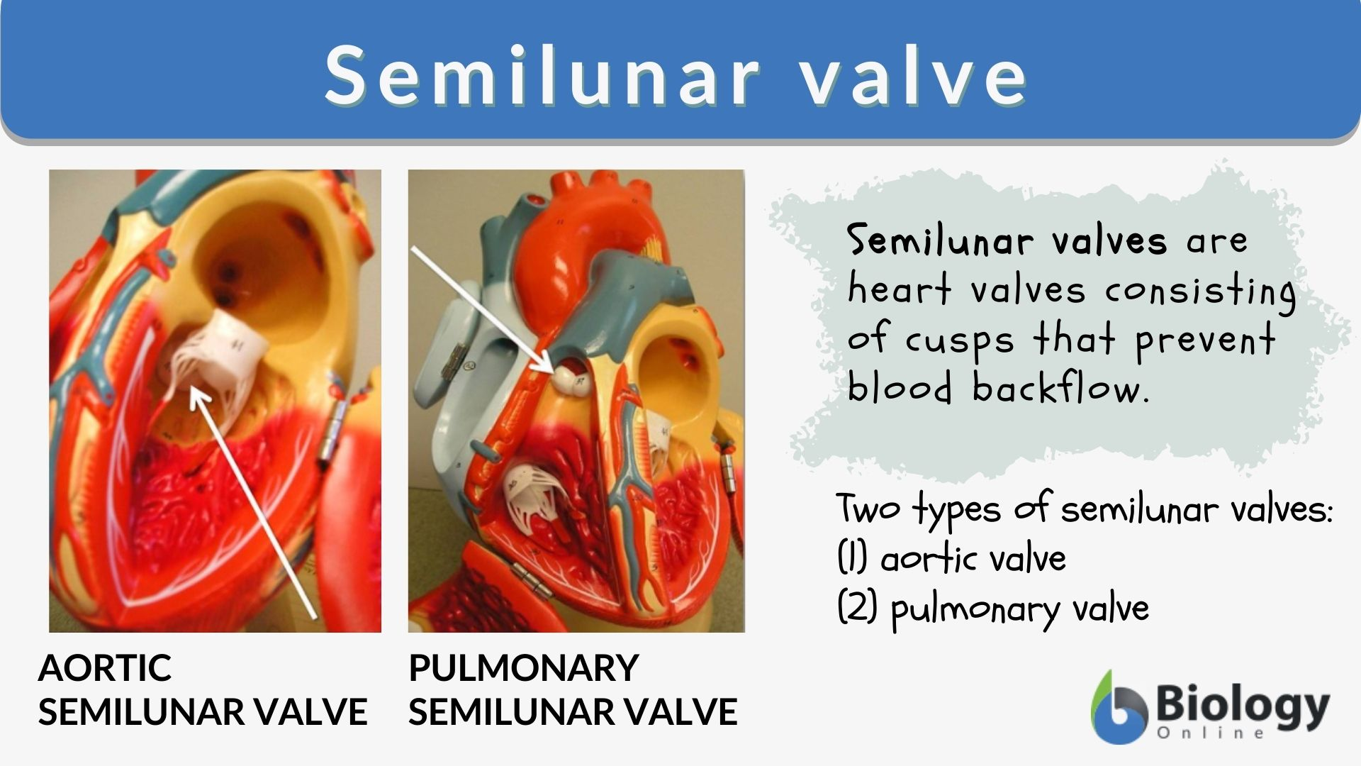
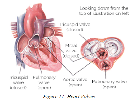
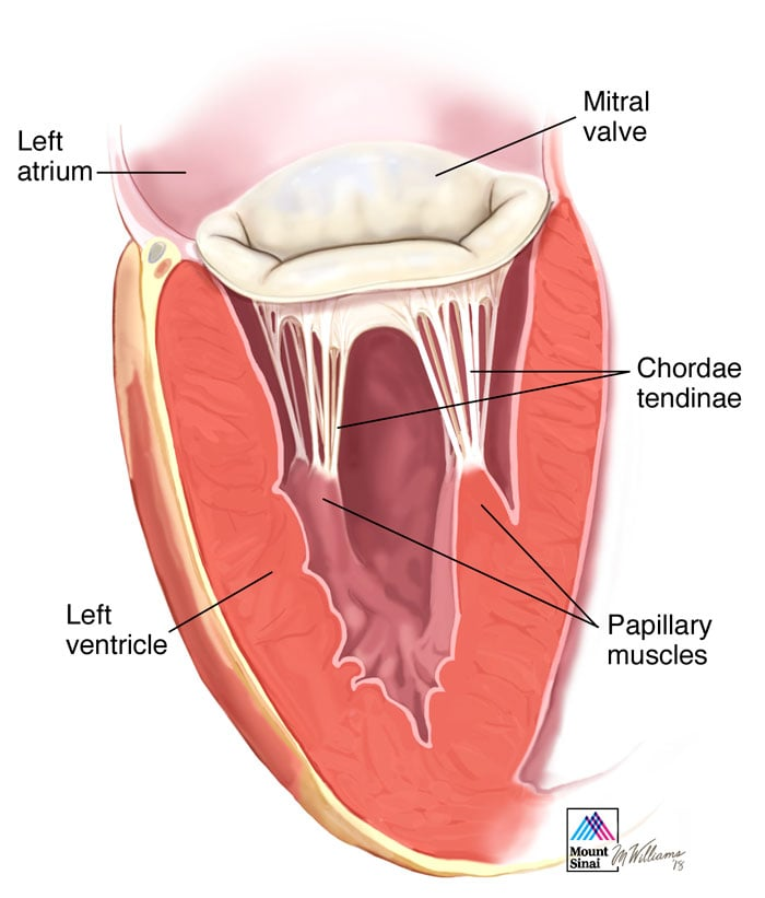
heart valves
Prevent backflow of blood
each valve has flaps of fibrous c.t. called cusps
Atrioventricular (AV) Valves – between atria and ventricles
Mammals: attached via Chordae Tendineae to Papillary Muscles
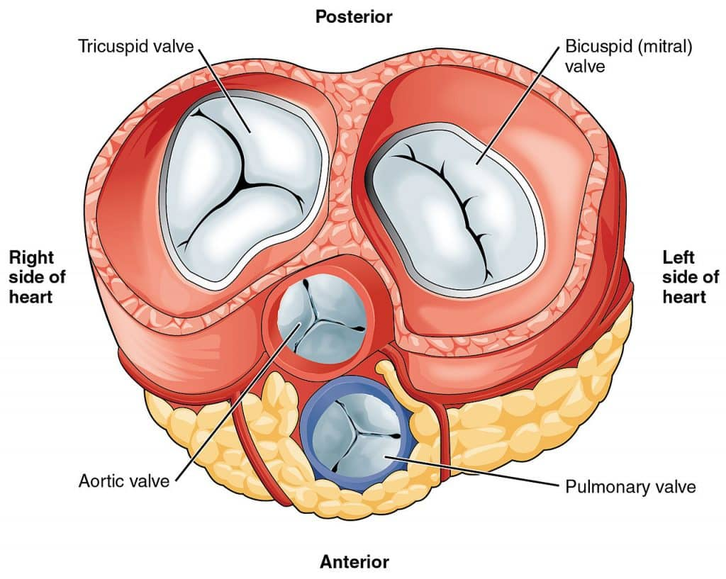
Semilunar Valves – at exits of ventricles
Prevent backflow from aorta and pulmonary trunk
atrioventricular valves
between atria and ventricles
Mammals: attached via c.t strands called Chordae Tendineae to Papillary Muscles (bundles of smooth muscle)

chordae tendinae
anchored c.t. strands in the heart that prevent backflow of blood by anchoring the atrioventricular (AV) valves

papillary muscles
connect to the mitral and tricuspid (atrioventricular) valves via tough fibrous cords called the chordae tendineae
bundles of smooth muscle
semilunar valves in amniotes
two semilunar valves are the aortic valve and the pulmonary valve
prevent the backflow of blood from the arteries back into the ventricles
semilunar valves in amniote vs fish
Fish:
Many small semilunar valves in the conus/bulbus arteriosus.
Prevent backflow into the single ventricle in a single-circuit system.
Amniotes:
Two major semilunar valves (aortic & pulmonary), each with three cusps.
Prevent backflow into ventricles in a double-circuit system.
innervation of heart
Cardiac muscle is naturally rhythmic and can beat on its own.
Specialized cardiac muscle, called nodal tissue, controls and coordinates the heartbeat.
The ANS (autonomic nervous system) adjusts heart rate based on the body’s needs.

nodal tissue
specialized cardiac muscle that controls and coordinates the heartbeat
ANS stimulates nodal tissue for needs

development of innervation in Fish, Amphibians, Reptiles
Keep the sinus venosus throughout life. ANS nerves grow into sinus venosus
ANS signals → received by sinus venosus → atria → ventricles → conus arteriosus (if present).
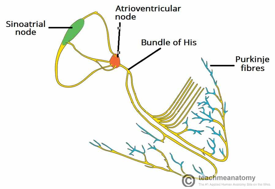
development of innervation in birds and mammals
Lose the sinus venosus, remains only as mass of nodal tissue called the Sinoatrial node in the right atrium!
SA node gets ANS input and fires:
Sends depolarization through both atria → they contract.
Also activates the Atrioventricular node.
Atrioventricular node sends depolarization through both ventricles → they contract.
Ventricles use special conduction fibers (including Purkinje fibers) to spread the signal quickly.
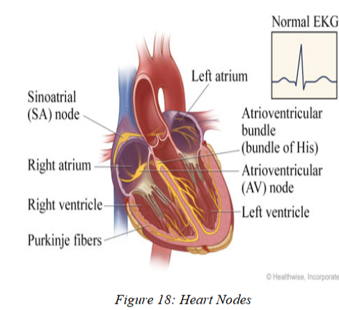
sinoatrial node
in BIRDS AND MAMMALS this is what’s left of sinus venosus and is in the right atrium
receives ANS innervation!
IS HEART’S PACEMAKER!
Sinoatrial node gets ANS input and fires:
Sends depolarization through both atria → they contract.
Also simultaneously activates the Atrioventricular node.

atrioventricular node
cluster of nodal tissue in right atrium
sends depolarization through both ventricles → they contract.
conducts electrical signals from the atria to the ventricles, ensuring coordinated contractions
stimulated by sinoatrial node
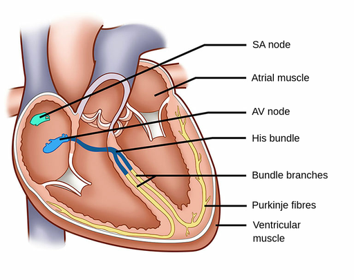
Purkinje fibers
specialized muscle fibers in the heart that rapidly conduct electrical impulses from the atrioventricular (AV) node to the ventricles, ensuring synchronized and efficient contraction
General Pattern of Arteries
Arteries carry oxygenated blood from the heart to body tissues.
Exception: Pulmonary arteries carry deoxygenated blood to the lungs.
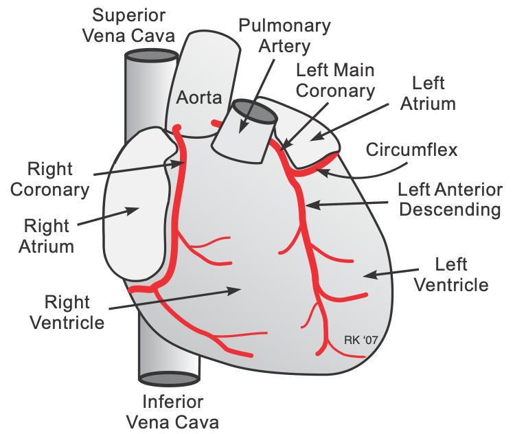
pulmonary arteries
only artery that carries deoxygenated blood instead of oxygenated blood cuz its carrying it to lungs
the artery carrying blood from the right ventricle of the heart to the lungs for oxygenation.
primitive gnathostomes arterial channels
A ventral aorta under the pharynx emerging from heart.
A paired dorsal aorta above the pharynx that becomes single farther back.
6 pairs of aortic arches connecting the ventral and dorsal aortae.
aortic arches in sharks
Ventral aorta grows forward and connects to developing arches.
6 pairs form, but changes occur:
1st arch disappears early, leaving spiracular arteries.
2nd arch forms first pretrematic arteries.
3rd–6th arches form buds → posttrematic arteries, which also form more pretrematic arteries.
Later, arches 2–6 develop occlusions (gaps):
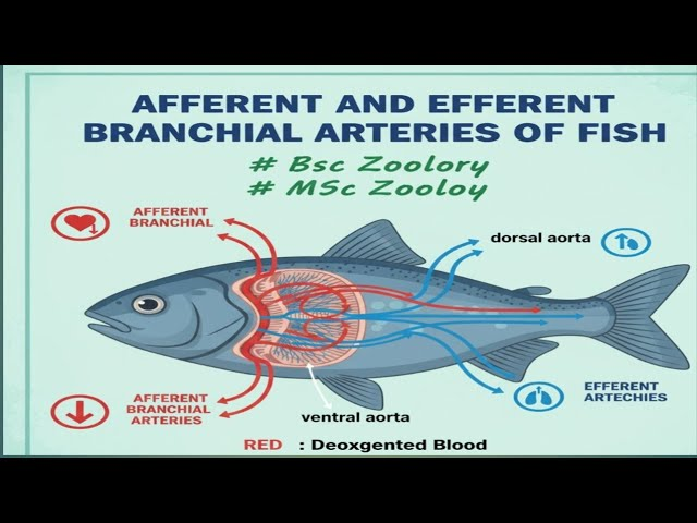
Ventral segments → afferent branchial arteries (carry blood to gills).
Dorsal segments → efferent branchial arteries (carry blood away from gills).
Gill capillaries form in demibranchs.

spiracular arteries in shark form from
first aortic arch
1st arch disappears early, leaving spiracular arteries.
first pretrematic arteries form from
form from second pair of aortic arches in sharks
postrematic arteries form from
from 3rd–6th aortic arches in sharks form buds → posttrematic arteries, which also form more pretrematic arteries.
afferent branchial arteries (fish)
arise from Ventral segments
(carry blood to gills)

efferent branchial arteries (fish)
arise from dorsal segments
(carry blood away from gills).
aortic arches in teleosts
Same basic pattern as sharks, but:
# of afferent/efferent branchial arteries depends on number of gills.
Usually arches 1 and 2 disappear.
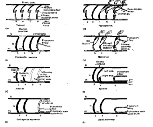
aortic arches in dipnoans
Pulmonary artery develops from the right & left 6th aortic arch
same pattern as tetrapods!
aortic arches in tetrapods
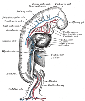
Embryos start with 6 pairs, like fishes.
1st and 2nd arches disappear early.
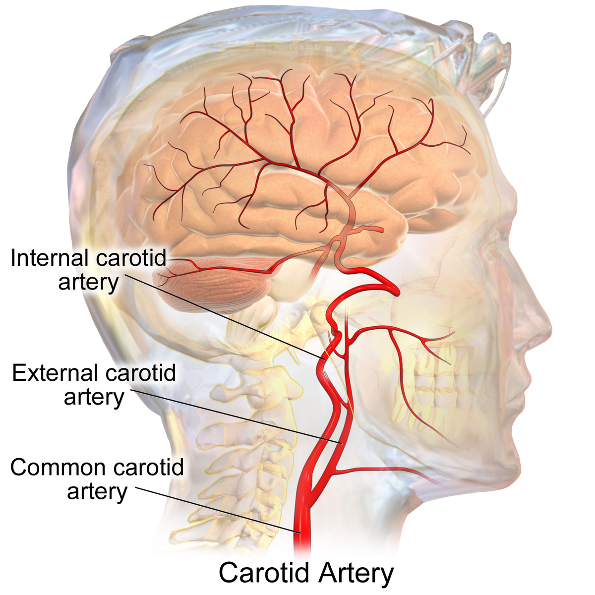
3rd arches + paired dorsal aorta → internal carotid arteries (“carotid arch”).
5th arch is lost in most amniotes.
6th arch → pulmonary arteries.(same as dipnoans)
internal carotid artery
develops from the 3rd aortic arch in tetrapods!
a major artery in the neck that supplies oxygen-rich blood to the brain, eyes, and face
dorsal aorta
a paired embryonic (not in adults) blood vessel that eventually fuses into the single, descending aorta, which is the body's largest artery
Embryonic vertebrates: dorsal aorta is paired.
Paired in the head → internal carotids.
single dorsal aorta becomes the main systemic artery and gives rise to almost all major arteries of the body.
-Somatic branches (to body wall & limbs):
Subclavian, axillary, brachial, radial, ulnar, vertebral, intercostal, lumbar, sacral, iliac, femoral, tibial, popliteal.
Visceral branches (to organs):
Single: celiac, superior mesenteric, inferior mesenteric
Paired: renal, gonadal, adrenolumbar
rete mirabilia (“wonderful maze”)
A specialized arterial structure where an incurrent artery branches into many small interconnecting vessels and drains into an excurrent artery.
Examples & functions:
Glomeruli (kidney filtration)
Shark pseudobranch networks
Red gland of swim bladders (regulates gas levels)
Oxygen storage in deep-diving mammals
Heat retention in mammals, birds, lamniform sharks, tuna
major venous channels in all vert.
General vertebrate venous system includes:
Cardinals (anterior, posterior, common)
Renal portal (kidneys)
Lateral abdominal
Hepatic portal
Hepatic sinus
Coronary veins
Tetrapods + Dipnoans also have:
Pulmonary veins (from lungs)
Postcava (drains kidneys → heart)