Biological female reproductive system and oogenesis
1/10
Earn XP
Description and Tags
week 9, lesson 3/5
Name | Mastery | Learn | Test | Matching | Spaced | Call with Kai |
|---|
No analytics yet
Send a link to your students to track their progress
11 Terms
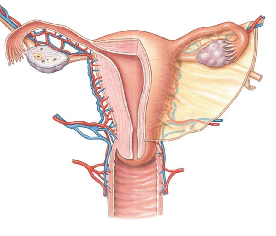
female anatomy of the uterus
Fimbrea
Uterine (Fallopian) tube
Uterus
Cervix
Vagina
Ovary

Fimbrea
Capture the oocyte after is it released by the ovary at ovulation. The fimbrea are finger-like projections that sweep the oocyte into the uterine (fallopian) tube.
Uterine (Fallopian) tube
this is where sperm and oocyte will meet and fertilization will occur. The uterine tube contains cilia that help to move the oocyte or embryo along the uterine tube to the uterus. The movement of the uterine tube is regulated, in part, by progesterone
Uterus
A muscular organ that accommodate and maintains a pregnancy. This is the site of normal embryo implantation into the endometrium. The development of the endometrium (the inner mucous membrane lining of the uterus) is regulated by estrogen, while the maturation of the endometrium is regulated by progesterone.
Cervix
The cervix forms the connection between the vaginal canal and the uterus. It secretes mucus that varies during the menstrual cycle from thin (to facilitate sperm entry) to thick (to prevent sperm entry). Higher estrogen levels causes cervical mucus to be thinner, while higher progesterone levels cause cervical mucus to be thicker.
Vagina
receives the penis and the sperm during copulation, allows for the discharge of fluid during menstruation, and the birth of the baby.
Ovary
The ovary is the site of the developing female gamete. It is responsive to FSH and LH, and it secretes estrogen and progesterone. The ovary will release the oocyte during ovulation.
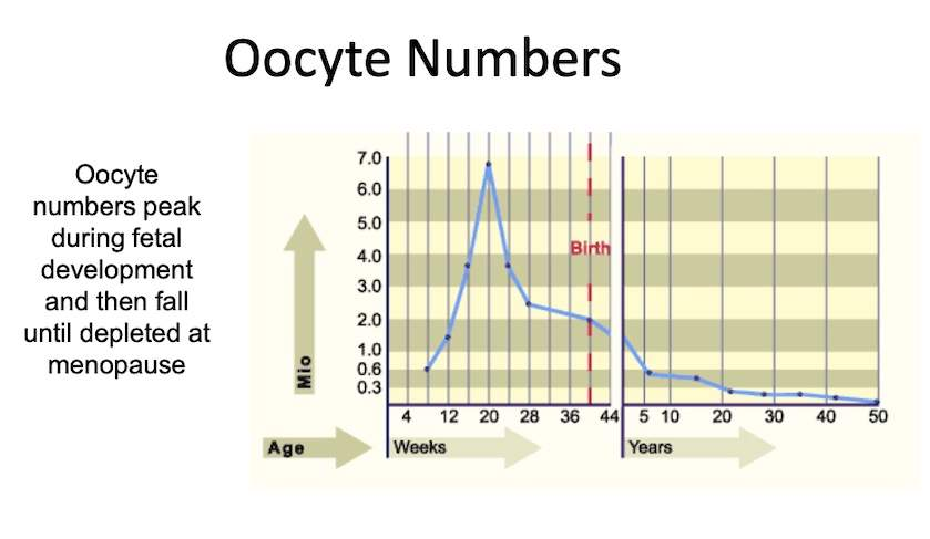
When does the oocyte reservoir develop and how does it change over the life course?
The oocyte that you came from first developed when your biological mother was in your biological grandmother's womb. At birth, there are usually around 1 to 2 million oocytes, however that number drops to approximately 300, 000 by puberty. The graph below depicts the number of oocytes generally present throughout life. The exact reason for the peak at 5 months of pregnancy is unclear, but what is clear, is that the ovary stockpiles the oocytes. One possible reason is that it allows the person to have enough oocyte, and healthy oocytes throughout their reproductive life. We also know that oocytes do not replace themselves, and eventually, oocyte number become depleted and signal the beginning of menopause.
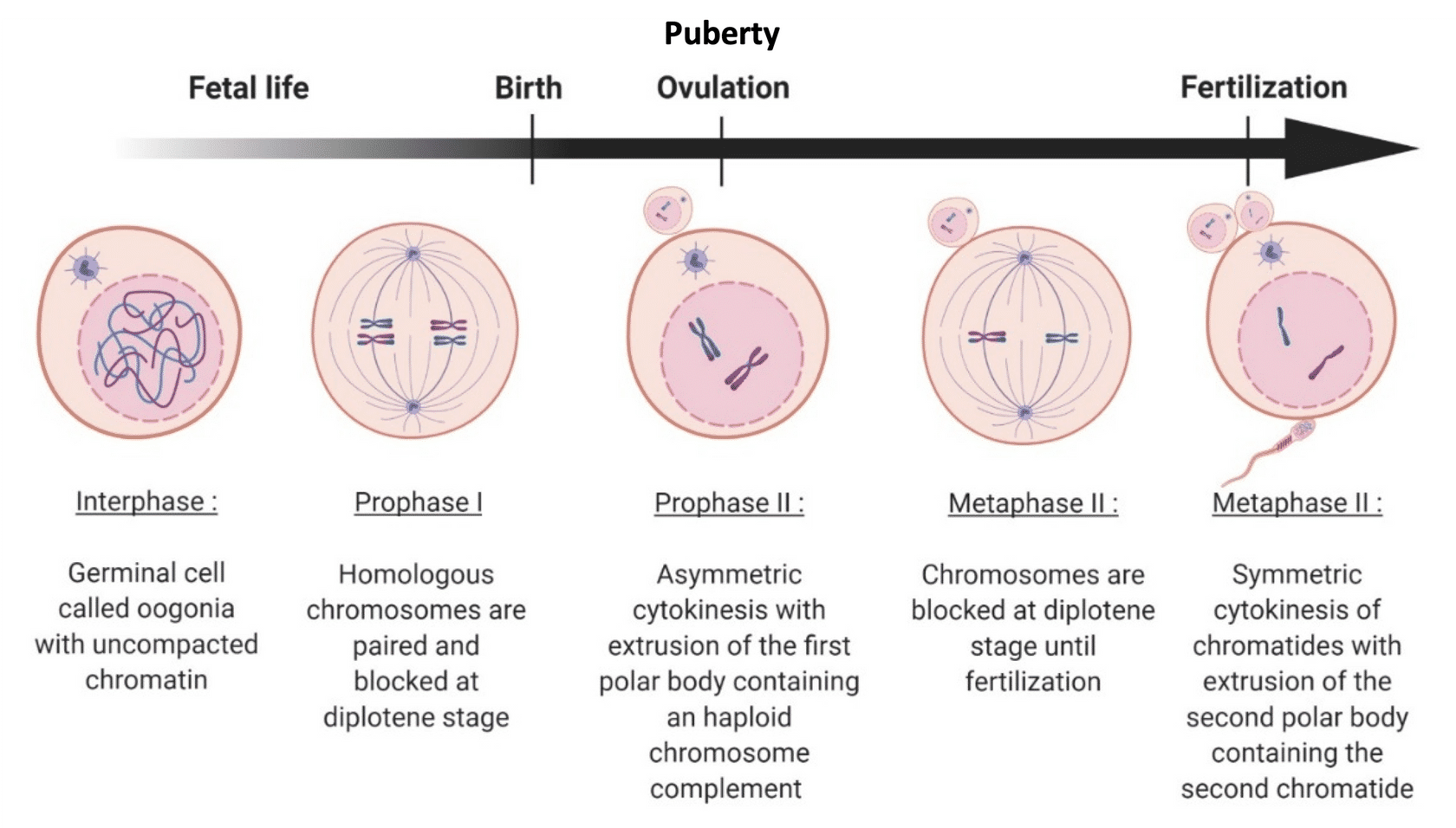
what stage are most oocyte blocked at?
Meiosis occurs in oocytes, but has a distinct path compared to spermatogenesis. Oocytes begin meiosis during gestation and then oocytes are arrested at the diplotene stage of prophase 1. Meiosis only resumes at puberty in oocytes that are recruited to begin maturation. Oocytes not recruited will remain dormant until they are perhaps recruited during a future menstrual cycles. This means that oocytes might stay blocked for 50-55 years, depending on the oocyte pool.
If an oocyte is recruited,
it will become unblocked and meiosis will resume. Oocyte do not divide evenly, this is distinct from spermatogenesis. Instead, the oocyte will undergo crossing over in metaphase 1, and separate the chromatids into one main cell, the oocyte, and a second, really tiny cell, called a polar body. The polar body cannot be fertilized, and is really just a small cell with the unneeded chromosomes and a little bit of cytoplasm. The oocyte then is blocked again in meiosis II, and will remain blocked until it is fertilized.
Fertilization is what triggers
meiosis II to resume, and another polar body is produced. This makes the oocyte a haploid cell just in time for the sperms DNA to be added to the oocytes DNA and form a zygote (1-cell embryo). The joining of the sperm DNA and oocyte DNA is called syngamy.