Physiology slide reading practice
5.0(1)
Card Sorting
1/66
Earn XP
Description and Tags
Last updated 4:51 PM on 10/6/22
Name | Mastery | Learn | Test | Matching | Spaced | Call with Kai |
|---|
No analytics yet
Send a link to your students to track their progress
67 Terms
1
New cards
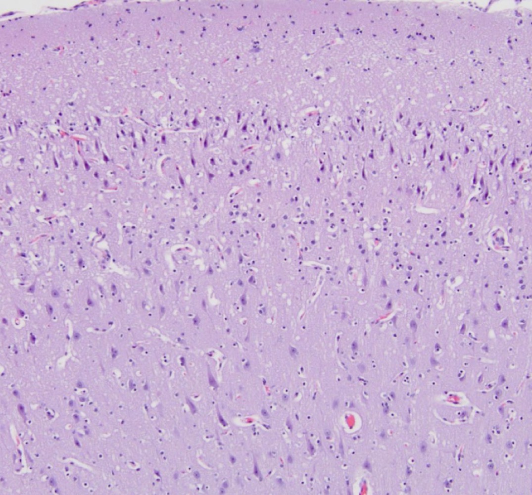
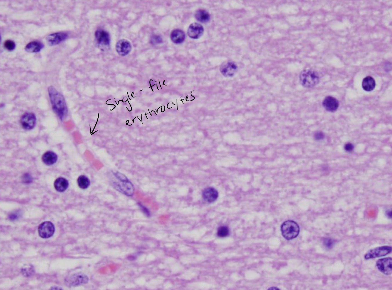
Cerebrum
- grey matter
- grey matter
2
New cards
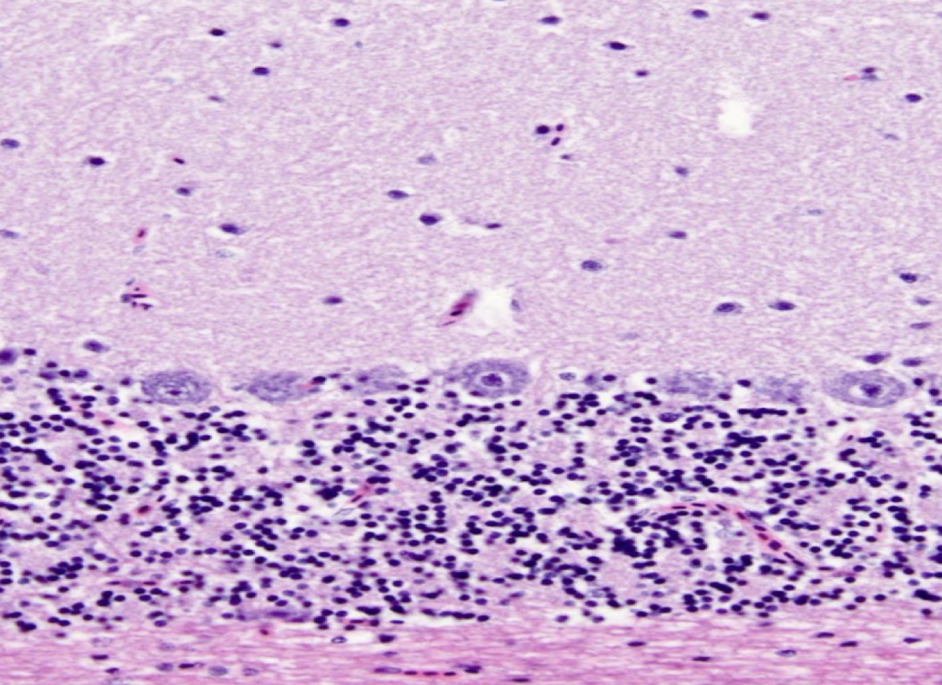
Cerebellum
- layering
- layering
3
New cards
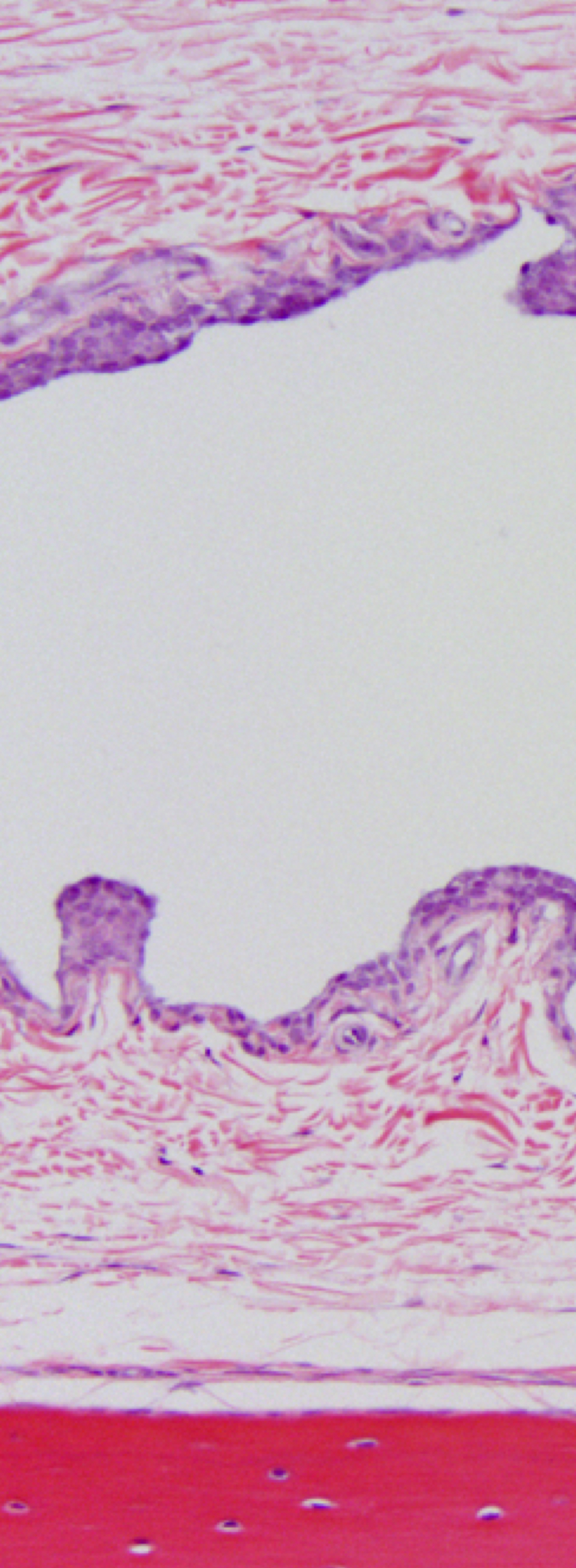
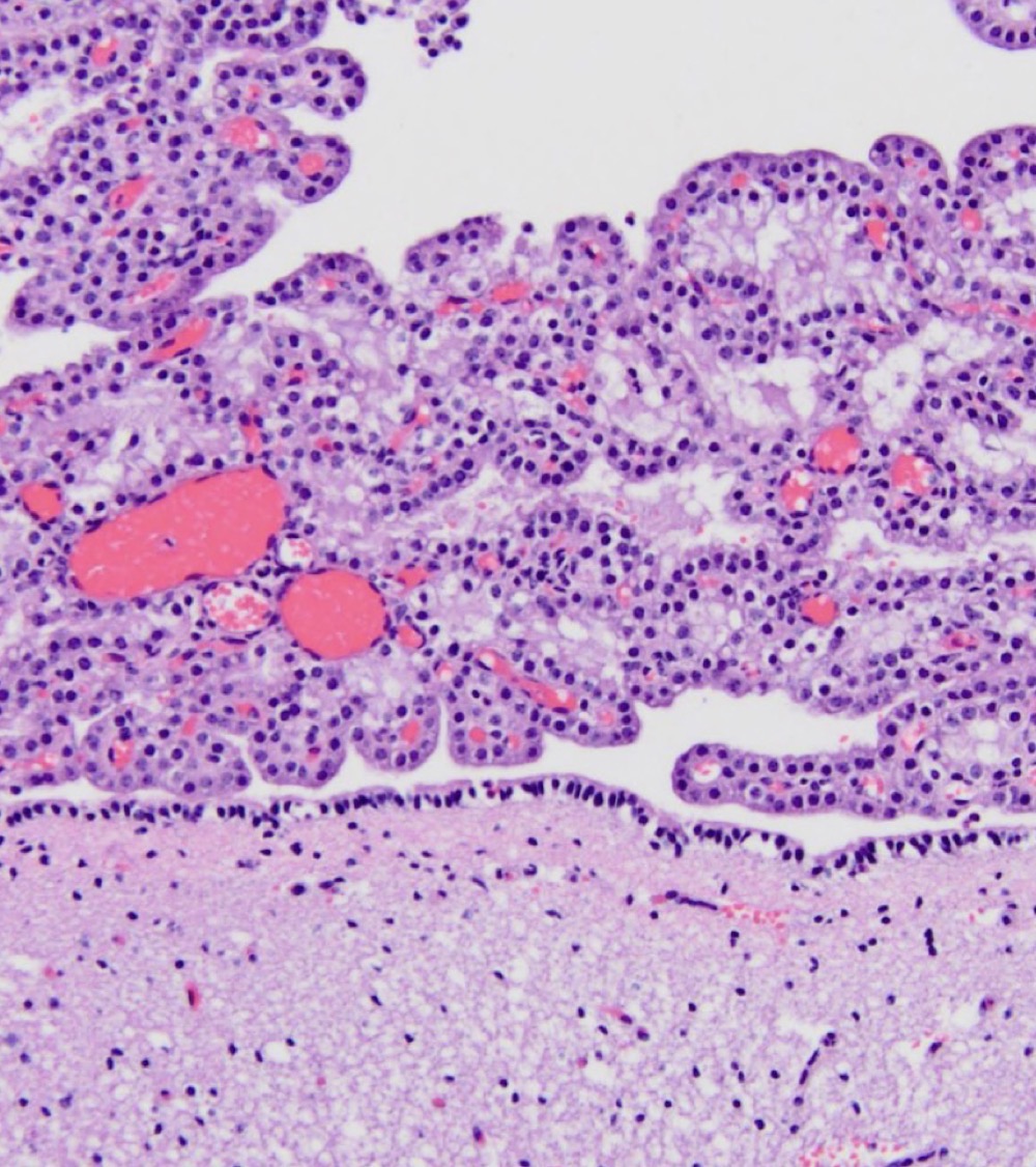
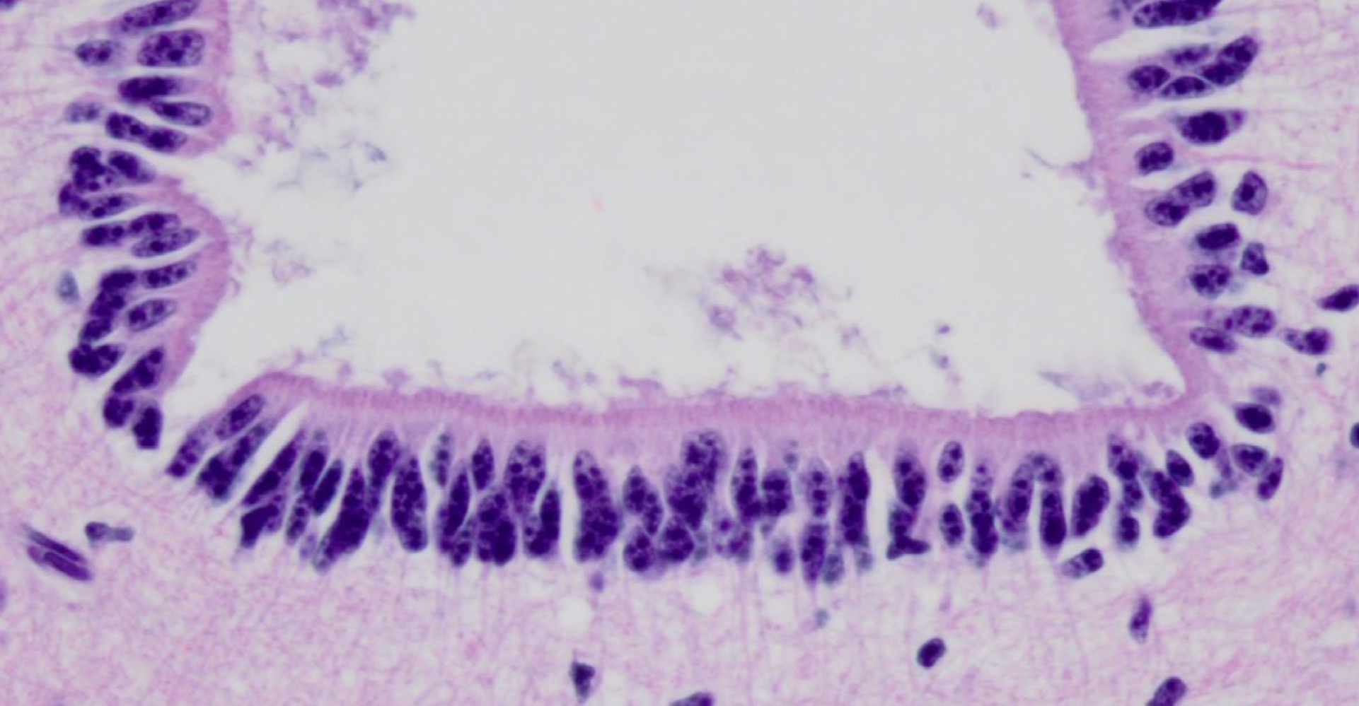
Choroid Plexus
- ependymal cells present
- ependymal cells present
4
New cards
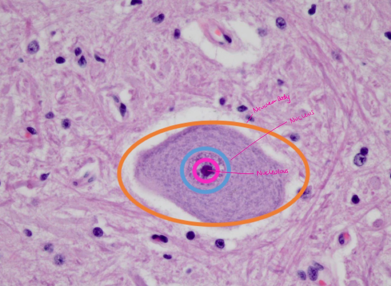
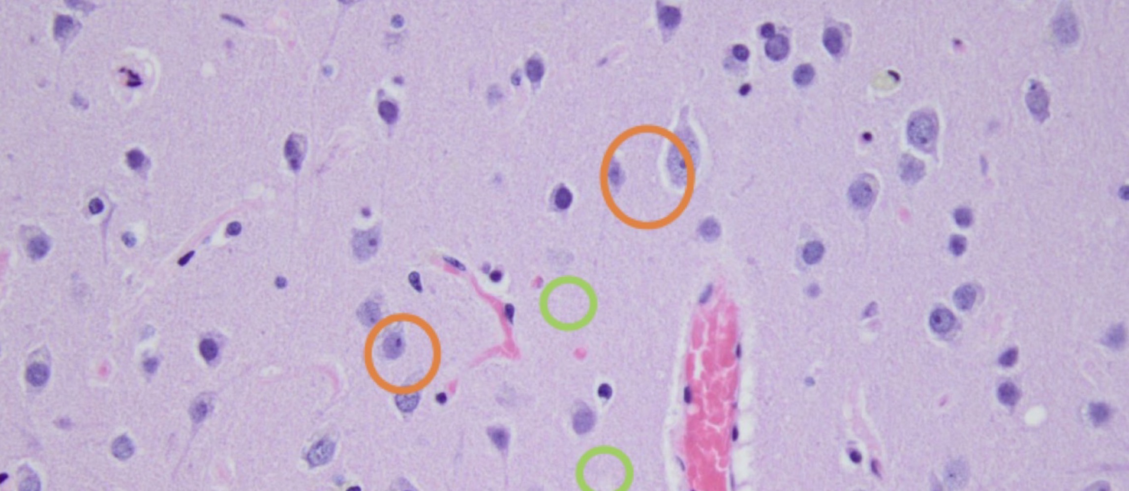
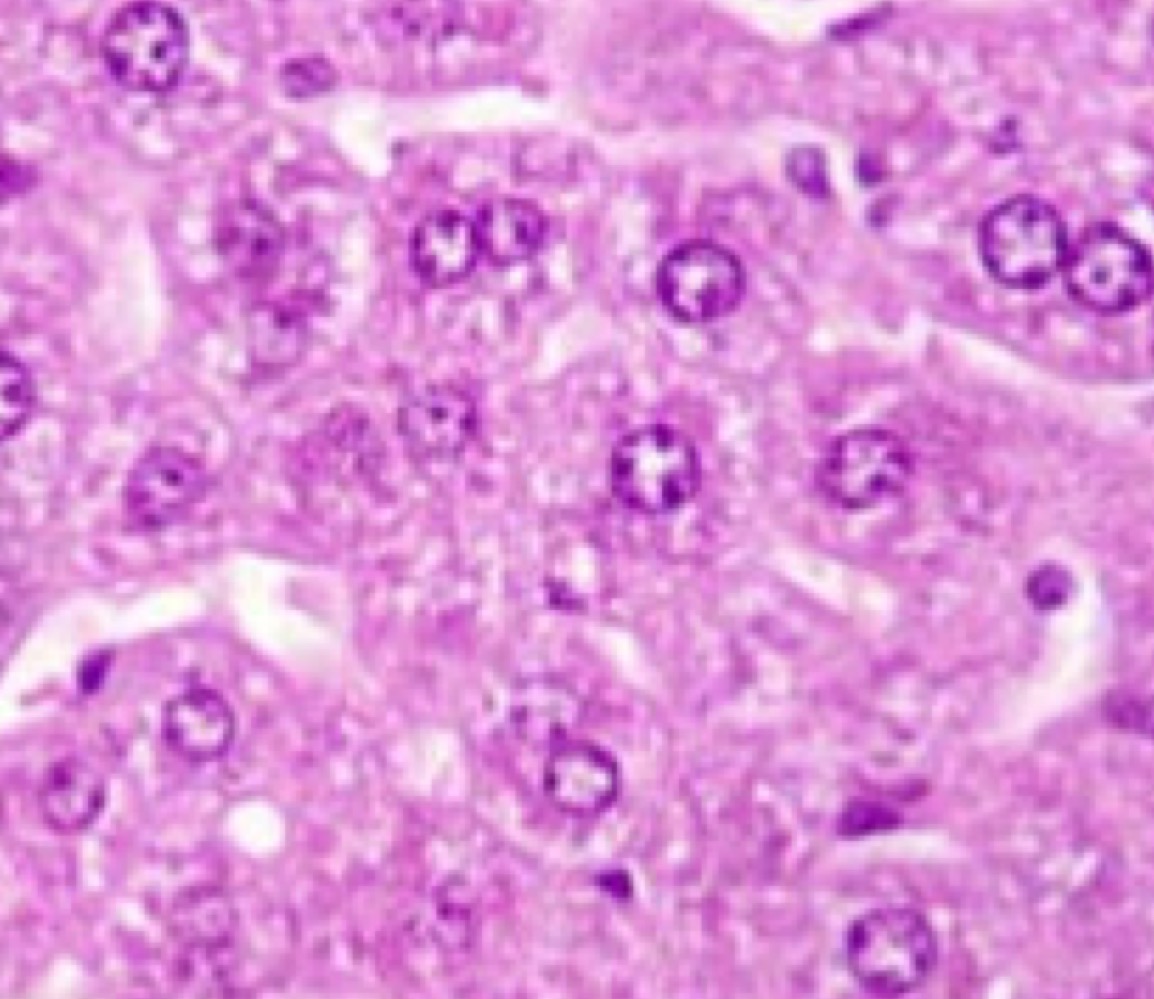
Neuron
- large, 3 Russian doll circles (nuclear body, nucleus, nucleolus)
- large, 3 Russian doll circles (nuclear body, nucleus, nucleolus)
5
New cards
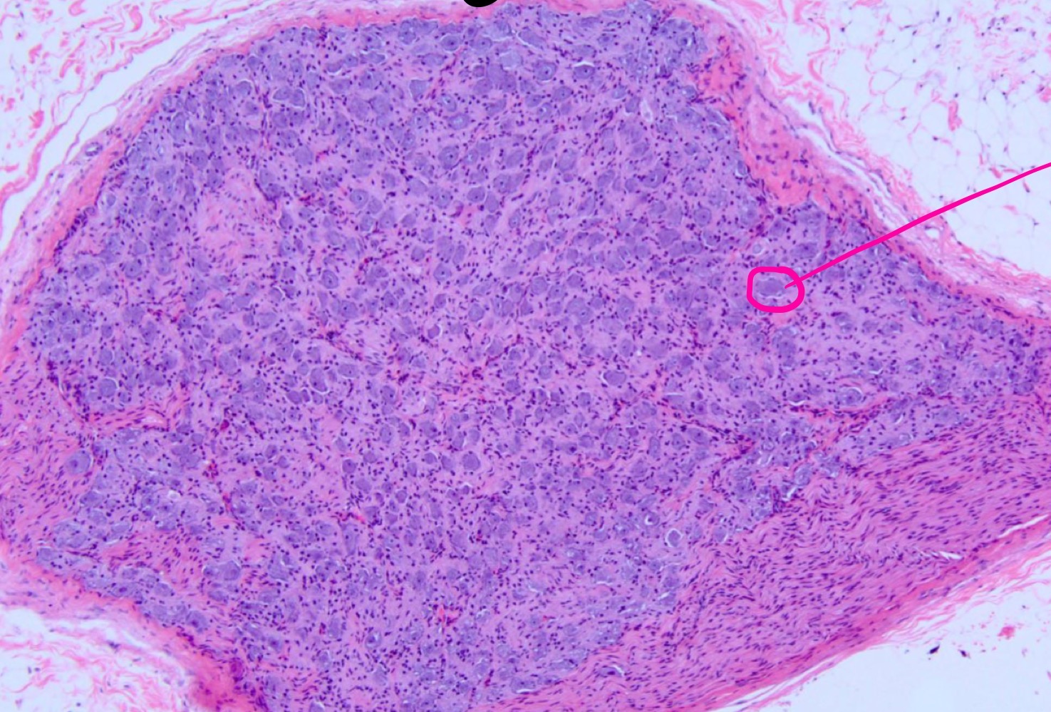
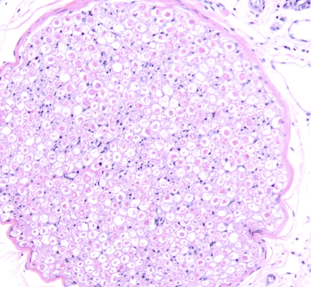
Ganglion
- lots of cell bodies surrounded by nothing else (bundle of neurons)
- lots of cell bodies surrounded by nothing else (bundle of neurons)
6
New cards
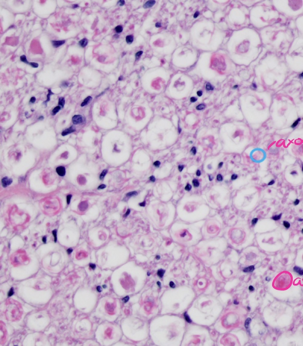
Oligodendrocyte
-white sections are the myelin
-white sections are the myelin
7
New cards
Oligodendrocyte
8
New cards
Ependymal cells
9
New cards
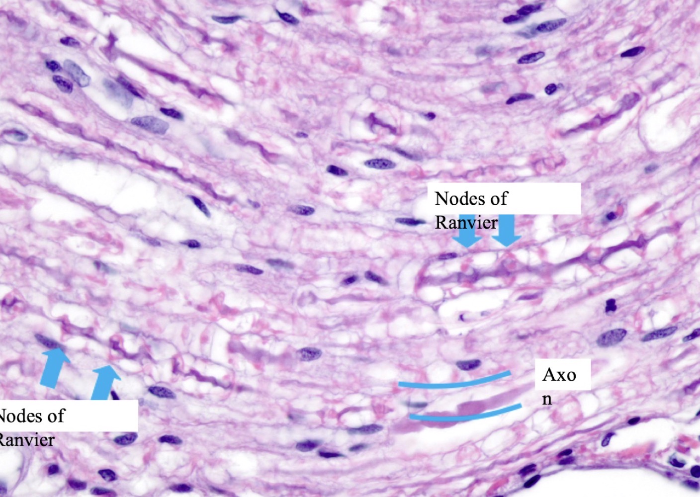
Nerve
10
New cards
Nerve
linear and TONS of nuclei ( even though it might looks like dense, regular connective tissue at first... there are too many nuceli).
linear and TONS of nuclei ( even though it might looks like dense, regular connective tissue at first... there are too many nuceli).
11
New cards
Astrocyte
-just cell body = just purple dots
-just cell body = just purple dots
12
New cards
Meninges
13
New cards
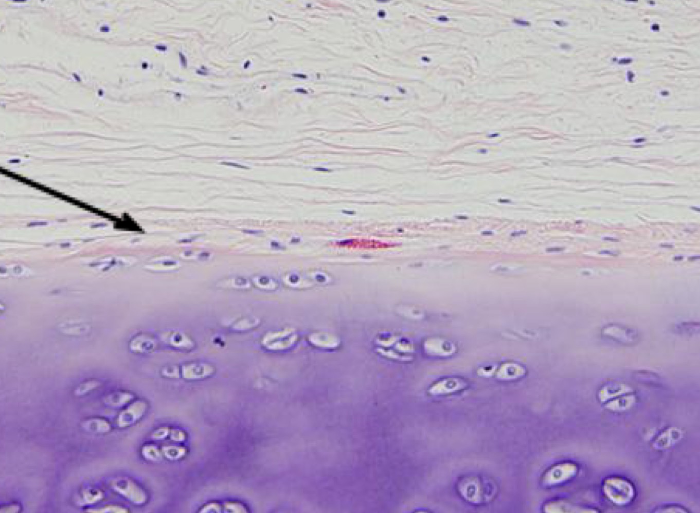
Appositional growth (perichondrium between connective tissue and cartilage)
14
New cards
Arteriole
-less than 3 fish layers/tunica media
-less than 3 fish layers/tunica media
15
New cards
Capillary
16
New cards
Capillary
17
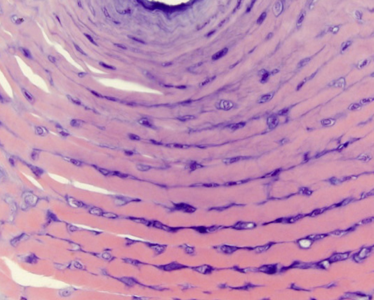
New cards
Elastin/elastic artery
18
New cards
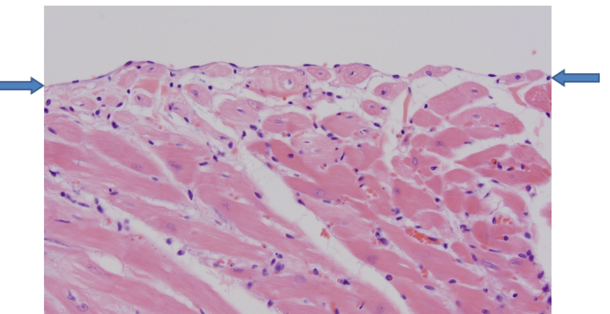
Endocardium
-single endothelial layer next to myocardium
-single endothelial layer next to myocardium
19
New cards
Fibrocartilage
20
New cards
Interstitial growth
21
New cards
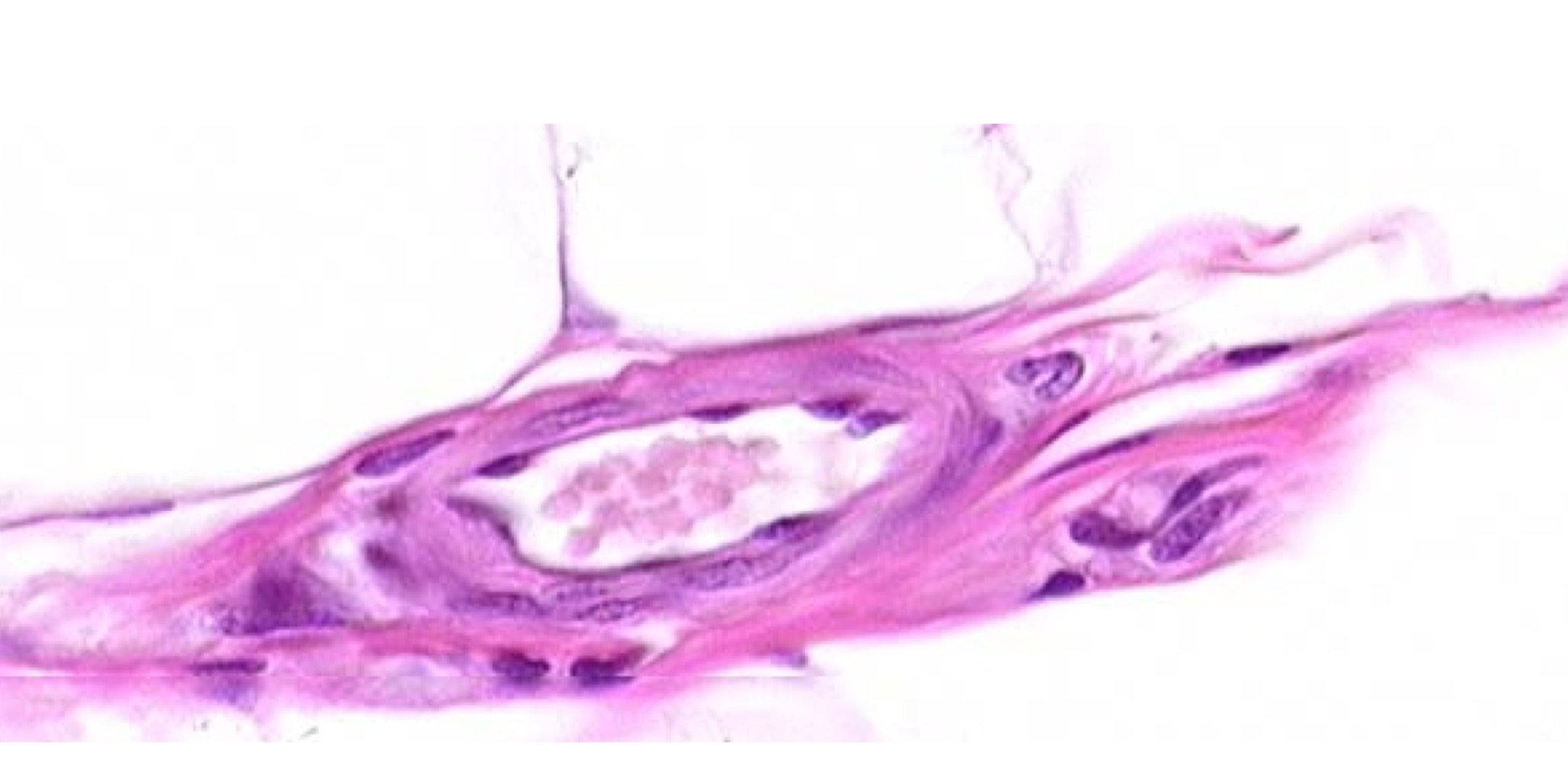
Lymphatic vessels
22
New cards
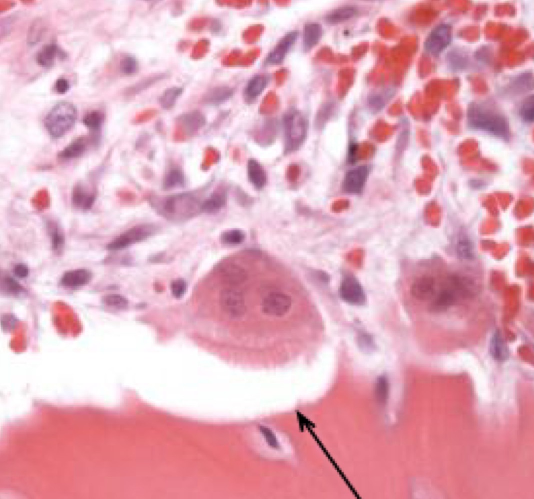
Osteoblast
23
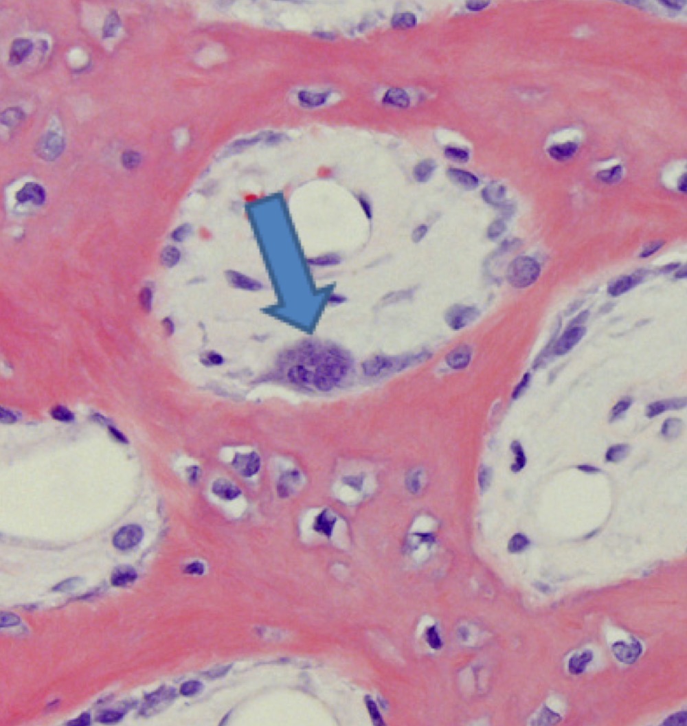
New cards
Osteoclast
24
New cards
Osteoclast
25
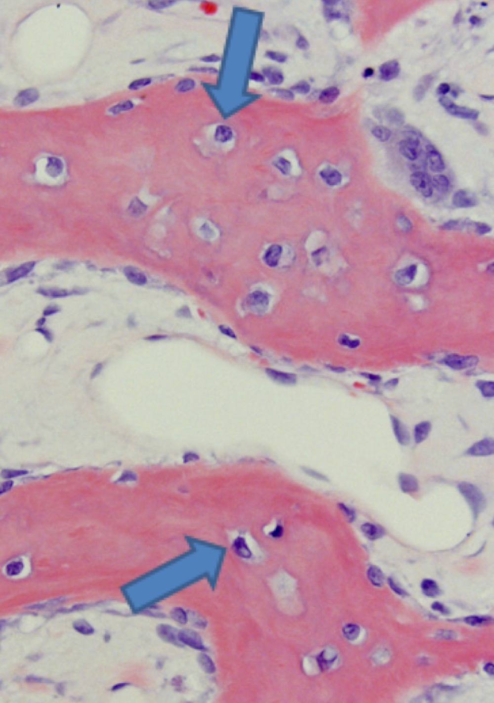
New cards
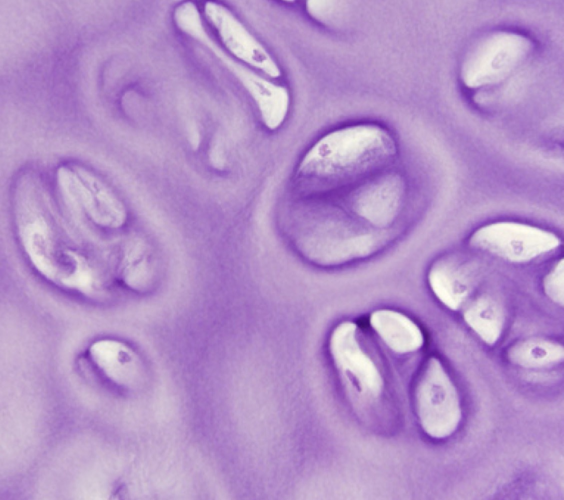
Osteocytes
26
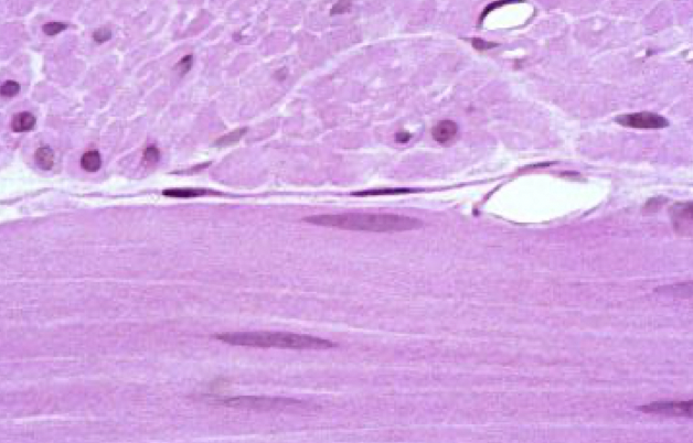
New cards
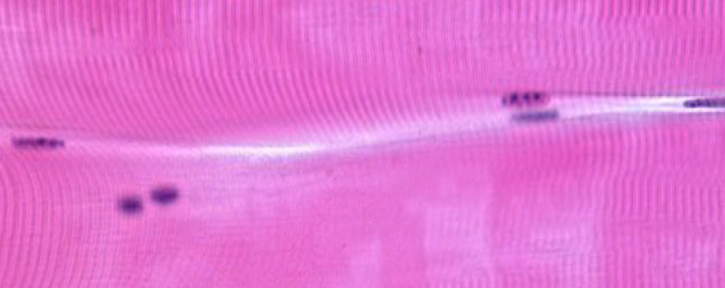
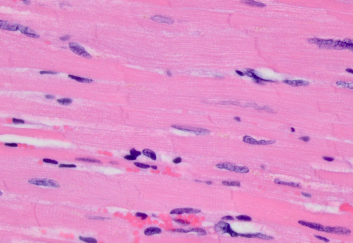
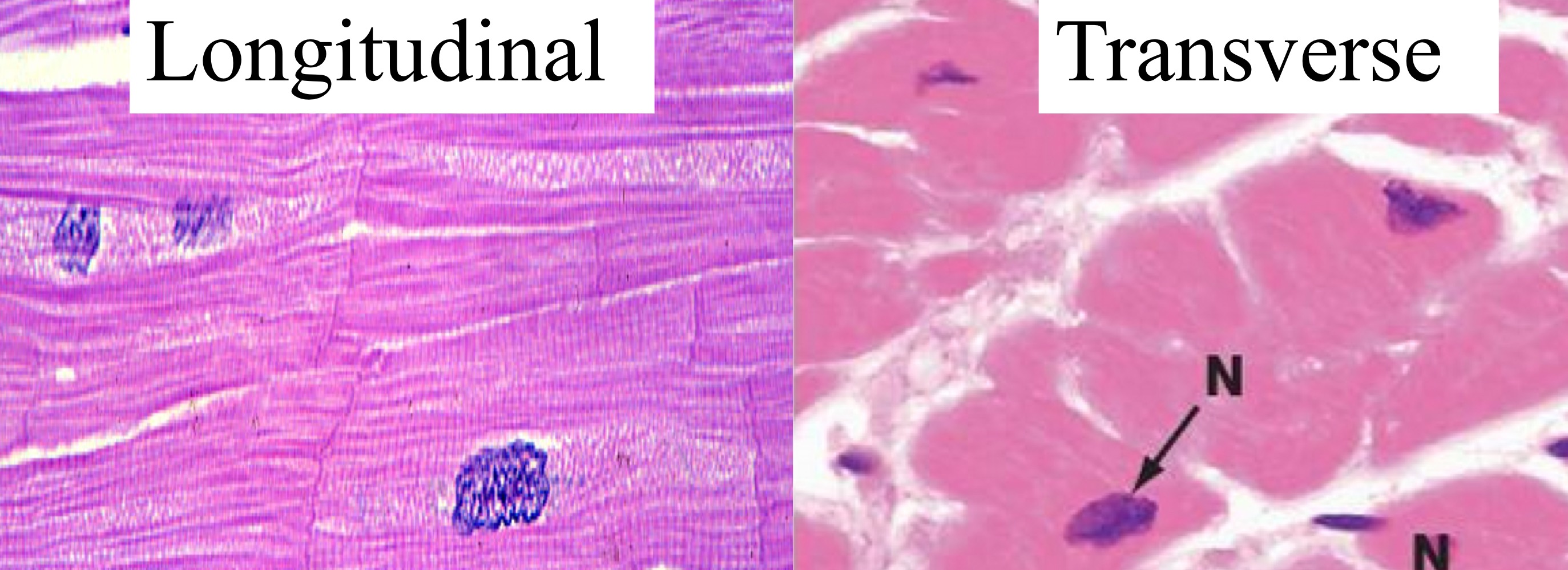
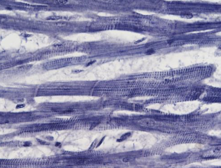
skeletal
-Striation, long parallel fibers, peripheral nucleus
-Striation, long parallel fibers, peripheral nucleus
27
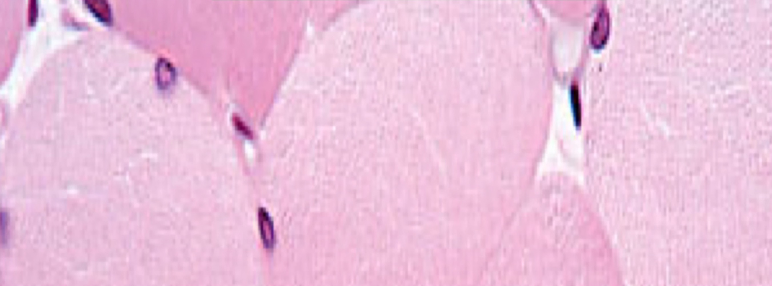
New cards
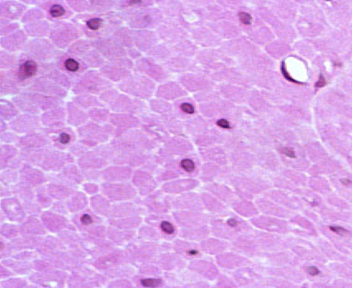
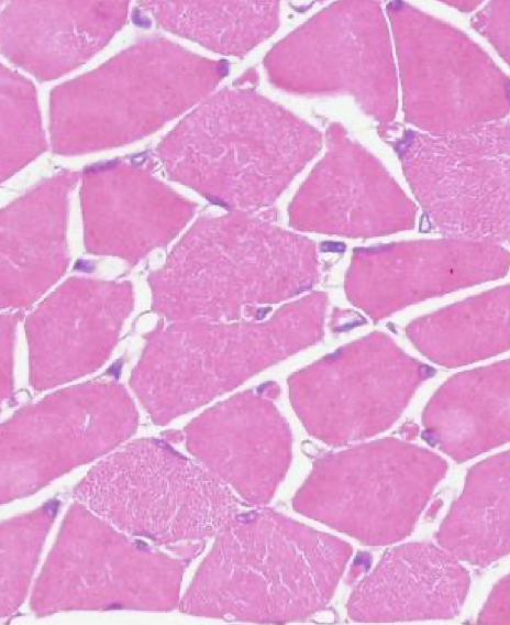
Skeletal
-Round fibers with peripheral nuclei
-Round fibers with peripheral nuclei
28
New cards
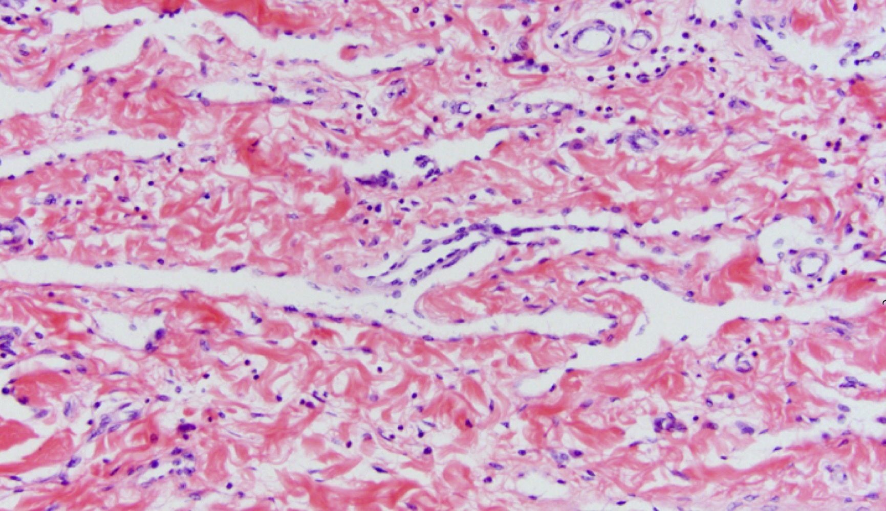
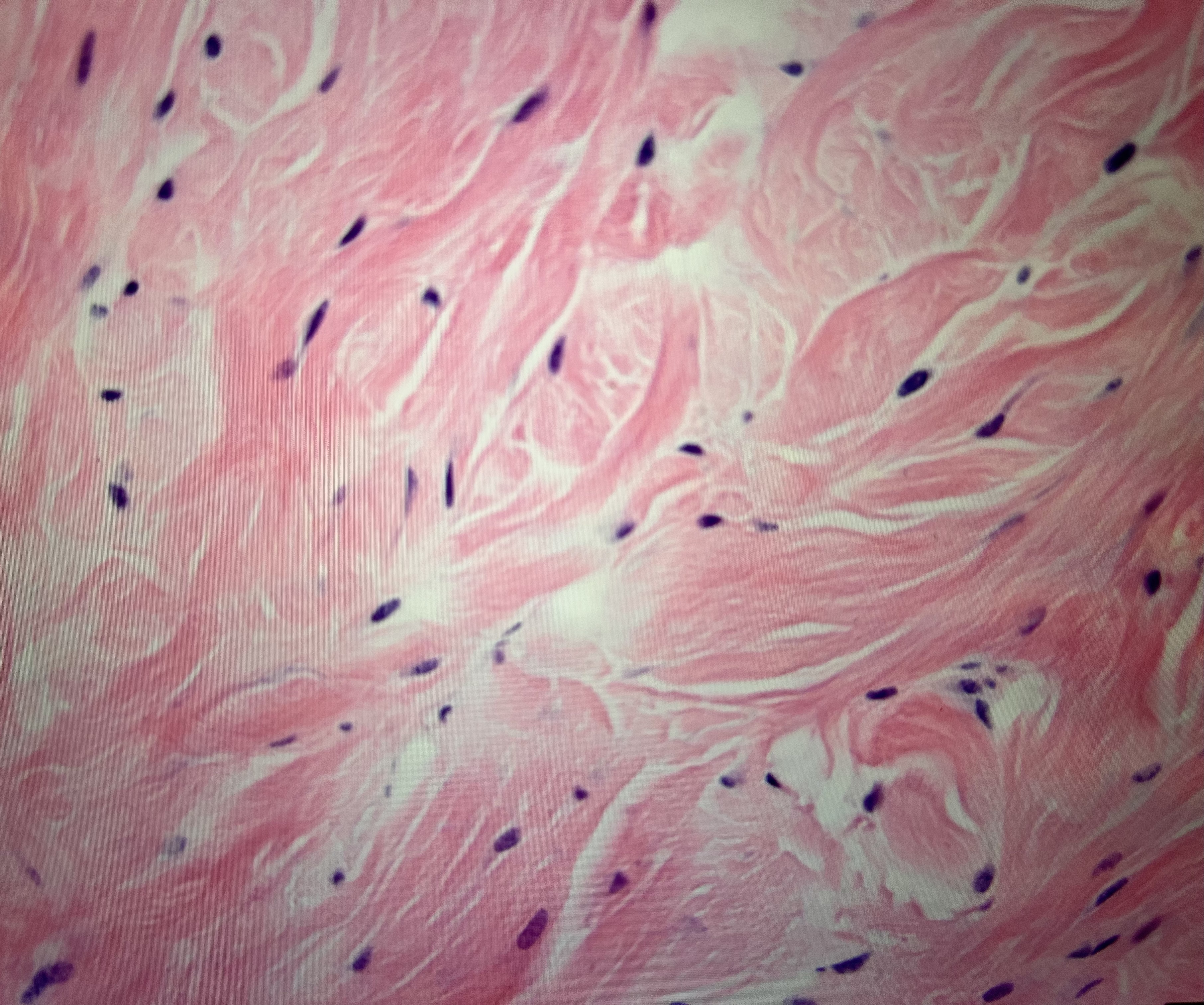
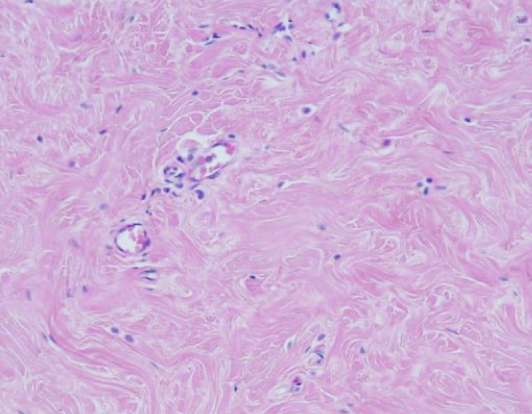
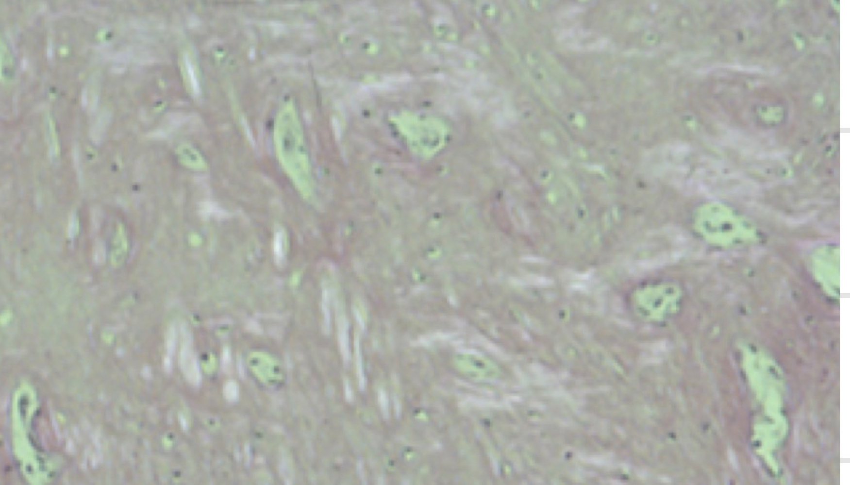
Dense, irregular connective tissue
-Nuclei sparse, tons of cytoplasm, random arrangement
-Nuclei sparse, tons of cytoplasm, random arrangement
29
New cards
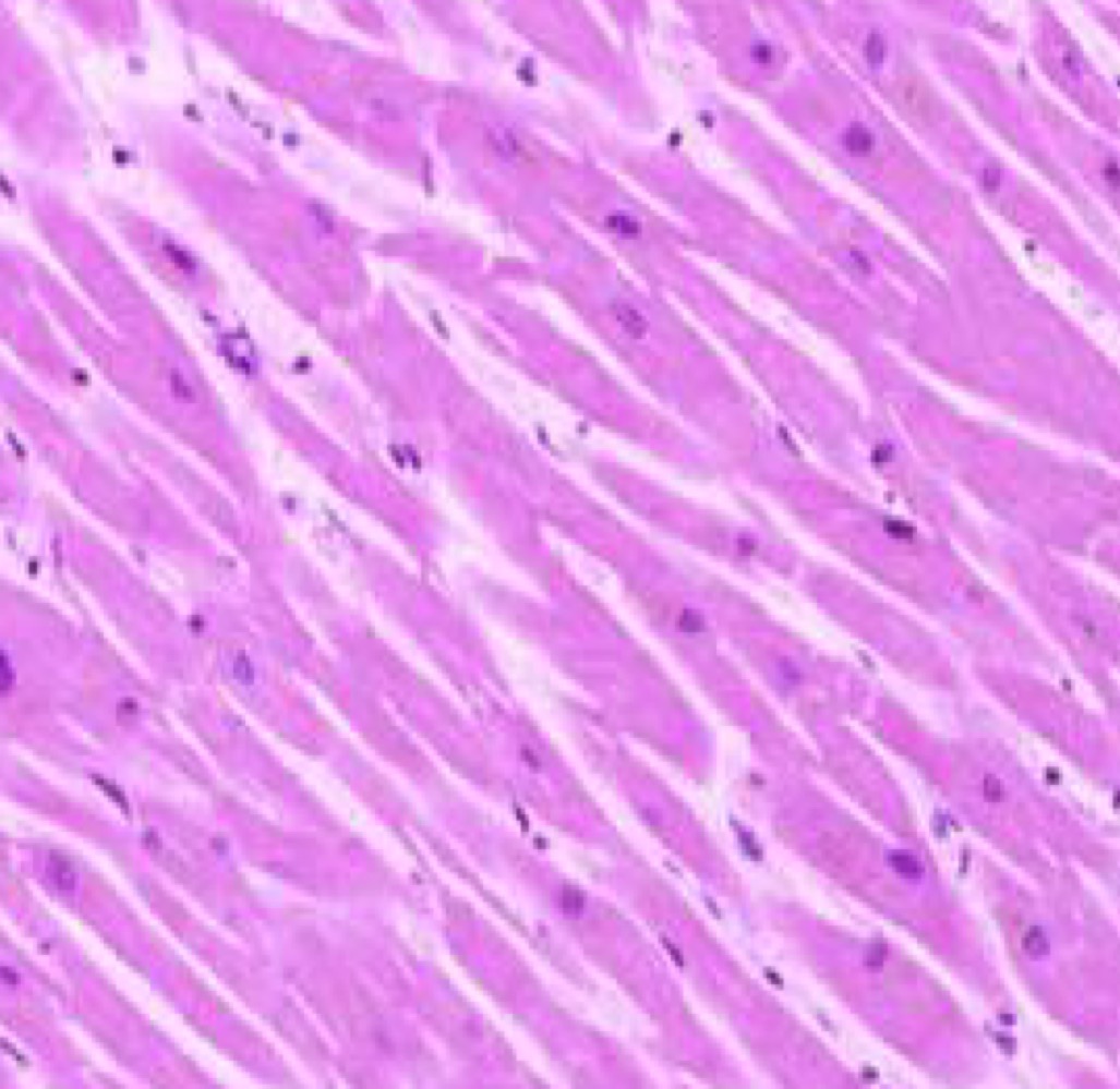
Cardiac
-branching with central nucleus
-branching with central nucleus
30
New cards
Dense, irregular connective tissue
-Nuclei sparse, tons of cytoplasm, random arrangement
-Nuclei sparse, tons of cytoplasm, random arrangement
31
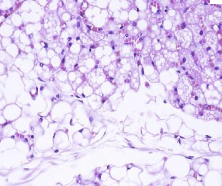
New cards
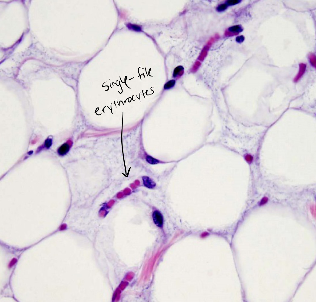
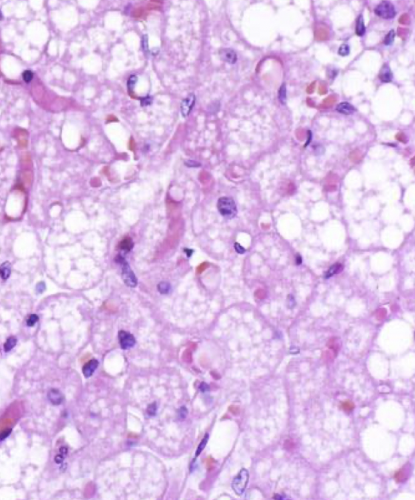
Brown adipose
-small lipid droplets
-small lipid droplets
32
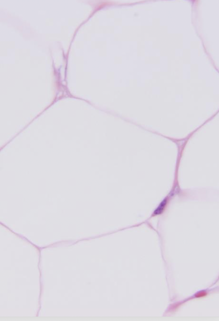
New cards
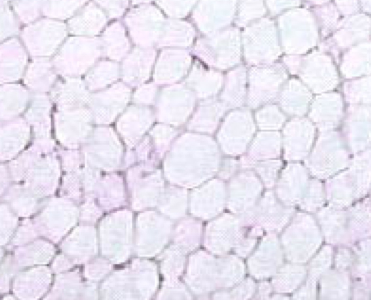
White adipose
-Looks like bubble wrap, one large lipid droplet, peripheral nucleus
-Looks like bubble wrap, one large lipid droplet, peripheral nucleus
33
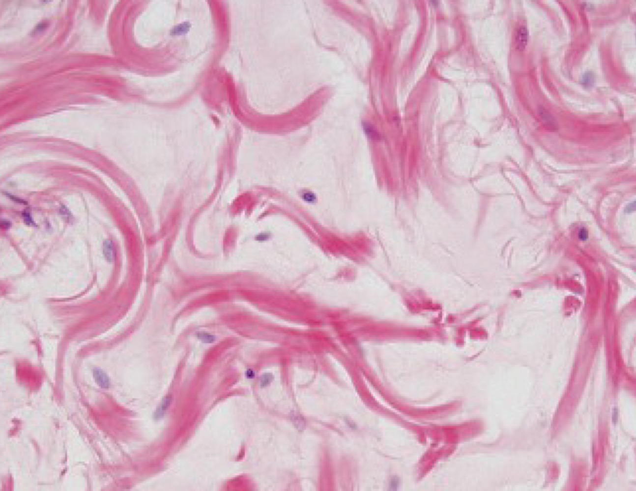
New cards
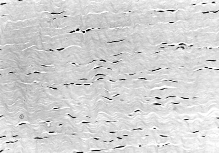
Elastin
34
New cards
Elastin
35
New cards
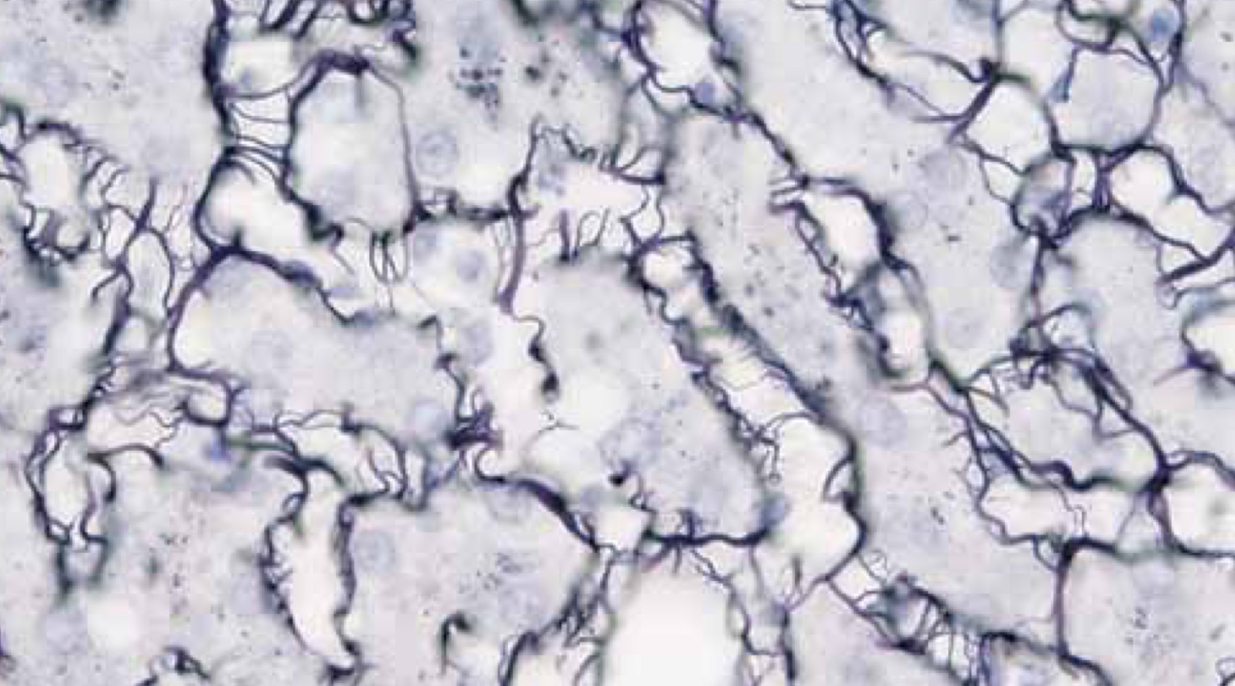
Recticulin
-Mesh work of thin fibers
-Mesh work of thin fibers
36
New cards
Recticulin
37
New cards
Loose connective tissue
-Whirl-like fibers, lots of white space
-Whirl-like fibers, lots of white space
38
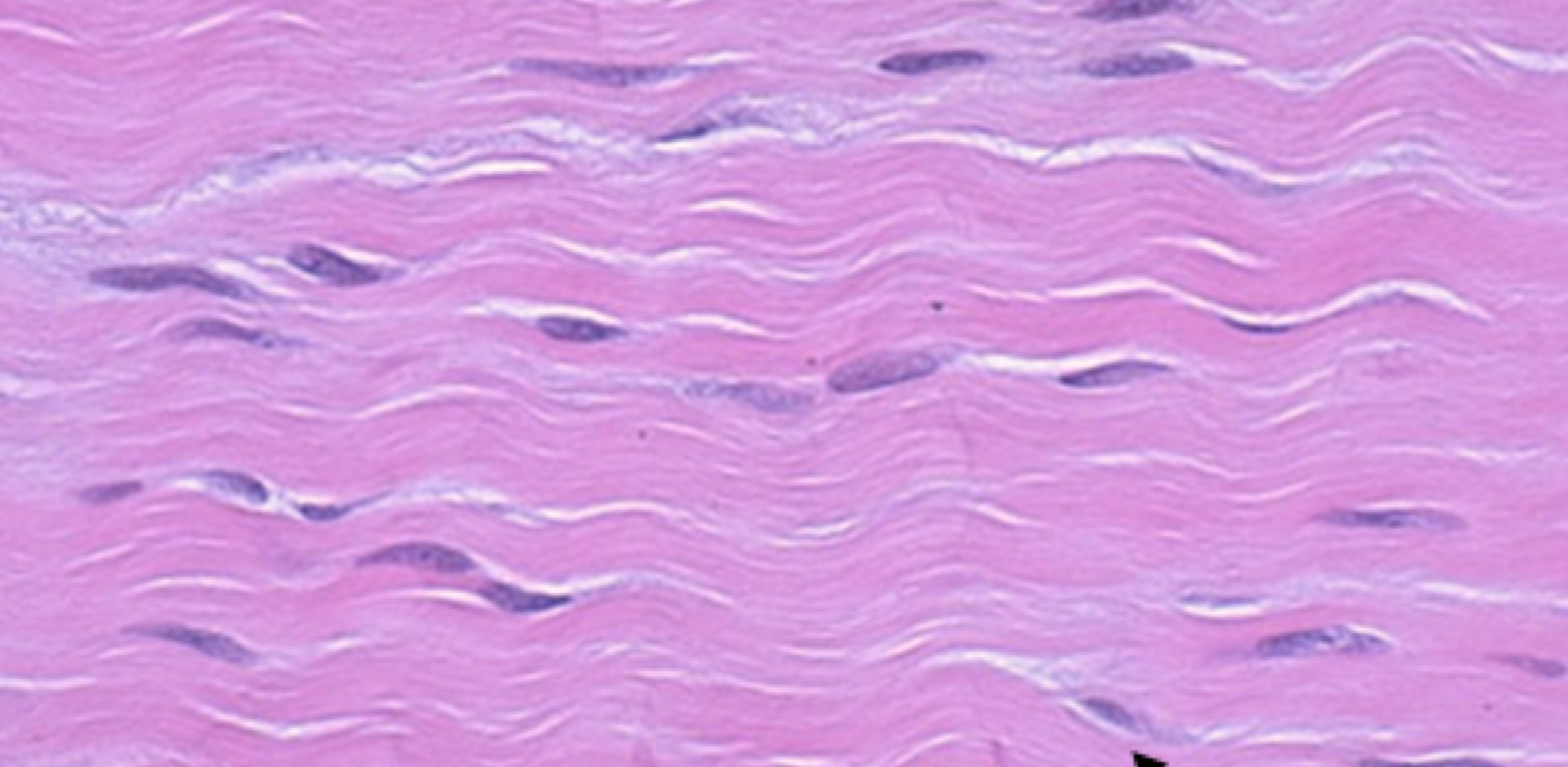
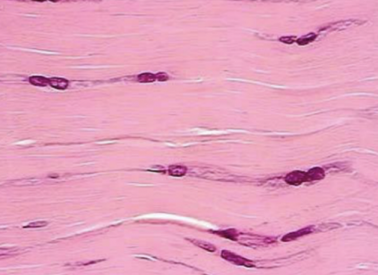
New cards
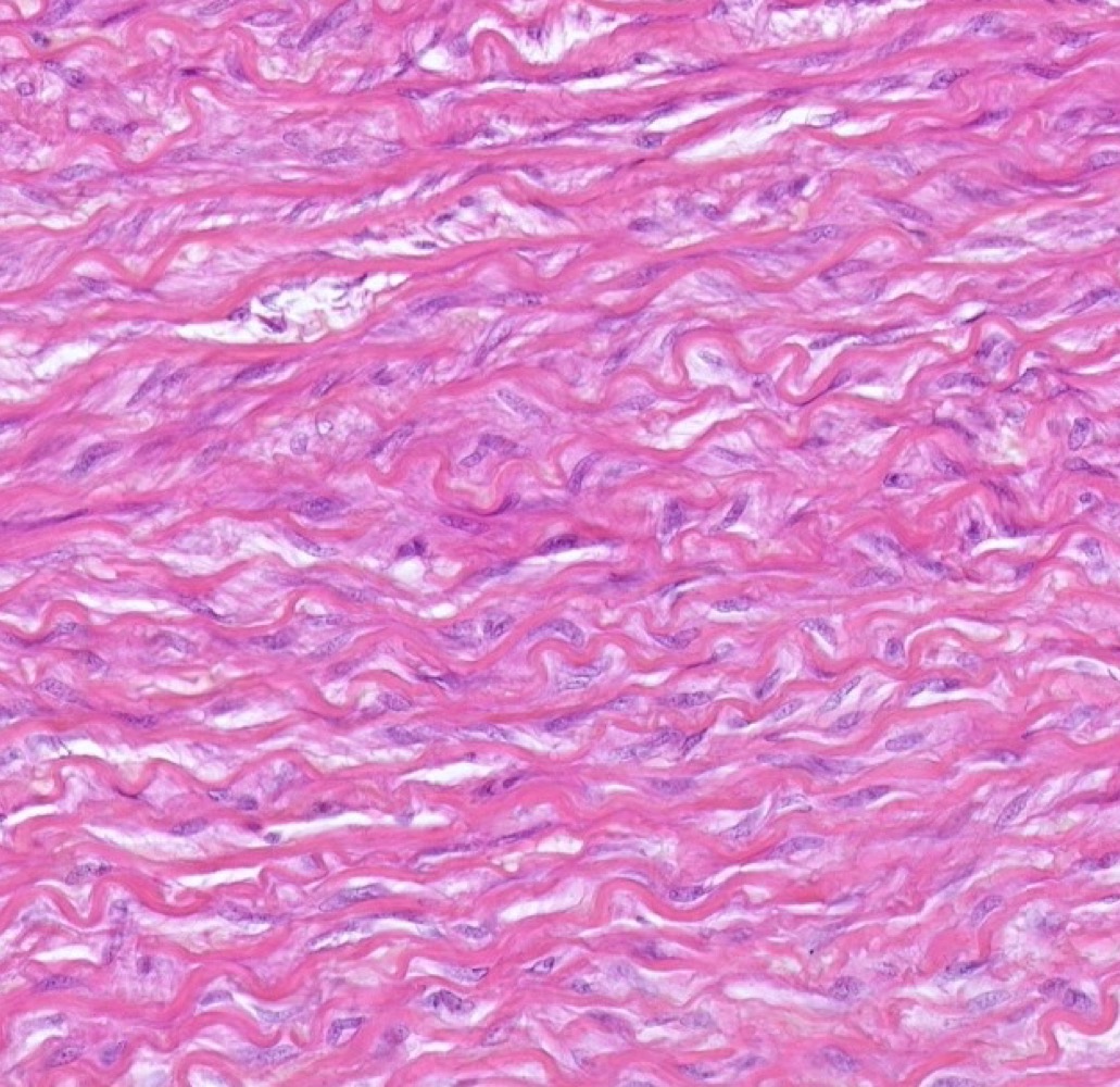
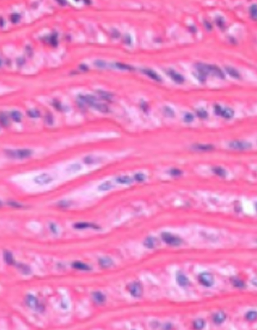
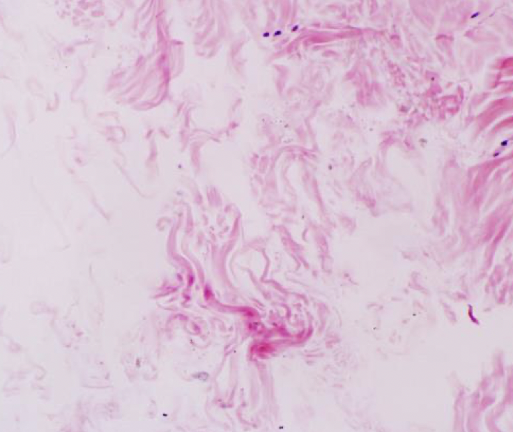
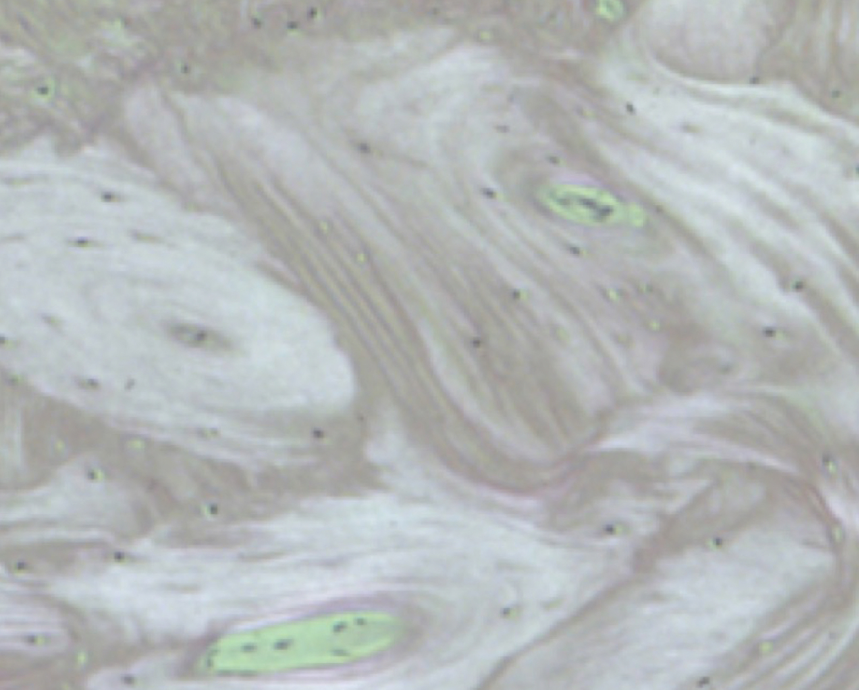
Dense Regular Connective Tissue
-mermaid hair
-linear fibers with oval peripheral nuclei
-mermaid hair
-linear fibers with oval peripheral nuclei
39
New cards
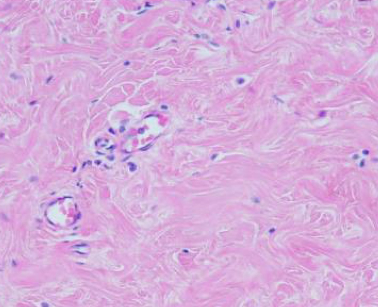
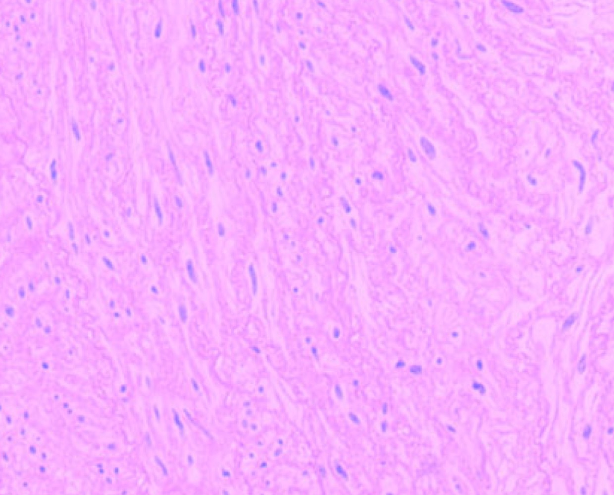
Dense, irregular connective tissue
-Sparse nuclei, lots of cytoplasm, random whirl-like appearance
-Sparse nuclei, lots of cytoplasm, random whirl-like appearance
40
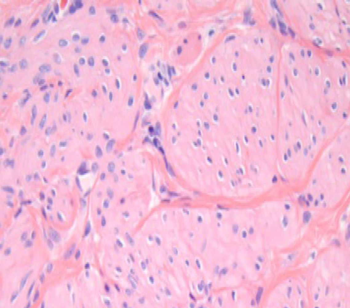
New cards
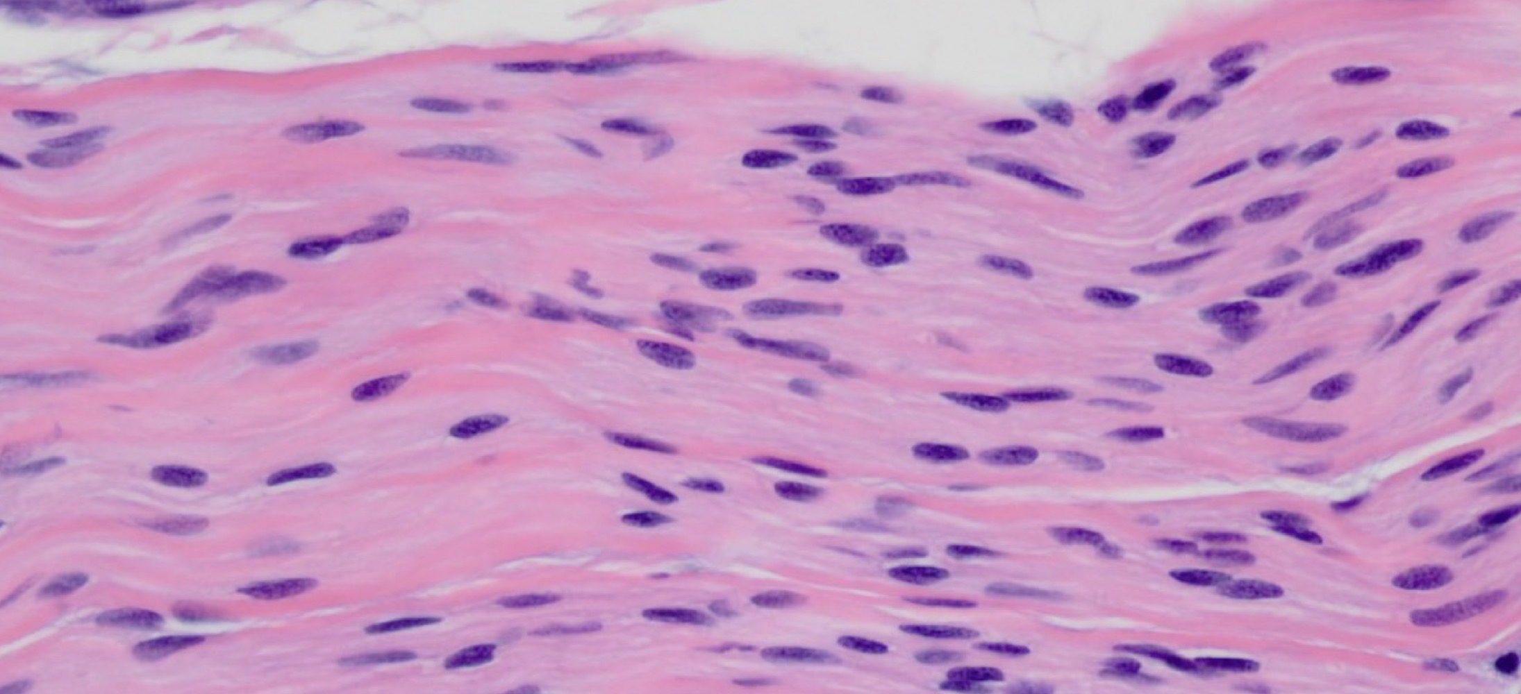
smooth muscle
-TONS of Oval nuclei, no striation
- you can tell cardiac and smooth apart because even though they both have central nuclei, smooth muscle will have a lot more nuclei
-TONS of Oval nuclei, no striation
- you can tell cardiac and smooth apart because even though they both have central nuclei, smooth muscle will have a lot more nuclei
41
New cards
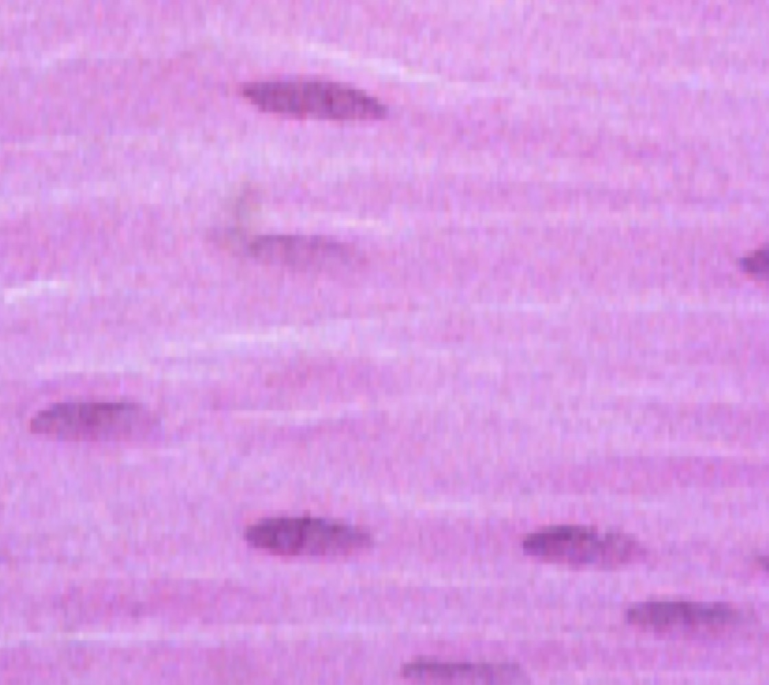
Smooth muscle
-
Tightly packed nuclei
-
Tightly packed nuclei
42
New cards
Smooth muscle
-Parallel oval nuclei, lots of nuclei, no striation
-Parallel oval nuclei, lots of nuclei, no striation
43
New cards
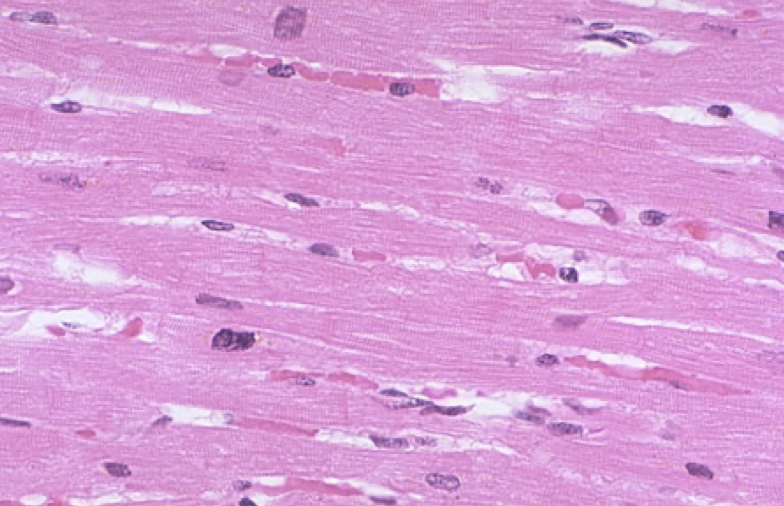
Cardiac muscle
-Striation, branching
-Striation, branching
44
New cards
Cardiac muscle
-Branching, striation, central nucleus, intercalated disc on longitudinal section
-Branching, striation, central nucleus, intercalated disc on longitudinal section
45
New cards
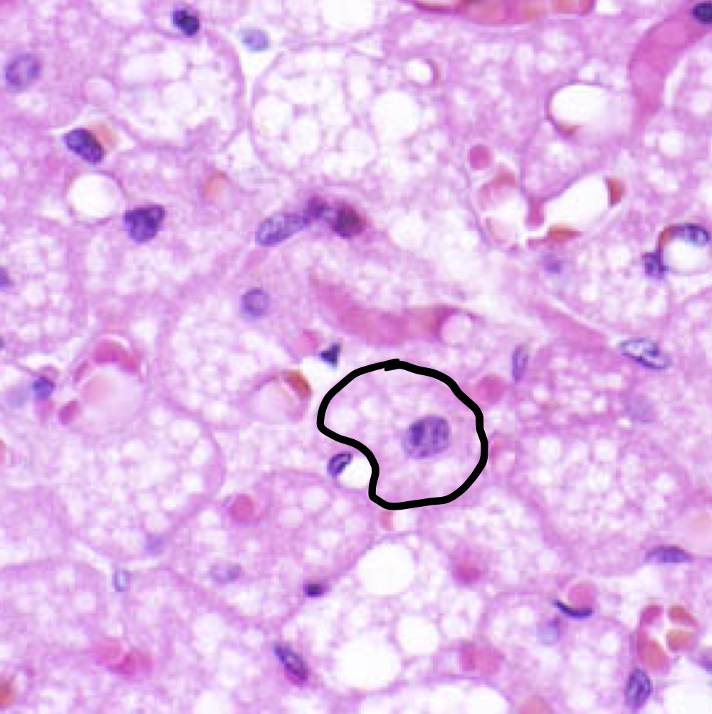
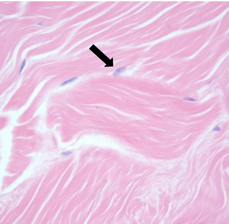
Nucleus of Fibroblast (fibroblasts are found in connective tissue)
What is the arrow pointing at and what is the cell type?
46
New cards
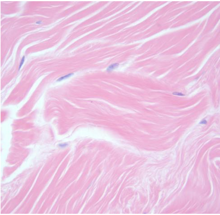
Collagen fibers within dense, irregular connective tissue
47
New cards
Dense, irregular connective tissue
48
New cards
Dense Regular Connective Tissue
-Dont mind the black/white (look at pattern)
-Dont mind the black/white (look at pattern)
49
New cards
Loose connective tissue
50
New cards
Cardiac muscle
-intercalated discs present
-intercalated discs present
51
New cards
Brown adipose
52
New cards
White adipose
53
New cards
White and Brown adipose
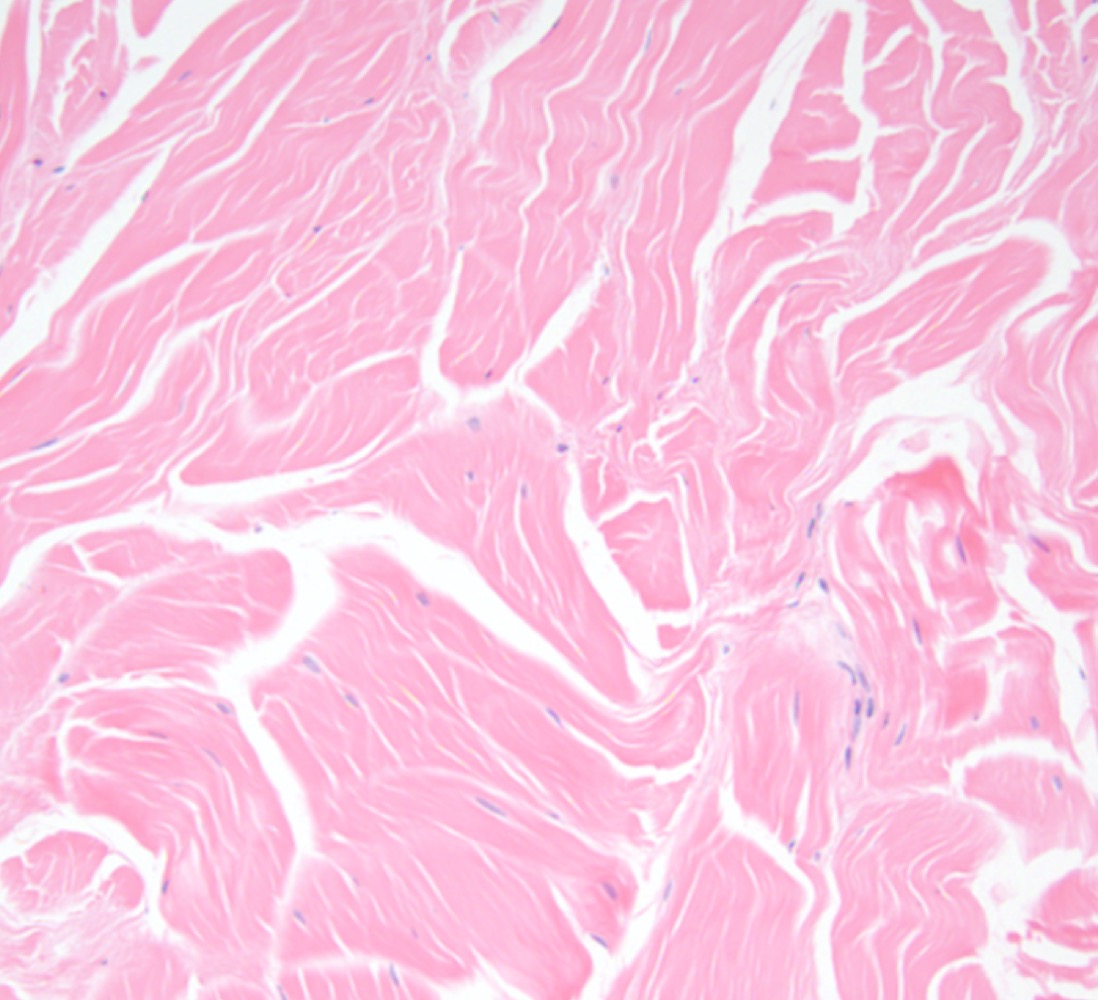
54
New cards
Dense, regular connective tissue
55
New cards
Smooth muscle
-both cross and longitudinal due to tubular organ
-both cross and longitudinal due to tubular organ
56
New cards
Cardiac muscle
57
New cards
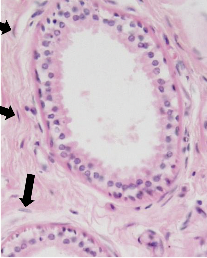
Fibroblasts
-within connective tissue
-within connective tissue
What cell is the arrow pointing at?
58
New cards
Skeletal muscle
59
New cards
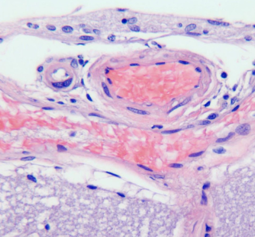
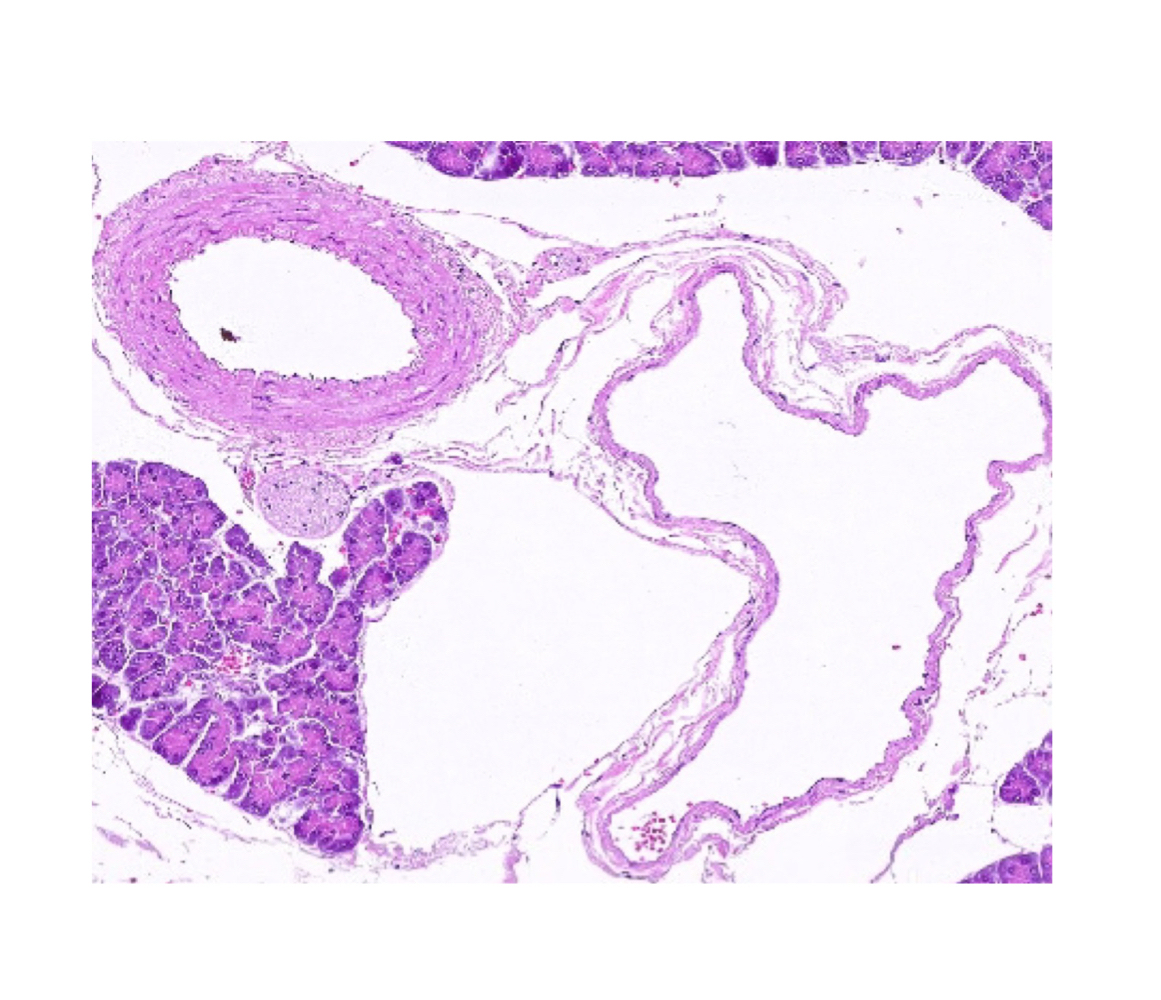
Vein and artery
What two structures are present?
60
New cards
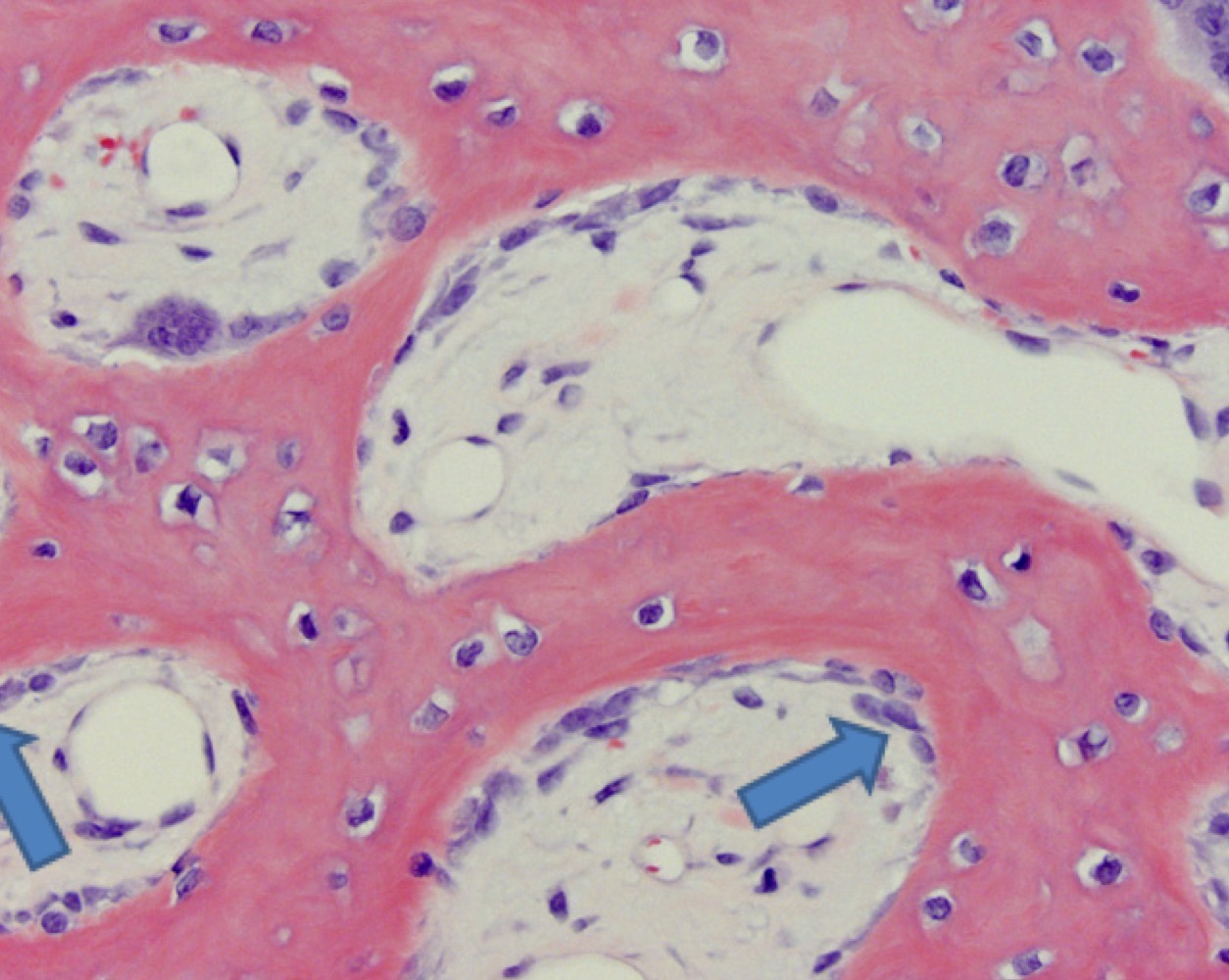
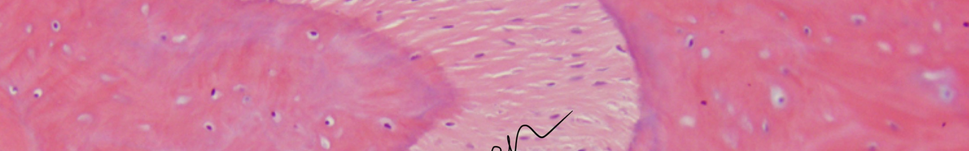
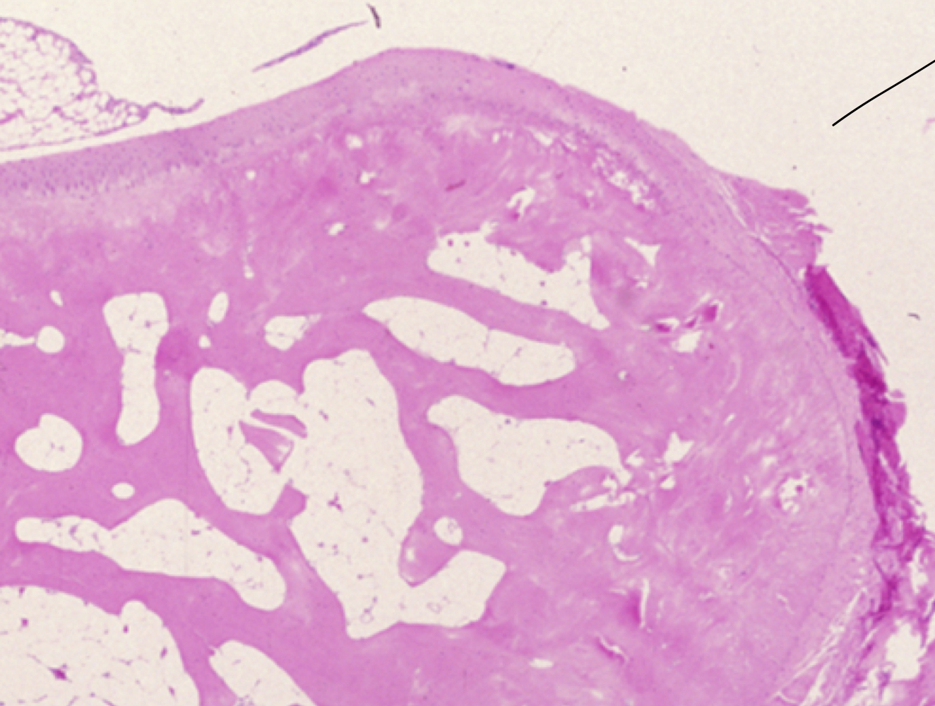
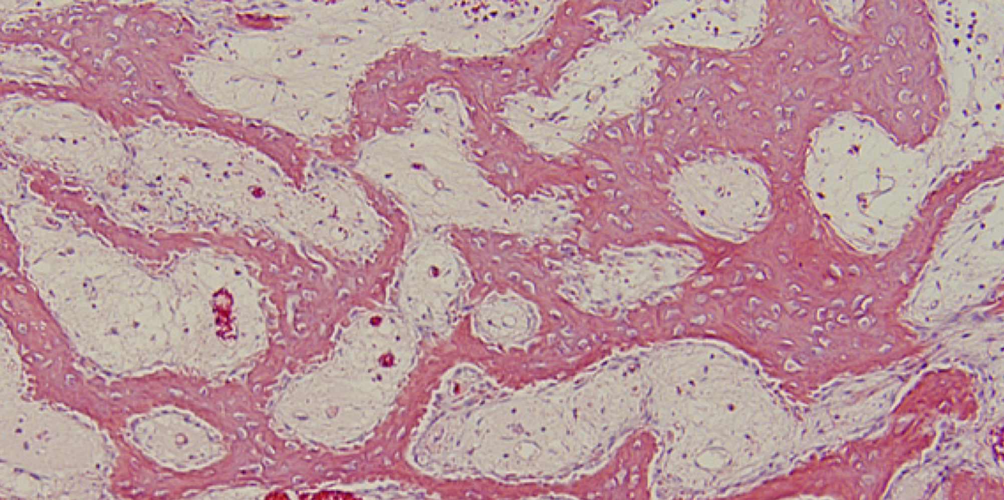
Woven Bone (cartilage)
61
New cards
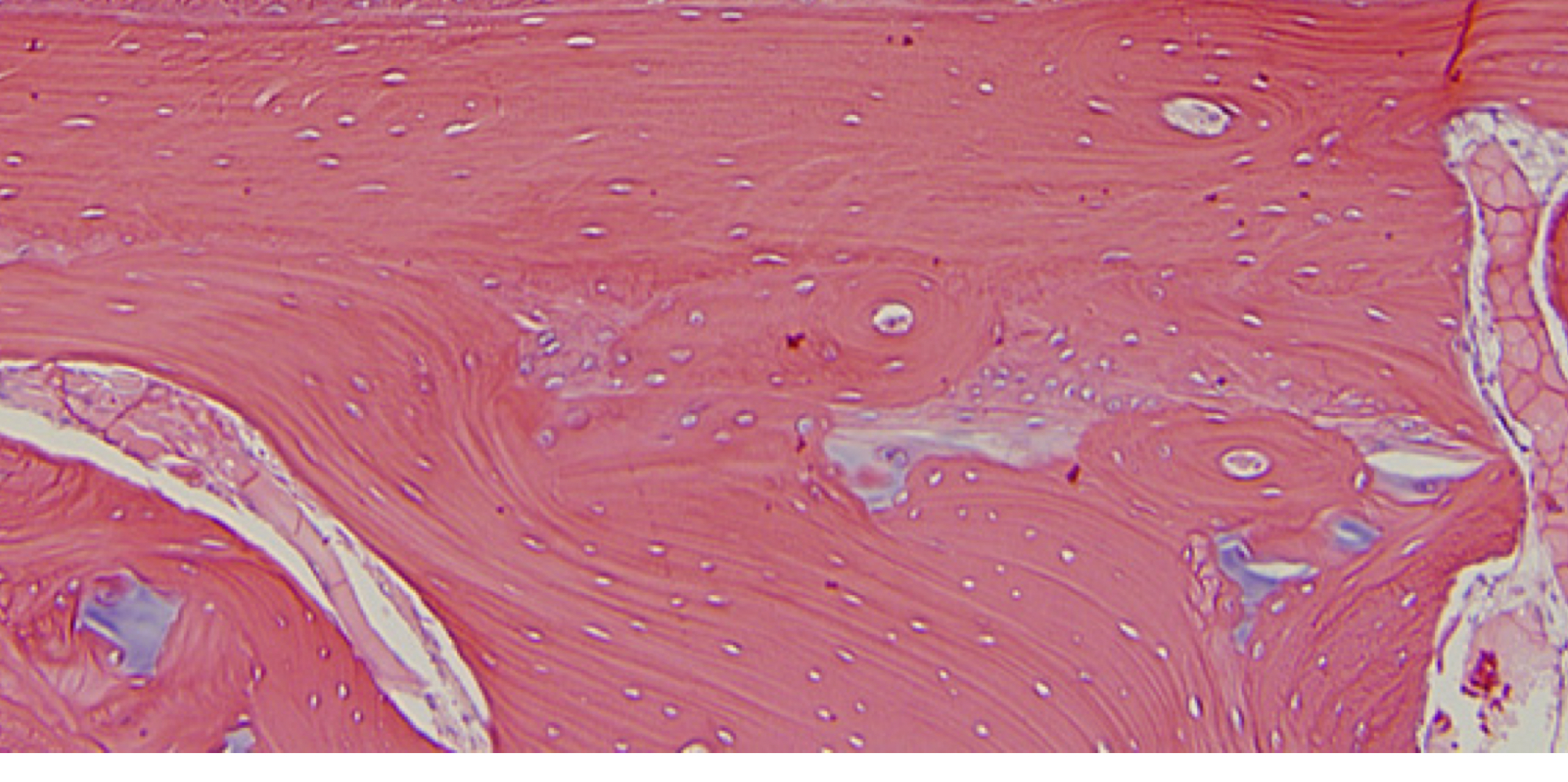
Lamellar Bone (cartilage)
62
New cards
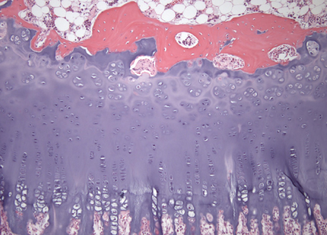
Endochondrial Ossification
63
New cards
Joint
-collagen connective tissue between two bones
-collagen connective tissue between two bones
64
New cards
Outside= compact
Inside= cancellous
Inside= cancellous
What are the outside and inside layers?
65
New cards
Woven Bone (cartilage)
66
New cards
Lamellar Bone (cartilage)
67
New cards
synovial joint
-white is synovial fluid
-cells lining are synoviocytes
-white is synovial fluid
-cells lining are synoviocytes