rad
1/25
There's no tags or description
Looks like no tags are added yet.
Name | Mastery | Learn | Test | Matching | Spaced | Call with Kai |
|---|
No analytics yet
Send a link to your students to track their progress
26 Terms
lateral cephalometric
evaluate facial growth and development, trauma, disease and developmental abnormalities; showing lateral bones of face and skull and soft tissues
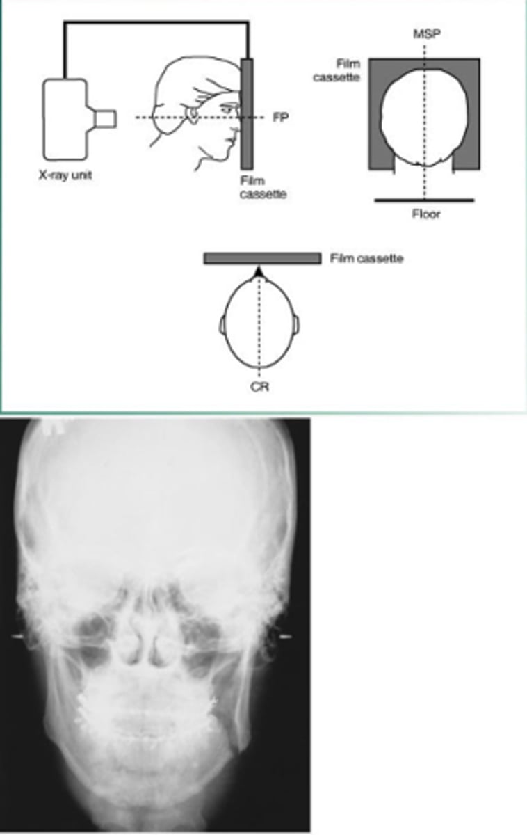
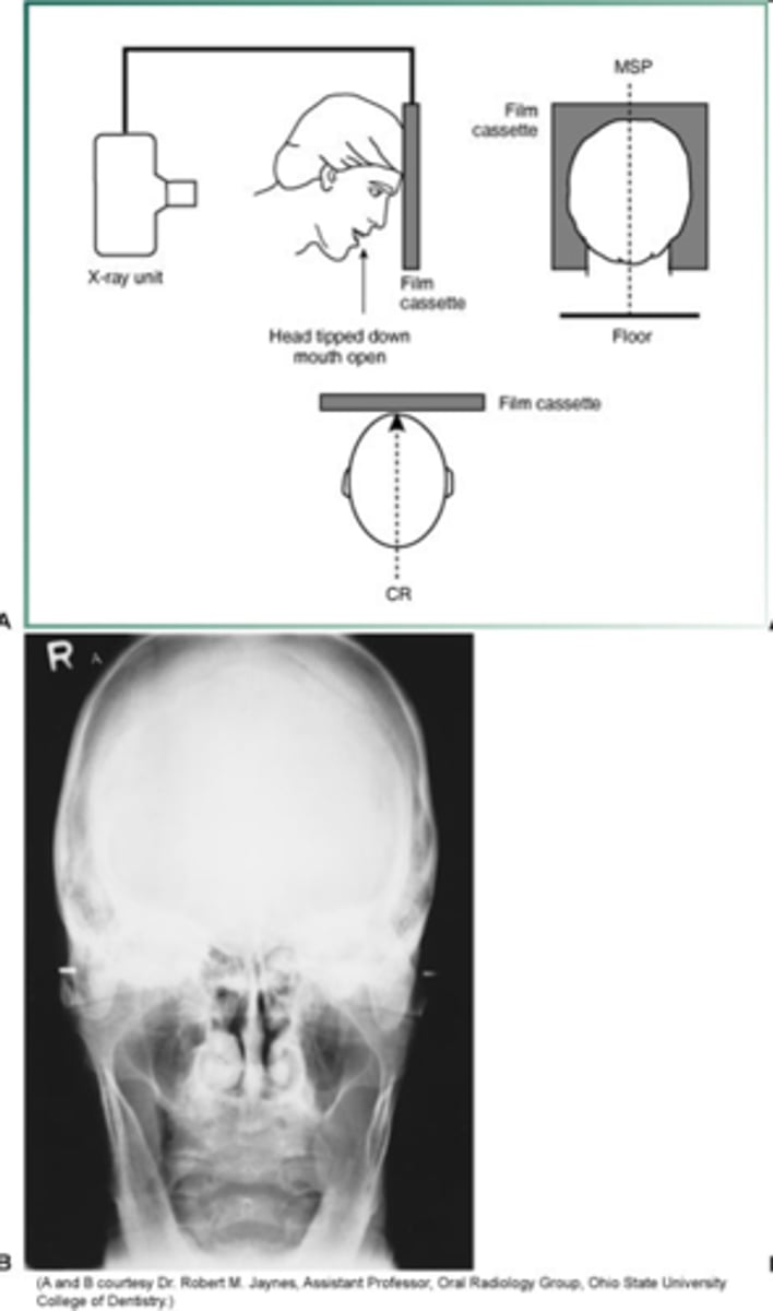
posteroanterior
evaluate facial growth and development, trauma, disease and developmental abnormalities; shows sinuses, nasal cavity, and orbits
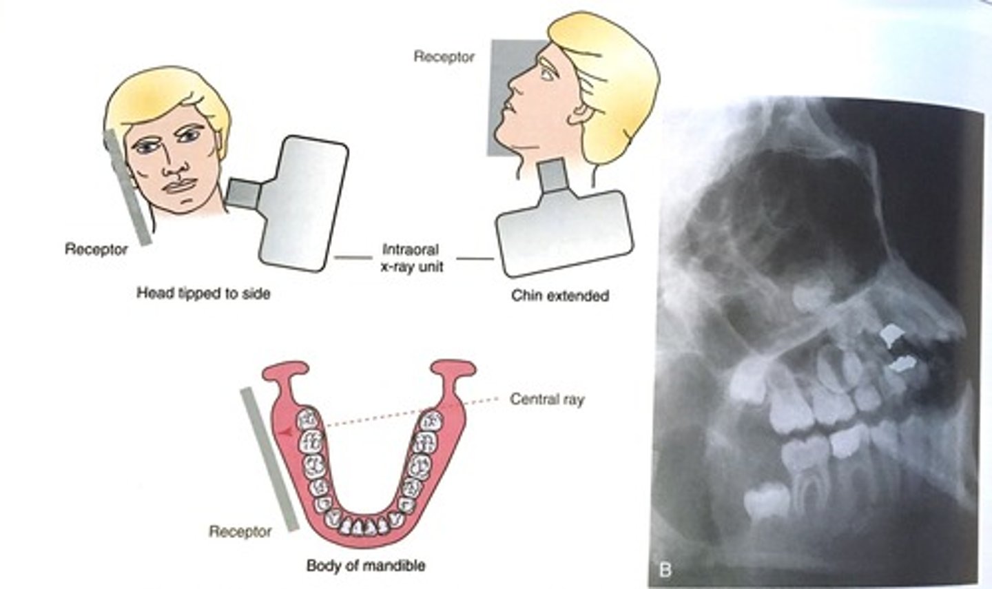
Lateral Jaw, body (mandible) receptor placement
-flat against cheek
-centered over body of mandible
Lateral Jaw, body (mandible) head position
-tipped 15 degrees towards side being imaged
- chin extended and elevated
lateral jaw body beam alignment
- below of mandible
- vertical angulation -15 to -20
Lateral Jaw Ramus of Mandible
evaluate impacted third molars, large lesions, and fractures in ramus
Lateral jaw projection-body of mandible
evaluate impacted teeth, fractures, and lesions in body
lateral jaw projections, ramus of mandible head position
- tipped 15 degrees towards side being imaged
- chin extended and elevated
Lateral jaw projection-ramus of the mandible beam alignment
- posterior to third molar area
- vertical angulation -15 to -20 degrees
posteroanterior projection receptor placement
long axis of receptor is vertical
posteroanterior projections head position
forehead and nose touch receptor
posteroanterior projection beam alignment
centered over receptor
parallel to receptor
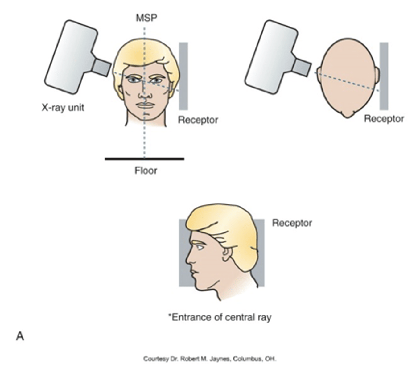
lateral cephalometric receptor position
long axis of receptor is horizontal
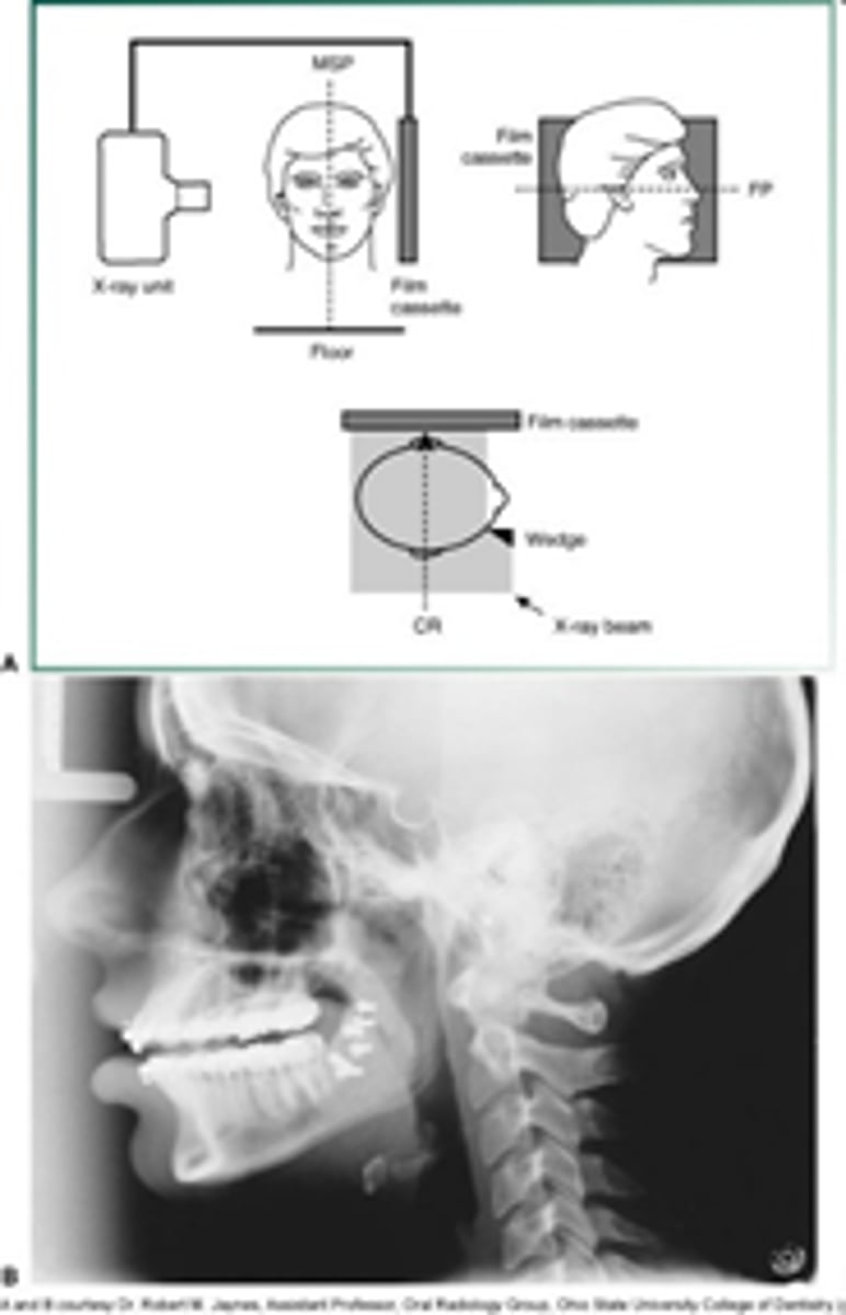
lateral cephalometric head position
left side of head is near receptor
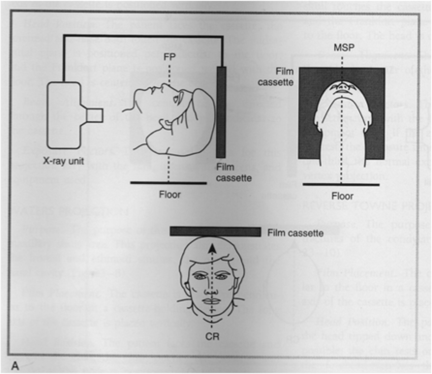
waters projection receptor placement
long axis of receptor is vertical
waters projection head position
- chin touches receptor
- tip of nose is 1-2 inches away from receptor
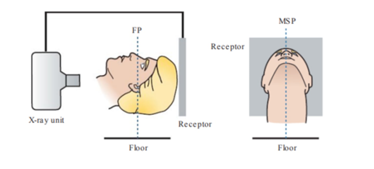
submentovertex projection receptor placement
long axis of receptor is vertical
submentovertex projection head position
- head tipped back
- top of head touches receptor
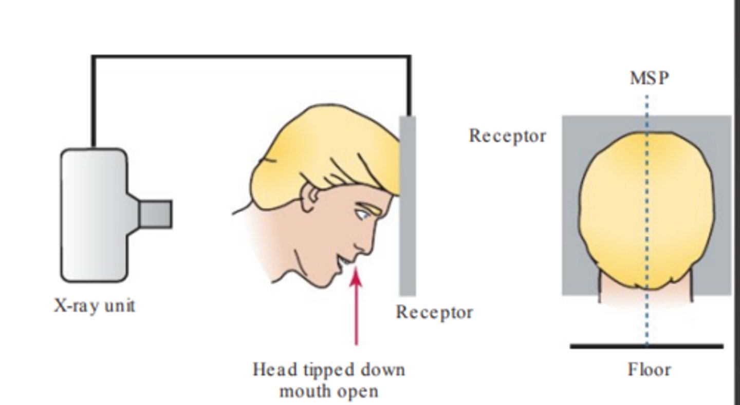
reverse towne projection head position
- head tipped down
- mouth open
- top of forehead touches receptor
transcranial projection receptor placement
-flat against ear
- centered over TMJ
waters projection
evaluate maxillary sinus area; shows sinuses, nasal cavity, and orbits
submentovertex
identify position of the condyles, base of the skull, and evaluate fractures of the zygomatic arch
reverse town projections
identify fractures of the condylar neck and ramus
temporomandibular joint tomography
- glenoid fossa, articular eminence of temporal bones
- condyle of mandible
transcranial projection beam alignment
- 2 inches above and 0.5 behind the ear canal opening
- vertical angulation +25degrees
- horizontal angulation 20 degrees
temporomandibular joint tomography purpose
estimate joint space and evaluate extent of movement of condyle when mouth is open