Exam 2 Review - Skeleton and Muscle Physiology
1/45
Earn XP
Description and Tags
Flashcards covering key terms and concepts related to the skeleton and muscle physiology for Exam 2.
Name | Mastery | Learn | Test | Matching | Spaced | Call with Kai |
|---|
No analytics yet
Send a link to your students to track their progress
46 Terms
Axial Skeleton
The part of the skeleton that consists of the skull, vertebral column, and rib cage.
Appendicular Skeleton
The part of the skeleton that includes the limbs and the girdles that support them, such as the scapula and pelvic girdles.
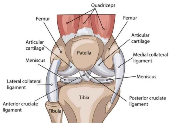
Bones of the knee joint
The femur, tibia, and patella.
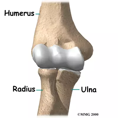
Bones of the elbow joint
The humerus, radius, and ulna.
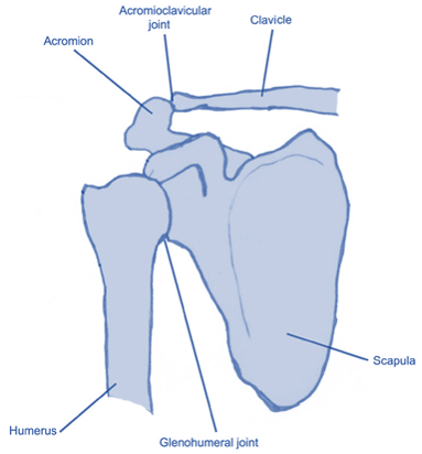
Pectoral girdle bones
The clavicle and scapula.
Orbital cavity bones
The frontal, sphenoid, zygomatic, maxilla, palatine, lacrimal, and ethmoid bones.
Medullary cavity
The central cavity of bone shafts where red and yellow bone marrow is stored.
Sutures of the skull
Fibrous joints that connect the bones of the skull.
Sphenoid bone
The bone that forms part of the cranial base and contributes to the orbit.
Sella turcica
The depression in the sphenoid bone that houses the pituitary gland.
Zygomatic arch
Formed by the zygomatic bone and the temporal bone.
Mandible joint with the skull
Temporomandibular joint (TMJ).
Gross anatomy of a vertebra
Includes components like the body, spinous process, transverse processes, and vertebral foramen.
Kyphosis
An exaggerated anterior curvature of the thoracic spine.
Lordosis
An exaggerated curvature of the lumbar spine.
Scoliosis
A lateral curvature of the spine.
Hyoid bone
The only free-floating bone in the body.
Sternum bones
Consists of the manubrium, body, and xiphoid process.
Cervical vertebrae names
The first two are called the atlas (C1) and axis (C2).
Scapula features
Includes the spine, acromion process, subscapular fossa, and glenoid fossa.
Humerus features
Includes the head, medial epicondyle, trochlea, and olecranon fossa.
Radius and ulna features
Includes the styloid processes, olecranon, head, and trochlear notch.
Constituent bones of the hand
Includes metacarpals and phalanges.
Major bones of the pelvis
Ilium, ischium, and pubis.
Joint between sacral and pelvic bones
Sacroiliac joint.
Femur features
Includes the head, neck, greater/lesser trochanters, and medial/lateral condyles.
Tibia features
Includes the medial malleolus and medial/lateral condyles.
Largest bone of the foot
The calcaneus, which is medial to other tarsals.
Striated appearance of muscle fibers
Results from the arrangement of actin and myosin filaments.
Functions of skeletal muscles
Include movement, posture maintenance, joint stabilization, and heat production.
Calcium storage in muscle cells
Occurs in the sarcoplasmic reticulum.
Connective tissue sheaths in muscles
Includes epimysium (around whole muscle), perimysium (around fascicles), and endomysium (around individual fibers).
Basic structures of a muscle cell
Includes myofibrils, sarcolemma, sarcoplasmic reticulum, and t-tubules.
Sarcomere bands and lines
Include A band, I band, Z line, and M line, distinguished by light and dark striations.
Proteins surrounding actin filaments
Include tropomyosin and troponin, which regulate contraction.
Thick and thin filament proteins
Thick filaments are primarily made of myosin; thin filaments are primarily made of actin.
Rigor mortis onset
Begins within a few hours after death and resolves after 24 to 48 hours.
Neuromuscular junction (NMJ) components
Consists of the synaptic cleft, acetylcholine in vesicles, and cholinergic receptors.
Muscle fiber interruption effect
Leads to paralysis or inability of muscles to contract.
Excitation-contraction coupling steps
Involves nerve impulse triggering calcium release, leading to sarcomere contraction.
Forms of tetanus vs. muscle twitches
Tetanus is a sustained contraction, while muscle twitches are single quick contractions.
Characteristics of muscle tissue
Include excitability, contractility, extensibility, and elasticity.
Tendon connecting gastrocnemius to calcaneus
Achilles tendon.
White band of tissue between abdominal muscles
Linea alba.
Large tendon connecting occipitofrontalis muscle
Galea aponeurotica.
Definitions of key movements in anatomy
Pronation: inward rotation; supination: outward rotation; elevation: lifting; depression: lowering; abduction: moving away from midline; adduction: moving toward midline.