Nervous System - CNS
1/48
There's no tags or description
Looks like no tags are added yet.
Name | Mastery | Learn | Test | Matching | Spaced | Call with Kai |
|---|
No analytics yet
Send a link to your students to track their progress
49 Terms
What are some emergent properties of neural networks?
Affective behaviors: Related to feeling and emotion (affecting someone)
Cognitive behaviors: Related to thinking
Plasticity: The restructuring of the brain networks in response to sensory input and experience
What does dorsal and ventral mean?
Dorsal: back
Ventral: abdominal
What does rostral and caudal mean?
Rostral: nose/front
Caudal: tail
Explain the different directions in neuroanatomy
**In the brain, dorsal = up and ventral = down. rostral = pointing outward (from the front) and caudral = pointing outward (from the back)
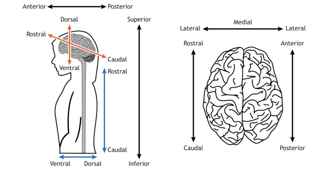
Directions in neuroanatomy
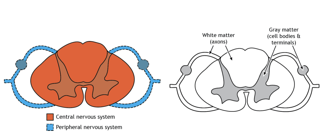
What is the difference between gray and white matter?
Gray matter:
Unmyelinated nerve cell bodies
Dendrites
Axon terminals
Layers of cell bodies
Clusters of cell bodies in the CNS are nuclei
White matter:
Myelinated axons
Axon bundles connecting CNS regions are tracts
How is the CNS protected?
Bone
Connective tissue
Fluid
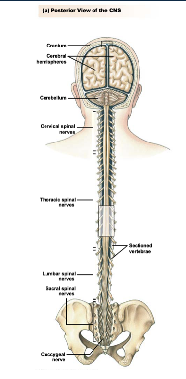
What components of the CNS are made of bone?
Brain: Encased in bony skull, or cranium
Spinal Cord: Runs through vertebral column
What is the connective tissue in the CNS?
Meninges lie between bone and tissues to stabilize neural tissue and protect from bruising (3 layers)
3 layers:
Dura matter
Arachnoid membrane
Pia matter
What is the cerebrospinal fluid?
The brain floats in it
Physical and chemical protection
Reduces pressure in CNS and protects your brain/head after you hit your head
Fills ventricles, subarachnoid space to surround and protect the CNS
Produced by choroid plexus in ventricles
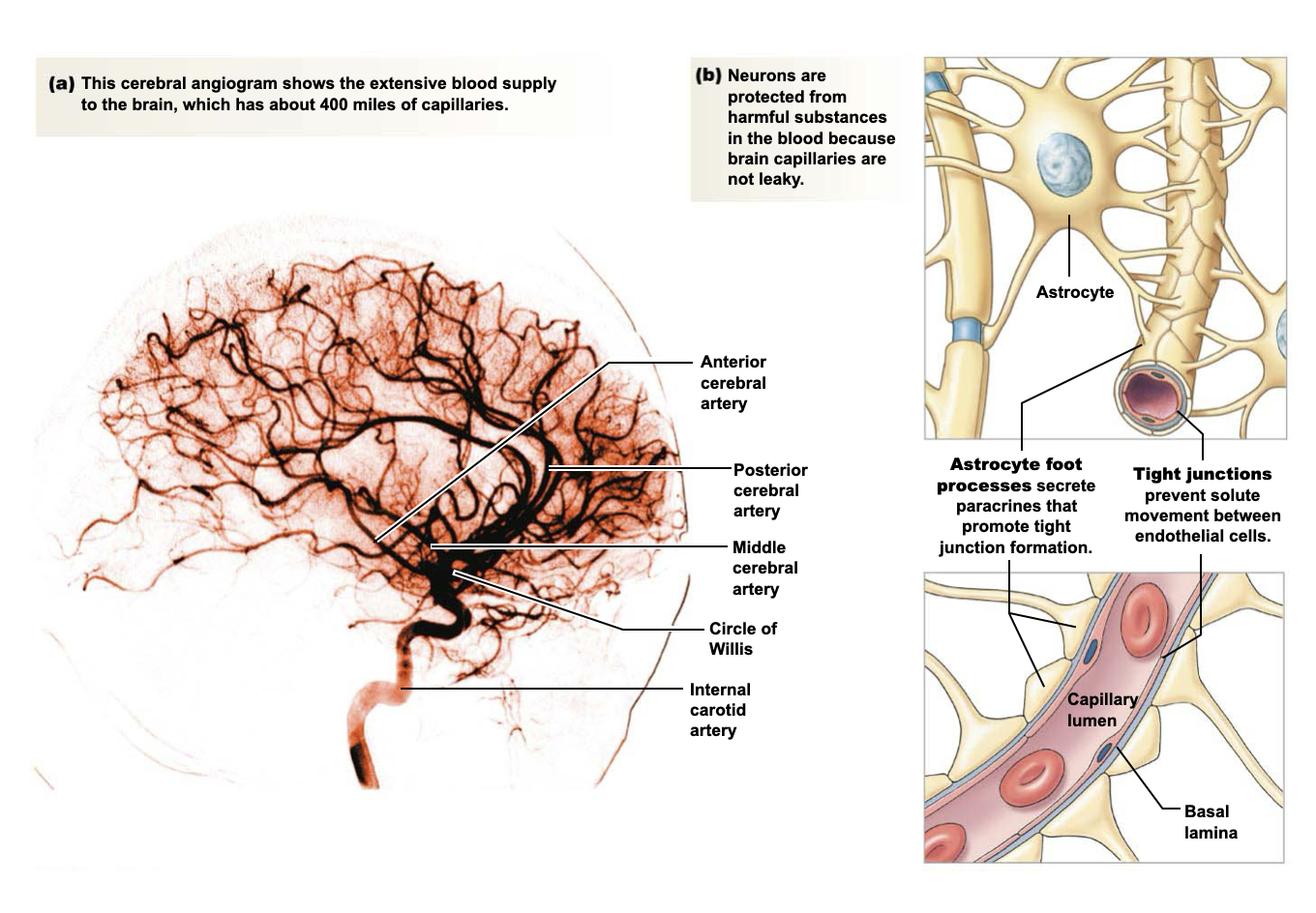
What is the blood-brain barrier?
Highly selective permeability (less leaky) of brain capillaries
Astrocytes helps promote tight junctions between endothelial cells
Protects brain from toxic water-soluble compounds and pathogens
Water, gases, and small lipid-soluble molecules can diffuse across the blood-brain barrier
Larger molecules can only cross if there is a transporter in the capillary endothelium
1) What is L-DOPA?
2) What is its relationship to the blood-brain barrier (BBB) and pharmacology?
1)
L-DOPA: Primary Parkinson’s disease treatment
2)
L-DOPA leads to dopamine production
Dopamine can’t cross the BBB but L-DOPA can
L-DOPA is converted to dopamine (DA) in the CNS and peripherally
Too much peripheral DA leads to many side effects
Carbidopa prevents conversion to DA and does not cross BBB
What are the metabolic requirements of the CNS?
Brain receives 15% of blood pumped by heart
Oxygen
Brain uses about 20% of body’s oxygen supply
Passes freely across BBB
Only a few minutes w/o oxygen causes brain damage (when blood flow stops)
Glucose
Brain is responsible for about half of body’s glucose consumption
Membrane transporters move glucose from plasma into CSF
Progressive hypoglycemia leads to confusion, unconsciousness, and death
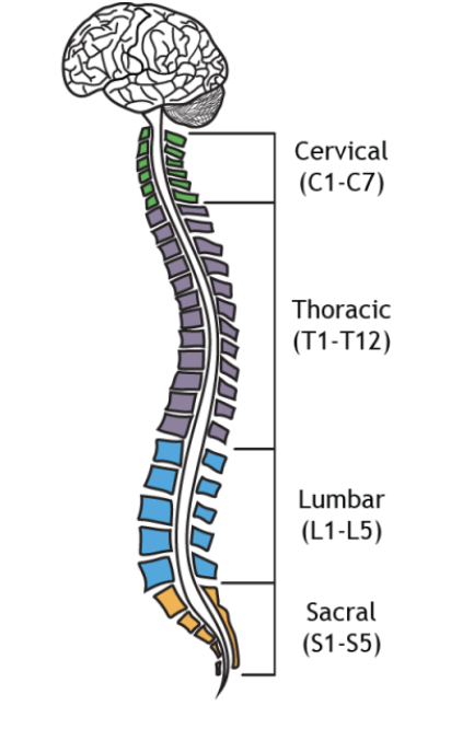
How is the spinal cord organized?
4 regions: cervical, thoracic, lumbar, sacral
Each region is divided into segments
Spinal nerves enter these segments
How is the spinal cord organized?
PNS contains fibers that enter/exit the spinal cord
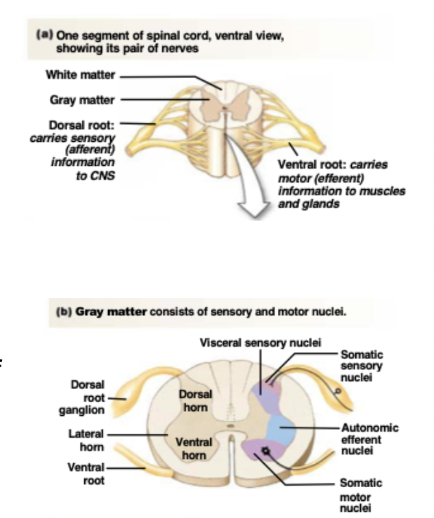
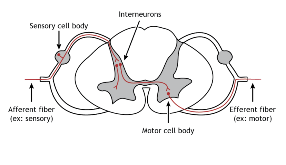
What are the structural grey matter components of the spinal cord?
Spinal nerves split into 2 roots (branches) before entering the spinal cord
Dorsal root neurons carry sensory info into the CNS
The cell bodies of these afferent neurons are located in the dorsal root ganglia, outside the spinal cordAfferent neurons connect with interneurons in the dorsal horns
Ventral roots carry info out from the CNS to muscles and glands
Ventral horns contain cell bodies of efferent neurons
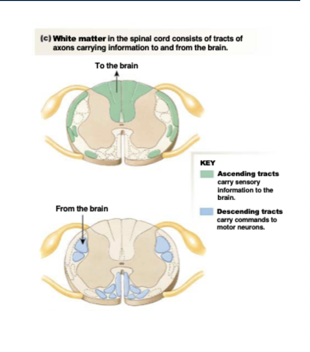
What are the structural white matter components of the spinal cord?
Ascending tracts take sensory info to the brain
Descending tracts carry signals from the brain
Propriospinal tracts stay in the cord
Spinal reflexes
What is the spinal cord made of?
Afferent neurons (sensory)
Sensory cell body
Interneurons
Motor cell body
Efferent neurons (motors)
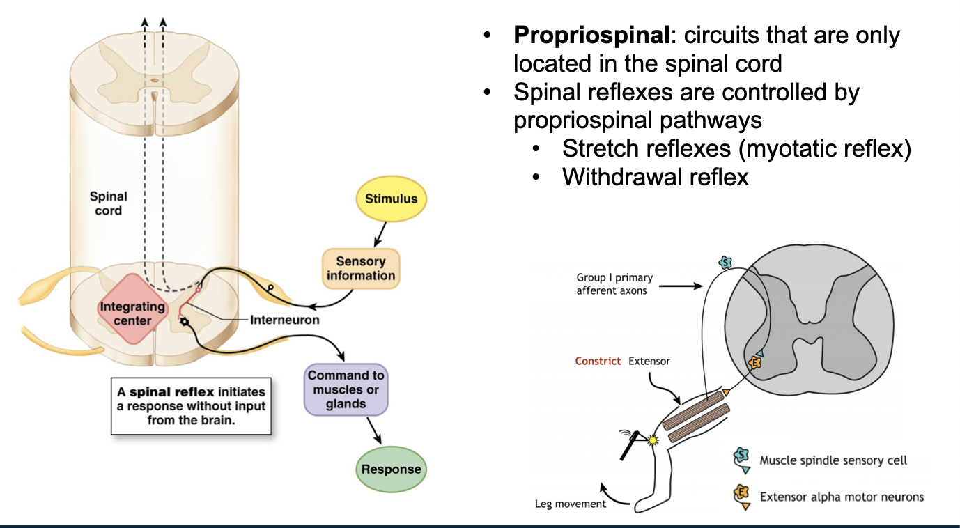
What is an example of a spinal reflex?
Propriospinal: Circuits that are only located in the spinal cord
Spinal reflexes are controlled by propriospinal pathways → initiates a response without input from the brain
Brain can increase or decrease brain strength
Stretch reflexes (myotatic reflex): Causes a muscle to contract in response to being stretched, helping maintain muscle tone and posture.
Withdrawal reflex: Automatically pulls a body part away from a painful stimulus, without needing the brain to initiate it.
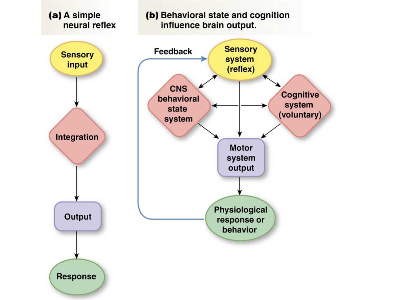
Reflex arc layout (left diagram)
Stimulus → sensory neuron → interneuron → motor neuron → response
Integrating center → spinal cord
Stretch (myotatic) reflex example (right diagram)
Muscle stretches
Muscle spindle detects the stretch
Afferent neuron carries sensory info to the spinal cord
Sensory neuron synapses in the spinal cord (release neurotransmitters → basically passing signal to next neuron)
Alpha motor neuron to the same muscle is activated
Response: Muscle contracts/knee-jerk
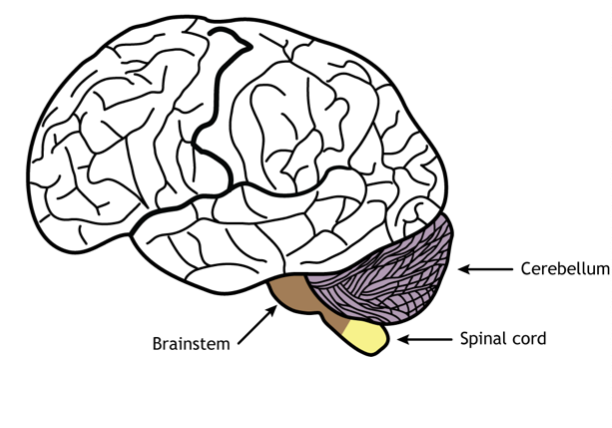
What is the brainstem?
Oldest region of the brain
Contains midbrain, pons, and medulla
Midbrain is made of tegmentum and tectum
All connections between brain and body
Regulation of consciousness and critical functions like heart rate and breathing
Has 12 cranial nerves (most start in the brainstem → except I and II)
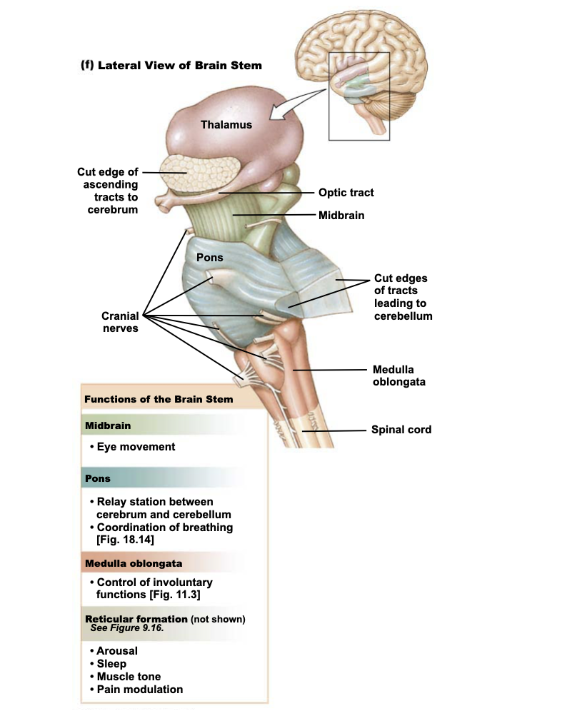
What is reticular formation (brainstem)?
Network brain stem nuclei that control wakefulness, sleep, muscle tone, pain modulation, coordination of breathing, blood pressure regulation
What is the medulla (medulla oblongata)?
Controls involuntary functions
White matter
Somatosensory (ascending) and corticospinal (descending) tracts
Pyramidal tracts: Paired white matter tracts of corticospinal and corticobulbar tracts
90% of corticospinal tracts cross in this region
Grey matter
Nuclei control involuntary functions: blood pressure, breathing, swallowing, vomiting
What are the pons?
Relay station (bridge) between cerebellum and cerebrum
Coordinates control of breathing
What is the midbrain (mesencephalon)?
Eye movement
Relays signals for hearing and seeing reflexs
Substantia nigra → produces dopamine
What is the cerebellum?
Movement coordination
Equilibrium and balance
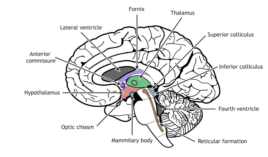
What are the components of the diencephalon?
Thalamus: Relay station and integration of sensory and motor info
Hypothalamus: Control of homeostasis, hunger, thirst, and influences endocrine function and the autonomic division
Pituitary gland: Hormones
Pineal gland: Melatonin production
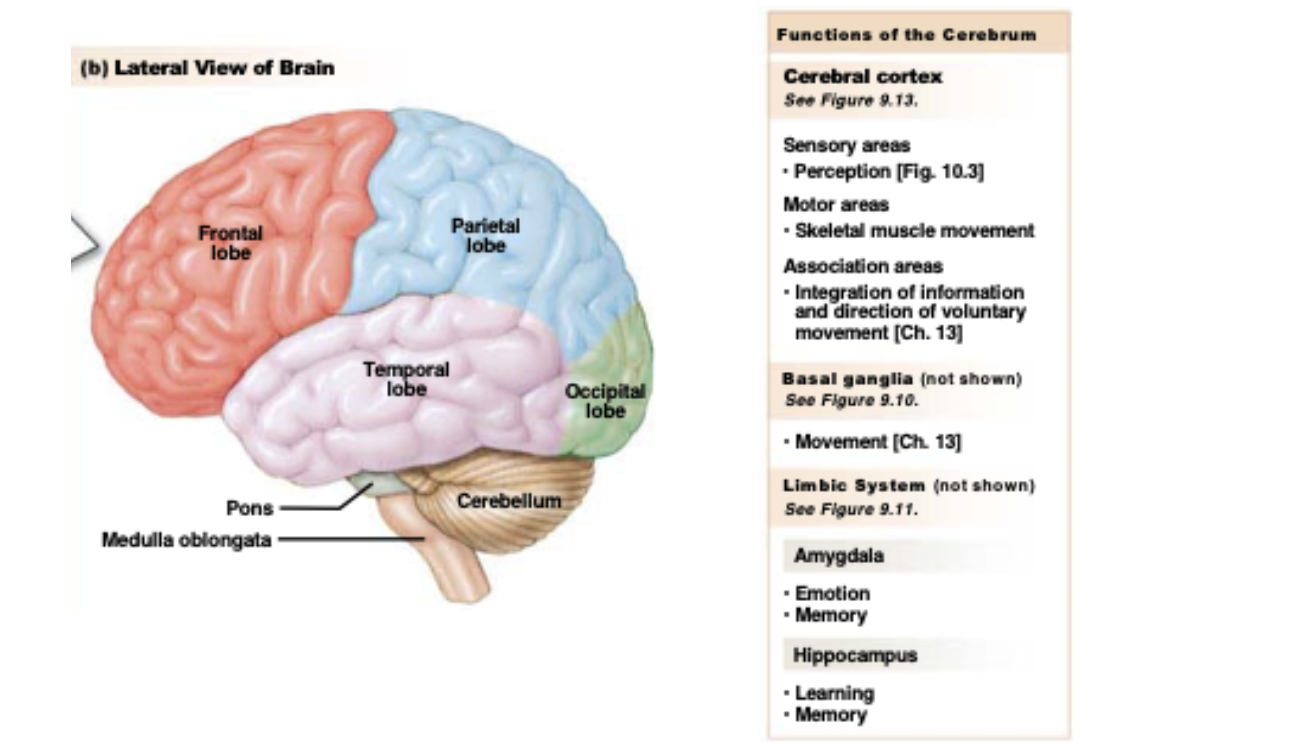
What is the cerebrum?
Consists of different structures + two hemispheres
Responsible for conscious thought, voluntary movement, sensation, memory, and language
Corpus callosum connects two hemispheres
4 lobes: frontal, parietal, occipital, temporal
Grey matter:
Cerebral cortex: Thinking, perception, decision-making
Basal ganglia: Control of movement
Limbic system: Link between cognitive functions and emotions
Amygdala:
Emotion (especially fear-related emotions) nand memory
Hippocampus:
Critical region for learning and memory
Affected in Alzhemier’s disease
White matter: Communication between brain regions
What is the basal ganglia, and what are its components?
Group of subcortical nuclei:
Striatum (caudate and putamen)
Subthalamic nuclei (STN)
Globus pallidus external and globus pallidus internal
Connected with substantia nigra (midbrain)
Control of voluntary movement via thalamic output to motor cortex
Disrupted in Parkinson’s disease and Huntington’s disease
What are the simple and complex pathways in the brain?
What are some functions of the brain?
Perception
Motor output
Behavioral state
Sleep
Circadian rhythms
Emotions, motivation, and moods
Learning and memory
Language
Personality
Movement
What are some examples of brain imaging techniques (and their functions)?
Electroencephalography (EEG): Brain electrical activity from how many neurons is measured by electrodes placed on the scalp
Positive emission tomography (PET): Glucose is tagged with a radioactive substance that emits positively charged particles. Metabolically active cells using glucose light up more
Functional magnetic resonance imaging (fMRI): Active brain tissue has increased blood flow and uses more oxygen. Hydrogen nuclei in water create a magnetic signal that indicates more active regions
What are some examples of brain injuries?
Split brain syndrome: Corpus callosum is severed
Deficits in tasks and learning that require coordination of both sides of the body
Parietal lobe injury: Problem with somatic sensory perception and coordination due to damage to primary somatic sensory cortex and sensory association area
Temporal lobe injury: Auditory and language issues
Occipital lobe injury:
Visual issues; blindness, blind spots, visual distortion, visual inattention, spatial analysis
What did Phineas Gage do?
Rod entered his skull by accident → dramatic change in personality afterwards → learned that frontal lobe controls personality
What are the three functional areas of the cerebral cortex?
3 functional specializations
Sensory areas: Receive sensory input and translate into perception (awareness)
Motor areas: Direct skeletal muscle movement
Association areas:
Integrate info from sensory and motor areas
Can direct voluntary behaviors
Cerebral lateralization:
Some functions are more concentrated on one side
Left brain → logical, analytical, and verbal processing (language, math, facts)
Right brain → intuitive, creative, and holistic thinking (arts, emotions, visualization)
This doesn’t mean we are “left-brained” or “right-brained”
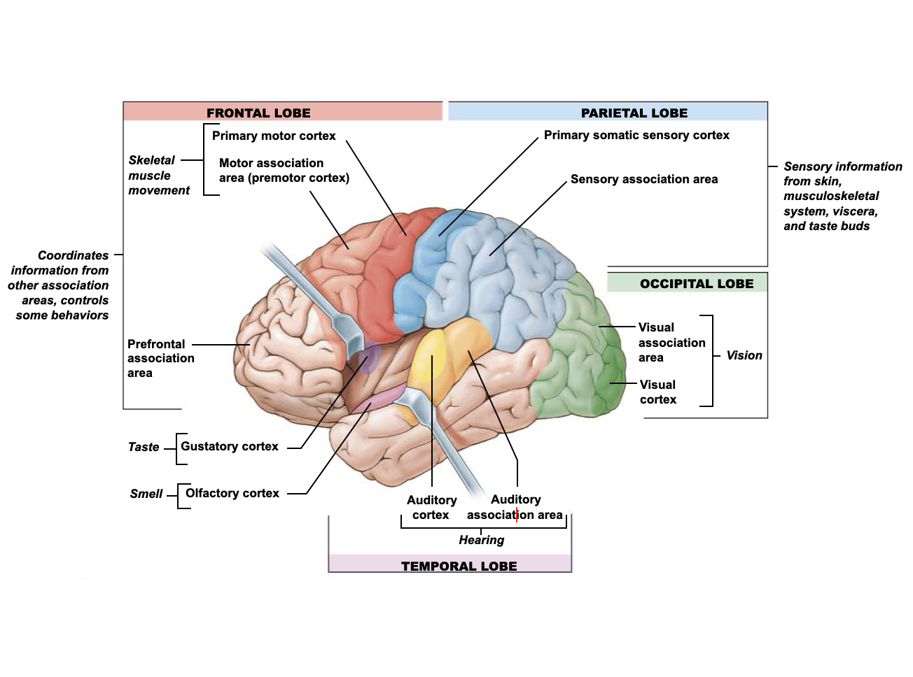
How are sensory information perceived by the brain?
Each sense has a devoted region:
Visual cortex, auditory cortex, olfactory cortex, gustatory cortex, proprioception (the "sixth sense" of body awareness, enabling the brain to understand the position, movement, and effort of limbs and trunk without visual input)
Primary somatic sensory cortex:
Skin, musculoskeletal system, and viscera
Somatosensory pathway (touch, temp, pain, itch)
Neural pathways extend from sensory areas to association areas, which integrate stimuli into perception
Integration of spinal reflexes
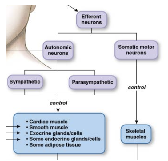
What are the three major types of CNS output?
Skeletal muscle movement: Somatic motor division
Neuroendocrine signals: Hypothalamus and adrenal medulla
Visceral responses: Autonomic division
Voluntary movement:
Primary motor cortex
Motor association areas
Neuroendocrine and visceral responses are coordinated in the hypothalamus and medulla
What is the behavioral state system?
Behavioral state neurons: Reticular formation of brain stem, hypothalamus, and limbic system
Modulator of sensory and cognitive processes, sleep/wake cycles, attention, arousal, modulation of muscle tone, and the ability to focus
Reticular activating system
14 brain nuclei
What are the four diffuse modulatory systems?
Originate in reticular formation in brain stem
Project axons to large areas of the brain
Noradrenergic (norepinephrine)
Serotonergic
Dopaminergic
Cholinergic
What are the components of the noradrenergic/norepinephrine diffuse modulatory system?
Functions: Attention, arousal, sleep-wake cycles, learning, memory, anxiety, pain, and mood
Neurons Originate: Locus coeruleus of the pons
Neurons Terminate: Cerebral cortex, thalamus, hypothalamus, olfactory bulb, cerebellum, midbrain, spinal cord
What are the components of the serotonergic/serotonin diffuse modulatory system?
Functions:
Lower nuclei: Pain, locomotion
Upper nuclei: Sleep-wake cycle, mood, and emotional behaviors, such as aggression and depression
Neurons Originate: Raphe nuclei along brain step midline
Neurons Terminate:
Lower nuclei project to spinal cord
Upper nuclei project to most of brain
What are the components of the dopaminergic/dopamine diffuse modulatory system?
What are the components of the cholinergic/acetylcholine diffuse modulatory system?
What are the components of sleep?
What is circadian rhythm?
Where are emotions regulated?
What is motivation?
What is mood?