Lecture 17 - Lipids and Membranes
1/49
Earn XP
Description and Tags
focus on membrane and proteins
Name | Mastery | Learn | Test | Matching | Spaced | Call with Kai |
|---|
No analytics yet
Send a link to your students to track their progress
50 Terms
How are lipids arranged in a membrane?
Lipids form a bilayer with hydrophobic tails inward and hydrophilic heads facing the aqueous environment.
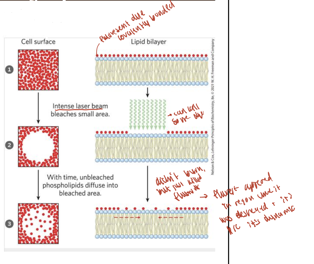
Is the lipid bilayer static or dynamic?
It is dynamic — lipids move around within each monolayer, allowing membrane flexibility and fluidity.
What affects membrane fluidity?
Cholesterol and saturated fatty acids make the membrane less flexible.
More unsaturated fatty acids increase fluidity.
What are the two main types of membrane proteins?
Peripheral proteins: on the membrane surface.
Integral proteins: embedded within the membrane.
Do both sides of a lipid bilayer have the same composition?
No, the two monolayers are different — the inside and outside of the membrane have distinct lipid and protein compositions.
What is the Fluid Mosaic Model of membranes?
A model describing membranes as fluid and flexible, where lipids and proteins move laterally within the bilayer like pieces in a mosaic.
How do lipids move within the membrane?
Lipids can diffuse laterally within a monolayer, contributing to membrane fluidity.
fluid mosaic model of membranes
What does the Fluid Mosaic Model say about lipid movement in membranes?
Individual lipids can move (diffuse) laterally within the lipid bilayer, making the membrane fluid and dynamic.
What type of lipid movement in membranes is fast and uncatalyzed?
Lateral diffusion (within the same leaflet) is fast and does not require enzymes.
Why is uncatalyzed transbilayer ("flip-flop") diffusion slow?
Because the polar head group must pass through the hydrophobic core, which is energetically unfavorable.
What enzymes catalyze lipid movement between leaflets of a membrane?
Flippase: moves lipids from outer → inner leaflet.
Floppase: moves lipids from inner → outer leaflet.
Scramblase: moves lipids in both directions to equalize distribution.
What are lipid rafts and what is their role in membranes?
Lipid rafts are long-lived microdomains in membranes, enriched in cholesterol and sphingomyelin.
They help compartmentalize membrane functions, and are involved in protein trafficking and signal transduction.
How do individual lipid molecules move in the plasma membrane?
Individual lipids can migrate laterally within the bilayer, contributing to membrane fluidity and dynamic organization.
What are peripheral membrane proteins and how are they removed?
Peripheral proteins are on the surface of the bilayer.
Held by weak interactions or covalent lipid anchors.
High salt can remove electrostatically bound proteins, but not covalently bound ones.
What are integral membrane proteins and how are they removed?
Integral proteins span the bilayer with hydrophobic regions inside the membrane and hydrophilic regions outside.
They are removed by organic solvents or detergents.
Can membrane proteins move within the bilayer?
Some membrane proteins can diffuse laterally, like lipids.
Others are anchored to the cytoskeleton and cannot move freely.
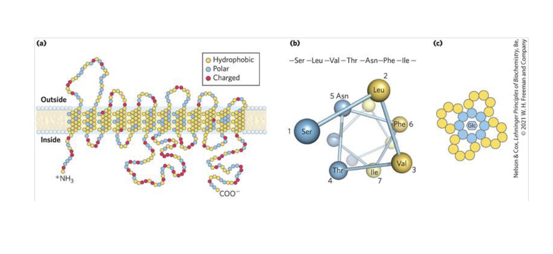
How do integral membrane proteins interact with the lipid bilayer?
Integral proteins have hydrophobic amino acids (like valine, leucine) that embed in the lipid tails of the membrane.
What are amphitropic proteins and how do they interact with membranes?
Amphitropic proteins associate with membranes only under certain conditions.
Their binding may involve lipid anchors, enzyme modification, or hydrophobic interactions.
Why do integral membrane proteins require detergents for removal?
Integral proteins have hydrophobic regions that interact tightly with the hydrophobic lipid tails in the membrane.
These interactions make them strongly embedded, so detergents (which have both hydrophilic and hydrophobic parts) are needed to disrupt the membrane and solubilize the proteins.
How do detergents help remove integral membrane proteins from the membrane?
The hydrophobic tail of the detergent binds to the protein's hydrophobic regions, replacing the lipid environment.
The hydrophilic head keeps the protein soluble in water, forming a protein–detergent complex.
What allows amphitropic proteins to associate with membranes?
Amphitropic proteins may have hydrophobic amino acids (e.g., valine, leucine) that can insert into the membrane.
Their membrane association is often reversible and can be regulated by enzymes or lipid modifications.
What are the main functions of membrane proteins?
Structural – Help maintain the integrity of the lipid-protein matrix.
Dynamic roles, including:
Transport proteins – move substances across membranes.
Enzymes – catalyze reactions at the membrane surface.
Receptor proteins – detect signals and trigger cellular responses.
Other specialized functions, depending on the cell type.
What are the structural classes of integral membrane proteins?
a) Single α-helix – spans the membrane once.
b) Multiple α-helices (4–12) – span the membrane multiple times (common in transporters & receptors).
c) β-barrel (8–16 β-strands) – form large pores or porins found in outer membranes (e.g., in bacteria and mitochondria).
Which types of molecules can cross the lipid bilayer without assistance?
Small, nonpolar molecules like O₂, N₂, CH₄, and some hydrophobic drugs can dissolve in the membrane and cross unassisted.
Which molecules need help crossing the membrane, and why?
Polar, charged, or large molecules cannot cross the membrane on their own.
They require protein-mediated transport to move through the hydrophobic bilayer.
What are the classes of membrane transport carriers?
Uniport – transports one solute in one direction.
Co-transport – transports two solutes together:
Symport – both solutes move in the same direction.
Antiport – solutes move in opposite directions.
What is the difference between symport and antiport co-transporters?
Symport: both solutes are transported in the same direction.
Antiport: solutes are transported in opposite directions.
What is facilitated diffusion and how do transporter proteins help?
Facilitated diffusion uses transporter or permease proteins to lower the activation energy (ΔG‡) for molecules crossing the membrane.
These proteins provide a pathway through the membrane for molecules that cannot cross freely.
What are the key characteristics of facilitated diffusion?
Moves molecules down their concentration gradient (high → low).
No energy (ATP) required.
Cannot concentrate molecules beyond the equilibrium position.
What is an example of facilitated diffusion involving glucose?
Glucose permease (GLUT1) in erythrocytes is a uniport transporter.
It has 12 α-helices forming a pore that transports glucose ~50,000 times faster than simple diffusion.
Allows glucose to move down its concentration gradient without energy input.
How does the structure of GLUT1 enable glucose transport and specificity?
GLUT1 is a highly organized protein with 12 α-helices forming a pore.
One side of the pore is hydrophobic (facing the membrane), while the interior lining has polar amino acids.
Glucose, with many polar hydroxyl (–OH) groups, interacts via hydrogen bonds with these polar residues.
These specific interactions stabilize glucose in the channel and allow selective transport — this is the basis for substrate specificity.
What is the function of the chloride-bicarbonate exchanger in erythrocytes?
It allows bicarbonate (HCO₃⁻) to leave and chloride (Cl⁻) to enter red blood cells.
This exchange maintains electrical neutrality.
It supports the transport of CO₂ (converted to HCO₃⁻) to the lungs.
Why is the chloride-bicarbonate exchanger important for CO₂ removal?
CO₂ is not very soluble in blood, but HCO₃⁻ is.
Carbonic anhydrase inside red blood cells converts CO₂ to HCO₃⁻ for easier transport.
In the lungs, the process reverses, and CO₂ is reformed and exhaled.
The exchanger helps move substrates/products in and out of red blood cells efficiently.
What is active transport across membranes?
Active transport moves solutes against their concentration gradient (low → high).
It is energetically unfavorable and therefore requires energy input.
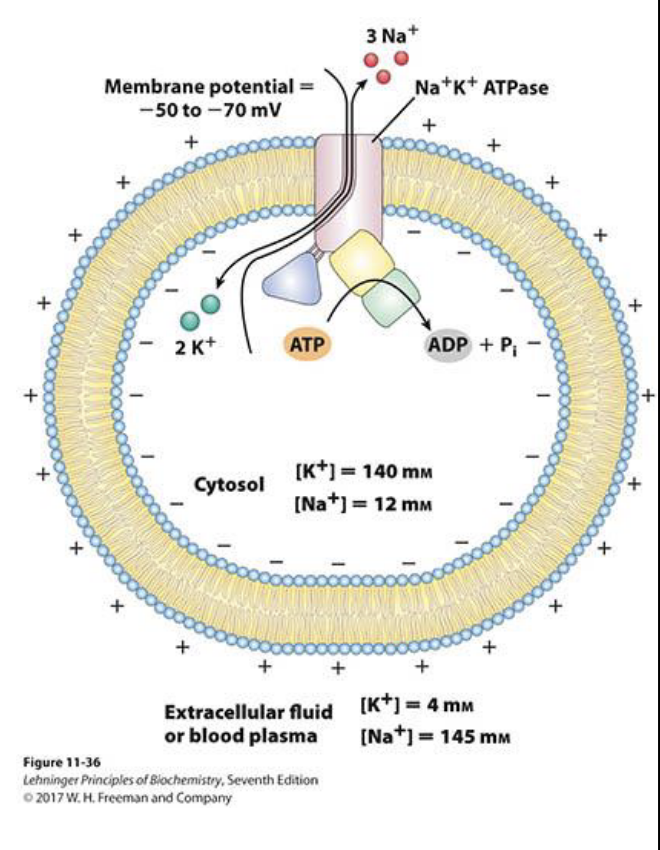
What is primary active transport?
It moves solutes against their concentration gradient (low → high), which is energetically unfavorable.
Energy is provided directly by an exergonic reaction, usually ATP hydrolysis:
ATP → ADP + PiExample: Na⁺/K⁺ ATPase pump in animal cells.
What is secondary active transport?
It uses an ion gradient (usually H⁺ or Na⁺), which was created by primary active transport.
As the ion moves down its concentration gradient, it releases energy.
That energy is used to cotransport another solute against its gradient (low → high).
This is an indirect use of ATP.
What does the Na⁺/K⁺ ATPase do?
It’s a primary active transport protein.
It moves 3 Na⁺ out of the cell and 2 K⁺ into the cell, using ATP.
This creates a net positive charge outside and helps establish a membrane potential.
Why is the Na⁺/K⁺ ATPase important?
It is essential for electrical signaling in neurons.
The Na⁺ gradient it creates is used to drive secondary active transport of other solutes.
Cells use up to 25% of their resting energy to maintain this gradient.3
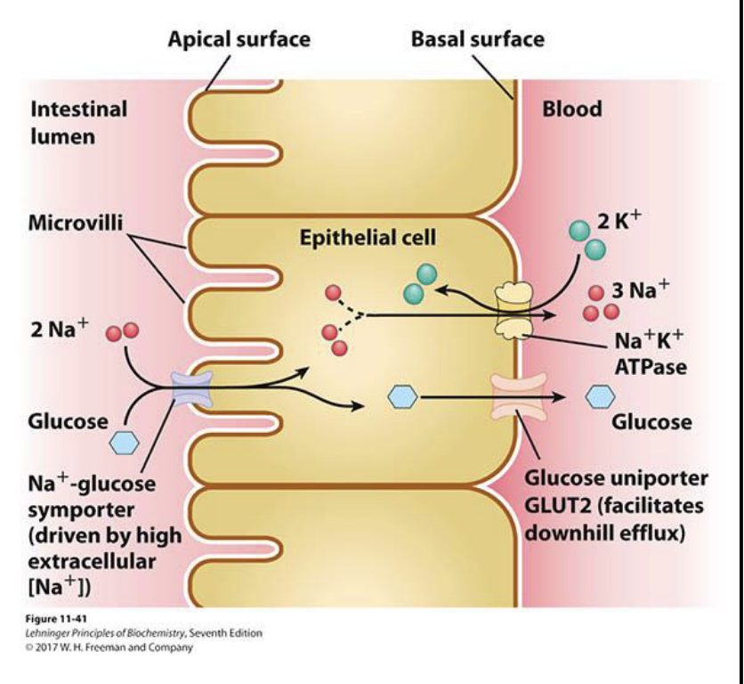
How is glucose absorbed from the intestine into the bloodstream using secondary active transport?
Na⁺/K⁺ ATPase creates a high Na⁺ concentration outside the intestinal epithelial cell.
Glucose and Na⁺ are co-transported (symport) into the cell from the gut lumen.
This is secondary active transport, powered by the Na⁺ gradient.
How does glucose move from the epithelial cell into the bloodstream?
Glucose exits the cell via GLUT2, a passive uniporter.
This is facilitated diffusion, moving glucose down its concentration gradient into the blood.
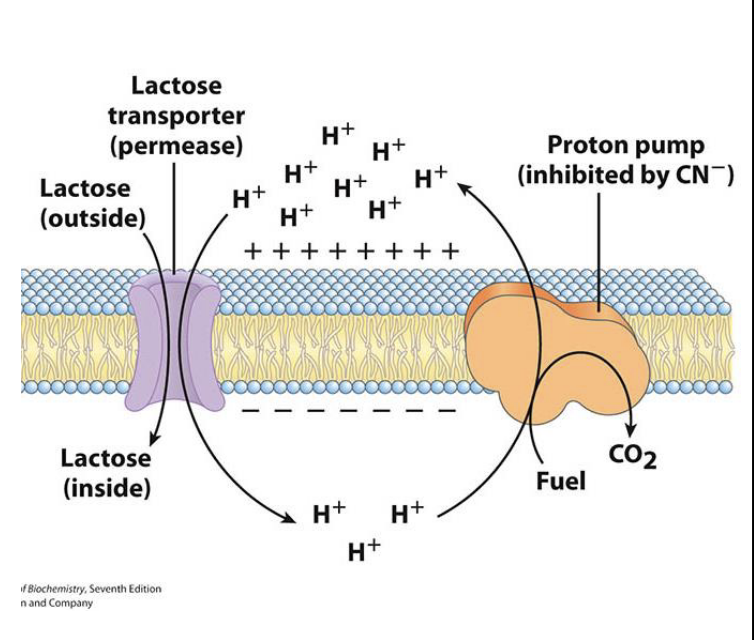
How is lactose taken up in E. coli using secondary active transport?
A proton pump uses energy from metabolic fuels to pump H⁺ out of the cell.
Lactose is co-transported into the cell with H⁺ as H⁺ flows back in down its gradient.
This allows lactose to enter the cell against its concentration gradient.
How do we know lactose uptake in E. coli is coupled to proton pumping
Cyanide (CN⁻) inhibits the proton pump.
When the pump is blocked, lactose transport stops, showing the process is energy-dependent and coupled to the H⁺ gradient.
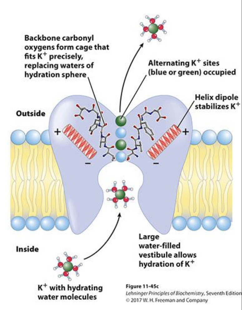
What are ion channels and what is an example of a non-voltage gated one?
Ion-selective membrane proteins that allow specific ions to cross the membrane.
Present in plasma membrane of all cells, they regulate ion flow for electrical and osmotic balance.
Example: A non-voltage gated K⁺ channel allows K⁺ ions to pass through selectively without needing a voltage signal.
How does a K⁺ ion pass through a non-voltage gated K⁺ ion channel, and how is selectivity achieved?
K⁺ enters the channel with its hydration shell.
The ion is desolvated (loses water) due to interactions with the channel protein.
Helix dipoles and backbone carbonyl groups stabilize the bare K⁺ ion.
The selectivity filter has four binding sites, each occupied by either K⁺ or water, allowing selective and efficient K⁺ transport.
How do voltage-gated ion channels open and close in response to membrane potential?
At rest, the cytosol is negative, pulling on positively charged Arg residues in the S4 helix, which keeps the channel closed.
When the membrane depolarizes (inside becomes less negative), repulsion between the now positive cytosolic charge and the Arg residues causes the channel to open, allowing K⁺ ions to exit the cell.
potassium ion channel
primary active transport
secondary active transport - glucose
secondary active transport - lactose