UT 505 Quiz 1 Study Guide
1/40
Earn XP
Description and Tags
UT 505 - MSK
Name | Mastery | Learn | Test | Matching | Spaced | Call with Kai |
|---|
No analytics yet
Send a link to your students to track their progress
41 Terms
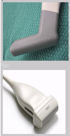
What types of TDRs are used for MSK exams?
“Hockey stick” TDR
L12 MHz
Long axis (LAX)
Longest plane of longest anatomy
Short axis (SAX)
Shortest axis of longest anatomy
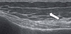
Anisotropy
Structure appears differently depending on the angle of insonation
Greatly affects tendons → can make normal tendon look pathogenic
Isotropy
Uniformity in all orientations
Articular surface
Two bones forming a joint opposite one another
Joint capsule
Fibrous tissue encompassing a joint
Abduction
Moving away from the midline
Adduction
Moving toward the midline
Floating
TDR is hovering over an abundance of acoustic gel to make “contact” while avoiding excessive pressure on structure
Enthesis
Site of attachment of a ligament or muscle to bone
Retinaculum
Band-like structure that binds tissue or organs to hold them in place
Muscle
Expands and contracts to create motion
3 types of muscles
Smooth (involuntary)
Cardiac striated
Skeletal striated
Appearance of muscles on US
LAX
Homogeneous with multiple parallel echoes
“Feather” appearance - striated pennate appearance
Hypoechoic muscle fibers arranged in columns interspersed by hyperechoic fibroadipose tissue
SAX
Homogeneous with punctuate echoes
Muscle pathology that can be seen on US
Contusion/hematoma/tear
Masses
Ligament
Attaches bone to bone, provides stability and strength, and aids in load distribution
Collagen fibers arranged in basket-weave pattern
US appearance of ligaments
Hyperechoic linear bands
Less prominent bands than tendons
No muscle component
Bursa
Synovial-lined pouches located at high friction joints
Aids in reducing friction between tendons/muscles and opposing structures
Potential space containing 1-3 mm of synovial fluid
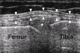
Types of cartilage
Fibrocartilage
Articular cartilage
Fibrocartilage
Found in the menisci (knee) and intervertebral disc spaces (back)
Function - shock absorption
Appearance - homogeneous and medium echogenicity
Articular cartilage
Found at terminal ends of bone at joints
Adheres to bone surface
Controls articular joint fluid level
Function - minimizes friction and aids in weight distribution and compression
Articular cartilage US appearance
Hypoechoic noncompressible layer in contact with bone surface
US appearance of subcutaneous fat
Heterogeneous with hypoechoic fat lobules
Subcutaneous fat pads
Location: articular surfaces where bone contacts adjacent bone surface
Ex: Hoffa fat pad at knee, Kager’s fat pad at heel
US appearance: hyperechoic and homogeneous
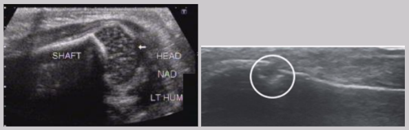
Bone
Most fundamental landmark when scanning
Attachment site for tendons, cartilage, ligaments, and joint capsules
US appearance of bone
Non-articular - sharp distinct hyperechoic line (bright reflector)
Articular - covered by cartilage (hypoechoic)
Should be smooth surface
Large gap may mean a break
Small gap is likely a nutrient channel
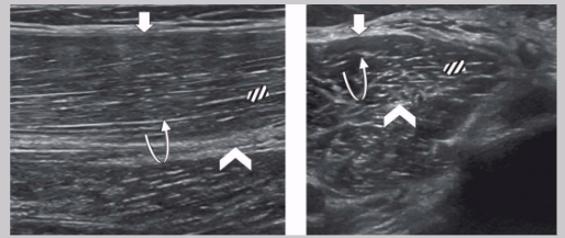
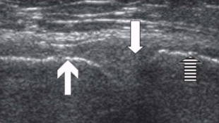
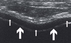
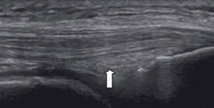
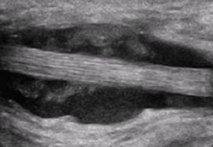
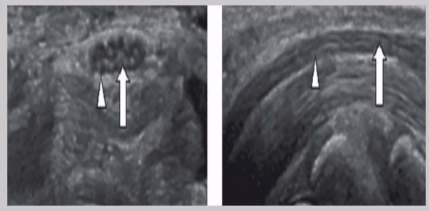
Tendon
Extension of muscles that attaches muscle to bone
Function - aid flexion and extension through lever mechanism
US appearance depends on insonation angle
> 10 degree off perpendicular → loss or lowering of echogenicity → false appearance of tendon pathology
US appearance of tendons
LAX
Linear band of hyperechoic strands and fibrillar pattern, interspersed with relatively hypoechoic connective tissue
SAX
“Whisk-broom” appearance
Hyperechoic foci throughout tendon distribution interspersed by hypoechoic connective tissue
Must maintain uniform thickness throughout except at site of insertion (might broaden)
Tendon pathology
Tendinopathy
Tendinosis
Tendinitis
Tenosynovitis
Tendinopathy
Generalized term for chronic tendon issues
Tendinosis
Progressive degeneration of tendon tissues → fraying/damage to tissue → tendon thickening
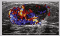
Tendinitis
Tendon-related pain and symptoms associated with inflammation → increased vascularity
Tenosynovitis
Inflammation of sheath surrounding tendon → presents as fluid accumulation around tendon
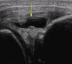
Nerves
Sensory and motor function by sending information throughout body
Bundle of nerve fibers - fascicle
Nerve appearance on US
Nerve fiber enclosed by hyperechoic epineurium
LAX
“Railroad” or “tram track”
Hypoechoic nerve fibers divided by hyperechoic perineurium
SAX
“Honeycomb” pattern
Hypoechoic nerve fibers surrounded by hyperechoic perineurium
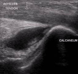
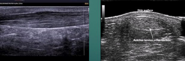
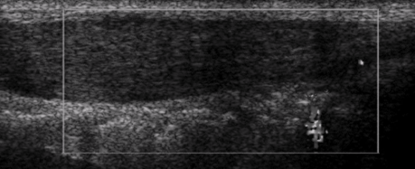
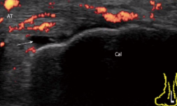
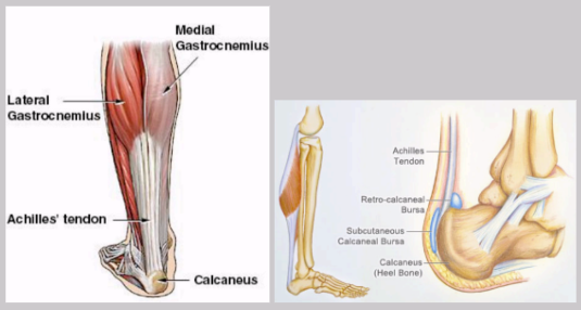
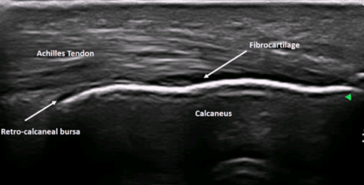
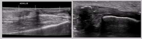
Achilles tendon
Strongest and thickest tendon in the body
Attachments
Proximal - gastrocnemius and soleus muscles
Distal - calcaneus (heel bone)
Normal US appearance of Achilles tendon
Common Achilles tendon pathology
Tendinopathy
Partial or complete tears
Retrocalcaneal bursitis
Partial or complete Achilles tendon tears
Retrocalcaneal bursitis