1. Cardiovascular system
1/34
There's no tags or description
Looks like no tags are added yet.
Name | Mastery | Learn | Test | Matching | Spaced |
|---|
No study sessions yet.
35 Terms
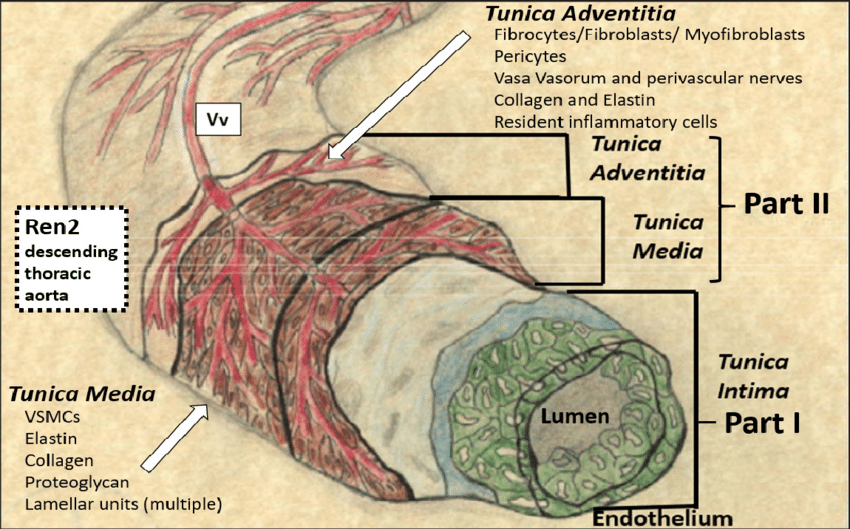
1. Fenestrated elastic membranes are typical for the arteries
of muscle type.
A.Yes
B.No
No
Fenestrated elastic membranes (also called elastic lamellae) are typical of elastic arteries, not muscular arteries.
Elastic arteries (e.g., aorta, pulmonary trunk) have multiple concentric layers of fenestrated elastic lamellae in the tunica media to help them stretch and recoil with each heartbeat.
Muscular arteries have a prominent internal elastic lamina and sometimes an external one, but they do not have multiple fenestrated elastic membranes in the media.
Fenestrations are small pores or openings in a structure — in histology, the term most often refers to pores in membranes, especially endothelial cells or elastic lamellae
The fenestrations allow:
Diffusion of nutrients and gases to smooth muscle cells in the tunica media.
Flexibility and elastic recoil without becoming too rigid.
2. Continuous capillaries consist of an endothelial layer, its underlying basal lamina and associated pericytes within the basal lamina.
A.Yes
B. No
No
3. Pericytes are associated cells within the basal lamina of the endothelial cells of capillaries.
A. Yes
B. No
Yes
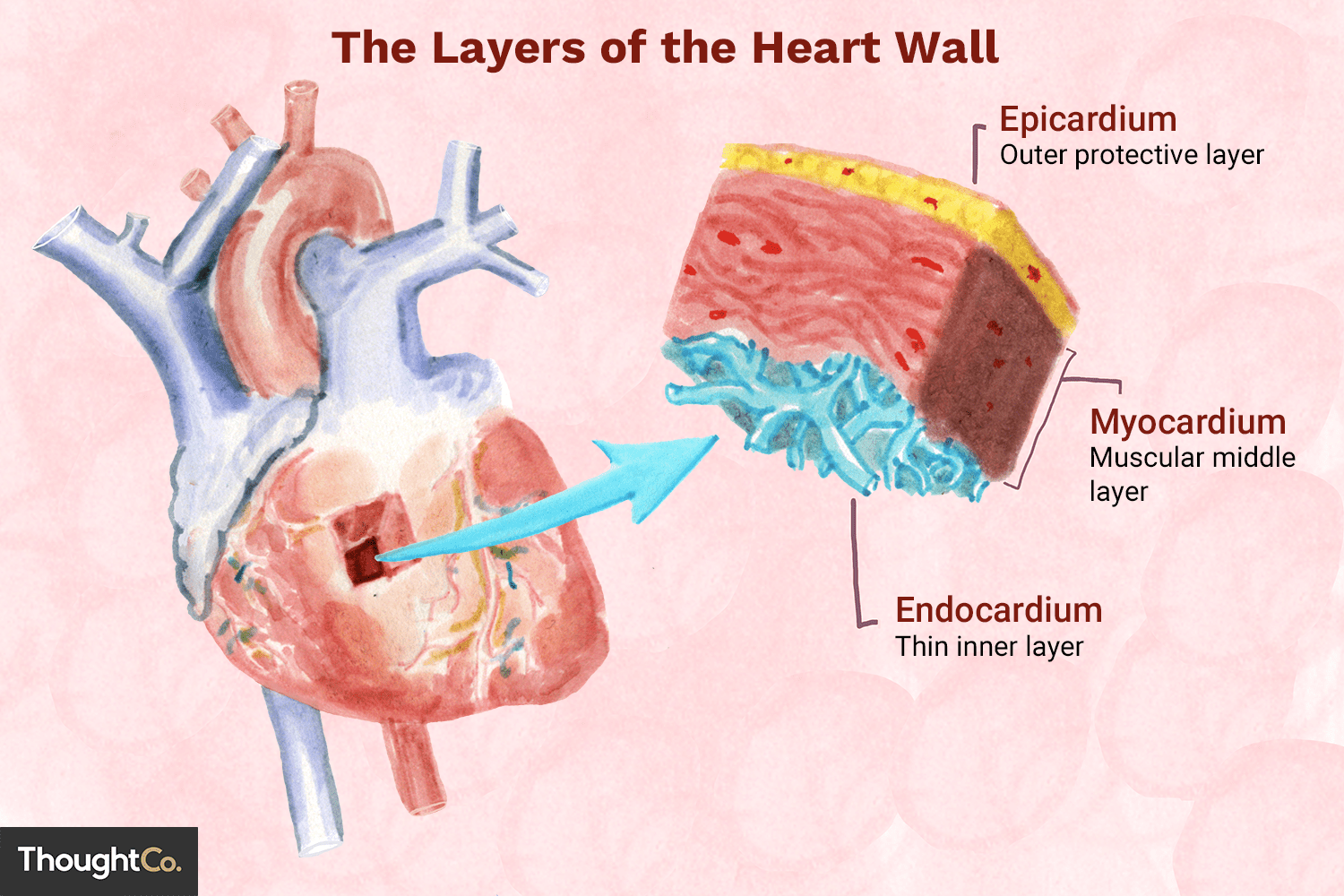
4. Adipose tissue is found in the epicardium.
A.Yes
B.No
Yes
5. In the region of fossa ovalis in septum interatriale there is no myocardium.
A. Yes
B. No
Yes
fossa ovalis is a depression in the interatrial septum — the wall between the right and left atria.
fossa ovalis is thin, with a fibrous composition
It is a remnant of the foramen ovale, an essential fetal structure.
In the fetus, the foramen ovale is an opening that allows blood to bypass the lungs by flowing from the right atrium to the left atrium.
This is crucial because fetal lungs are non-functional, and oxygen comes from the placenta.
After birth, when the baby starts breathing, pulmonary resistance drops, and left atrial pressure rises.
This increased pressure forces the septum primum against the septum secundum, functionally closing the foramen ovale.
This fused area forms the fossa ovalis
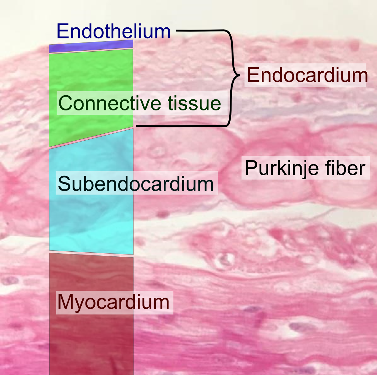
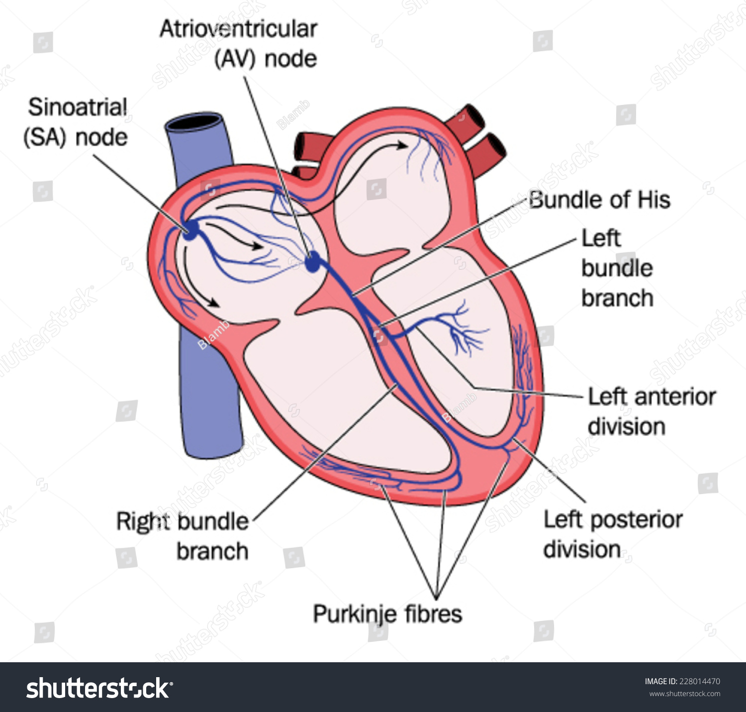
6. The atrioventricular node is situated in its subendocardial location.
A. Yes
B.No
Yes
atrioventricular (AV) node is located in the subendocardial layer of the interatrial septum, near the opening of the coronary sinus
“Subendocardial" means just beneath the endocardium - inner lining of the heart chambers.
7. The valves of the veins are double folding of tunica intima.
A.Yes
B.No
Yes
8. The right ventricle of the heart at cross section has semilunar shape.
A.Yes
B. No
Yes
9. There is adipose tissue in the heartepicardium.
A. Yes
B. No
Yes
10. The left ventricle of the heart at cross section has semilunar shape.
A. Yes
B.No
No
left ventricle in cross-section has a circular or oval shape, not semilunar.
It has thicker walls than the right ventricle due to higher pressure needed to pump blood through the systemic circulation.
The right ventricle, on the other hand, has a more crescent (semilunar) shape in cross-section because it wraps partially around the left ventricle.
11. There is adipose tissue in the heart endocardium.
A. Yes
B. No
No
endocardium is the innermost layer of the heart wall and consists mainly of endothelial cells and connective tissue — not adipose tissue.
Adipose tissue is typically found in the epicardium (outer layer), especially along the coronary vessels.
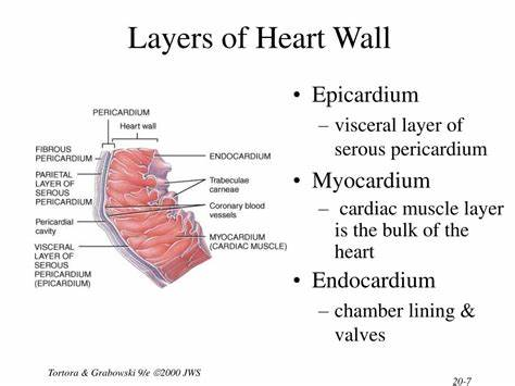
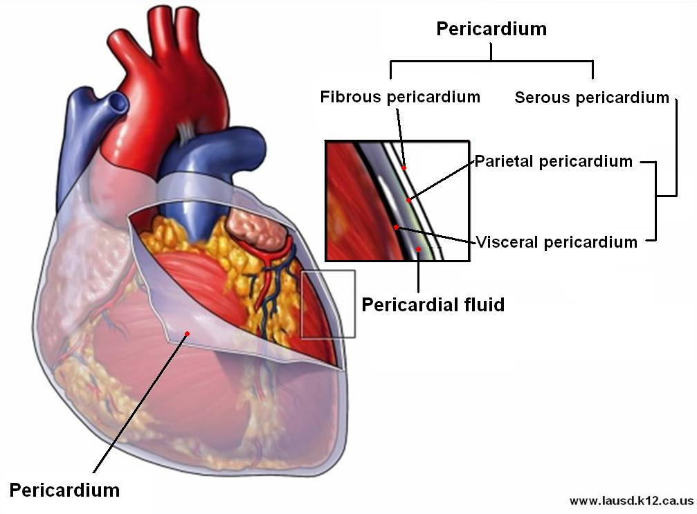
12. The lining of the inner walls of the heart's chambers is termed the visceral pericardium.
A. Yes
B. No
No
lining of the inner walls of the heart chambers is called the endocardium
Endocardium:
Lines the inside of the heart chambers
Made of endothelium and connective tissue
Continuous with the lining of blood vessels
Visceral pericardium (also called the epicardium):
Covers the outer surface of the heart
Part of the pericardial sac
Contains fat, vessels, and mesothelium
13. Subendothelium of blood vessels is:
A. loose connective tissue without blood vessels
B. loose connective tissue with blood vessels
C. loose connective tissue with glands
D. muscle tissue
A. loose connective tissue without blood vessels
14. Tunica media of the aorta is made of:
A. muscle tissue
B. elastic and muscle tissue
C. fenestrated elastic membranes
C. fenestrated elastic membranes
The tunica media of the aorta (an elastic artery) is primarily composed of multiple concentric layers of fenestrated elastic membranes (lamellae).
These elastic membranes allow the aorta to stretch during systole and recoil during diastole, maintaining continuous blood flow.
While smooth muscle cells are present, the dominant feature in the aorta’s tunica media is the fenestrated elastic lamellae, especially in the proximal aorta.
15. If the heart's natural pacemaker fails to fire, then:
A. no blood would enter the atria
B. no blood would enter the ventricles
C. the node on the floor of the right atrium would act as a secondary pacemaker
D. the node on the floor of the left ventricle would act as a secondary pacemaker
E. the person would die within minutes
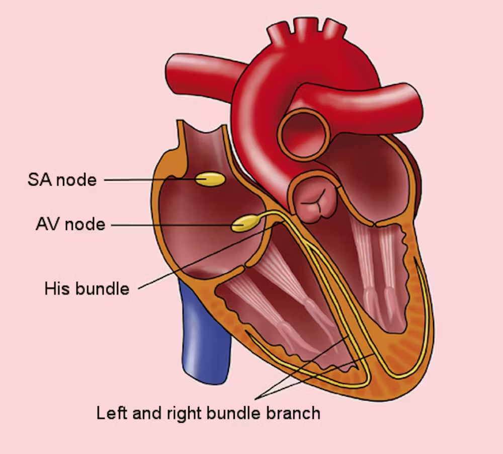
C. the node on the floor of the right atrium would act as a secondary pacemaker
If the sinoatrial (SA) node — the heart's natural pacemaker — fails to fire, the next in line is the atrioventricular (AV) node, which is located on the floor of the right atrium, near the opening of the coronary sinus.
The AV node can generate impulses on its own, though more slowly (~40–60 bpm).
This acts as a backup pacemaker, helping maintain cardiac rhythm, though less efficiently.
16. Which tunic of an artery contains endothelium?
A. tunica interna/intima
B. tunica media
C. tunica externa
A. tunica interna/intima
17. The exchange of gases and nutrients between blood and tissues is a major function of:
A. arterioles
B. arteries
C. capillaries
D. veins
C. capillaries
18. Which of the following statements best describes arteries?
A. all arteries carry oxygenated blood towards the heart
B. all arteries contain valves to prevent the back-flow of blood
C. all arteries carry blood away from the heart
D. only large arteries are lined with endothelium
C. all arteries carry blood away from the heart
19. The circulatory pathway that carries blood from the digestive tract towards the liver is termed the:
A. coronary circuit
B. cerebral circuit
C. hepatic portal circuit
D. pulmonary circuit
C. hepatic portal circuit
20. Immediately following strenuous and vigorous exercise, which of the following is most likely to occur?
A. blood will be rapidly diverted to the digestive organs
B. the skin will be cold and clammy
C. capillaries of the active muscles will be engorged with blood
D. blood flow to the kidneys quickly increases
C. capillaries of the active muscles will be engorged with blood
the body prioritizes increased blood flow to active skeletal muscles to supply oxygen and nutrients and remove waste products like CO₂ and lactate.
Capillaries in active muscles dilate and become engorged with blood — this improves oxygen delivery and metabolic exchange
21. The lining of the innerwalls of the heart's chambers is termed the:
A. visceral pericardium
B. serous pericardium
C. epicardium
D. myocardium
E. endocardium
E. endocardium
The endocardium is the innermost layer of the heart wall that lines the inner surfaces of all four chambers and the valves.
It is composed of endothelium (simple squamous epithelium) and a thin layer of connective tissue.
It’s continuous with the endothelium of blood vessels.
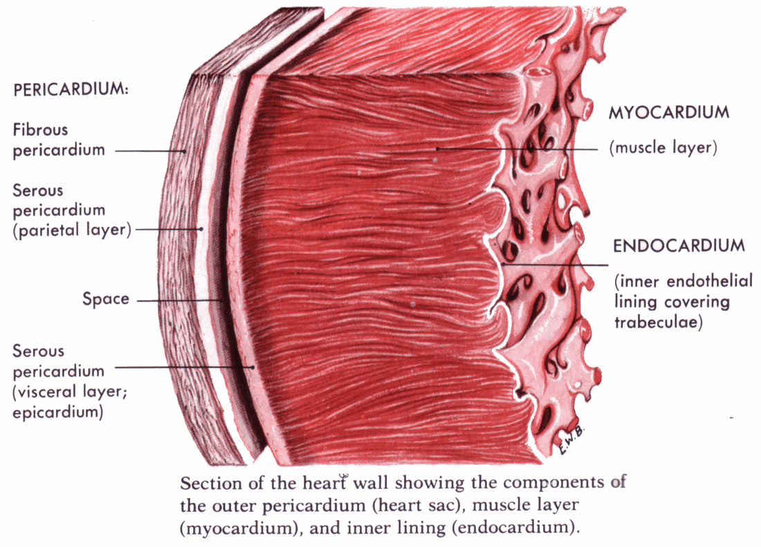
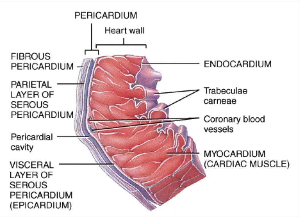
22. The outermost layer of the heart's serous pericardium is termed the:
A. visceral pericardium
B. parietal pericardium
C. epicardium
D. myocardium
E. endocardium
B. parietal pericardium
The serous pericardium has two layers:
Parietal pericardium – outermost layer
Lines the inner surface of the fibrous pericardium
This is the correct answer
Visceral pericardium (aka epicardium) – inner layer
Directly adheres to the surface of the heart
Between them is the pericardial cavity, filled with fluid for frictionless movement.
23. The heart's natural pacemaker is termed the:
A. sinoatrial node
B. atrioventricular node
C. bundle of His/atrioventricular bundle
D. left and right bundle branches
E. Purkinje fibers
A. sinoatrial node
located in the right atrium and is known as the heart’s natural pacemaker
generates electrical impulses that initiate each heartbeat.
impulses spread through the atria, causing them to contract.
AV node (B): Delays the impulse before passing it to the ventricles.
Bundle of His (C), bundle branches (D), and Purkinje fibers (E): Conduct impulses through the ventricles, but do not initiate the heartbeat
24. What is most responsible for propelling blood in the arterial system during cardiac diastole?
A. skeletal muscle contraction and breathing
B. hydrostatic blood pressure arising from ventricular contraction
C. elastic recoil of conducting (elastic) arteries
D. venous return of blood
C. elastic recoil of conducting (elastic) arteries
25. The basal membrane of the fenestrated capillaries is ••••••
continuous
The basement (basal) membrane of fenestrated capillaries is continuous, even though the endothelial cells have pores (fenestrations).
These fenestrations allow for rapid exchange of small molecules (e.g., in kidneys, intestines, endocrine glands).
However, the basal lamina remains intact and continuous, acting as a selective filter and structural support.
26. The layers of the endocardium are:
A.
B.
C.
D.
A. Endothelial
B. Subendothelial
C. Myoelastic
D. Subendocardial
27. The capillaries are:
A.
B.
C.
A. Continuous
B. Fenestrated
C. Discontinuous
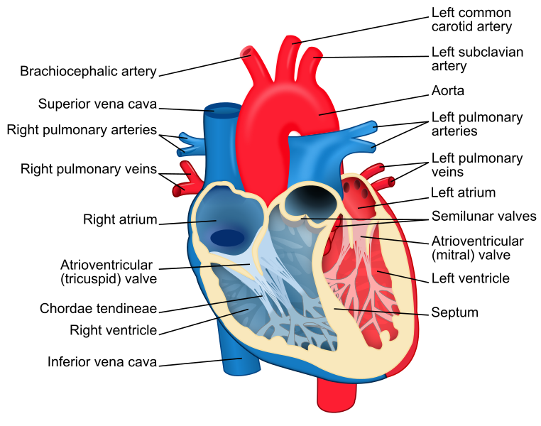
28. The valves between the atria and ventricles in the heart are:
A.
B.
A. Bicuspidal or mitral
B. Tricuspidal
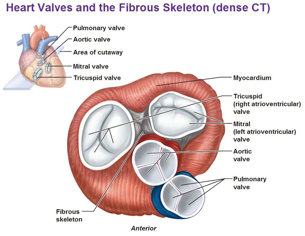
29. The fibrous skeleton of the heart includes:
A.
B.
C.
D.
A. Right fibrous ring
B. Left fibrous ring
C. Right fibrous trigone
D. Left fibrous trigone
30. The arteries which supply the heart wall with blood are:
A.
B.
A. Left coronary artery
B. Right coronary artery
T/ F
31. Typical for myocardium is:
A. 3 types of muscle tissue cells-cardiac myocytes, Purkinje cells, Myocardial endocrine cells
B. Myocardium of the ventricles is organized in 2 layers
C. Myocardium of the ventricles is organized in 3 layers
31. Typical for myocardium is:
A. 3 types of muscle tissue cells-cardiac myocytes, Purkinje cells, Myocardial endocrine cells TRUE
✅ True
The myocardium includes:Cardiac myocytes (contractile cells)
Purkinje fibers (specialized for conduction)
Myocardial endocrine cells (mainly in the atria, secrete ANP)
B. Myocardium of the ventricles is organized in 2 layers FALSE
❌ False
The ventricular myocardium is organized in 3 layers:Superficial (spiral)
Middle (circular, mainly in the left ventricle)
Deep (longitudinal)
C. Myocardium of the ventricles is organized in 3 layers TRUE
T/ F
32. The elements of the fenestrated capillaries are:
A. Endothelial cells
B. Basal membrane - interrupted
C. Basal membrane - non-interrupted
D. Pericytes
T/ F
32. The elements of the fenestrated capillaries are:
A. Endothelial cells TRUE
B. Basal membrane - interrupted FALSE
C. Basal membrane - non-interrupted TRUE
D. Pericytes TRUE
Match
33.
A. Fossa ovalis
B. Chordae tendinae
C. Trabeculae carneae
D. Mm. Papillares
E. Mm. Pectinati
//
1. Atrium
2. Ventricles
Atrium
A. Fossa ovalis
Located in the interatrial septum (right atrium side)
E. Mm. Pectinati
Parallel ridges found mainly in the right atrium, especially the auricle
Ventricles
B. Chordae tendinae
Fibrous cords that connect AV valve leaflets to papillary muscles
C. Trabeculae carneae
Irregular muscular ridges on the ventricular walls
D. Mm. Papillares
Muscular projections that anchor chordae tendineae
Match
34.
A. Ostium of v. cava inferior
B. Ostium of vv pulmonales
C. Conus arteriosus
D. Ostium aortae
//
1. Right ventricle
2. Left ventricle
3. Right atrium
4. Left atrium
A. Ostium of v. cava inferior // Right atrium
inferior vena cava opens into the right atrium, bringing deoxygenated blood from the lower body.
B. Ostium of vv pulmonales // Left atrium
pulmonary veins open into the left atrium, delivering oxygenated blood from the lungs
C. Conus arteriosus // Right ventricle
conus arteriosus (infundibulum) is the outflow tract of the right ventricle leading to the pulmonary trunk
D. Ostium aortae // Left ventricle
aortic orifice is where blood leaves the left ventricle to enter the aorta
Match
35.
A. Endothelial cells
B. Basal membrane - interrupted
C. Basal membrane - non-interrupted
D. Pericytes
E. Fenestrae
//
1. Fenestrated capillaries
2. Continuous capillaries
3. Non-continuous, interrupted capillaries
A. Endothelial cells // Fenestrated capillaries + Continuous capillaries + Non-continuous, interrupted capillaries
B. Basal membrane - interrupted // Non-continuous, interrupted capillaries
(aka sinusoidal)
Found in liver, spleen, bone marrow — allows large molecules and even cells to pass.
C. Basal membrane - non-interrupted // Fenestrated capillaries + Continuous capillaries
Endothelium has pores, but basement membrane remains continuous.
D. Pericytes // Fenestrated capillaries + Continuous capillaries
contractile support cells are associated primarily with continuous capillaries.
E. Fenestrae // Fenestrated capillaries
Fenestrae = pores in the endothelial cells for faster exchange (e.g., kidney, endocrine glands).