The External Skull
1/5
There's no tags or description
Looks like no tags are added yet.
Name | Mastery | Learn | Test | Matching | Spaced | Call with Kai |
|---|
No analytics yet
Send a link to your students to track their progress
6 Terms
Describe the anatomical division of the skull into Neurocranium and Viscerocranium.
Neurocranium
Bony case enclosing the brain, meninges, proximal cranial nerves and cerebral vasculature
Superior portion “calvaria” - consists of mostly flat bones
Floor “basicranium'“ - composed of irregular bones
Viscerocranium
Facial skeleton
Maxilla provides sockets for upper teeth
Mandible (only moveable skull bone) provides sockets for lower teeth
Mandible articulates with cranial base at TMJ
Describe the bony walls of the orbit, listing which bones contribute to each wall.
Roof: F-L “Flying Low”
Frontal and Lesser wing of sphenoid
Lateral Wall: Z-G “Zero Gravity”
Zygomatic and Greater wing of sphenoid
Medial Wall: M-L-E-S “My Little Eyes See”
Maxilla, Lacrimal, Ethmoid, Sphenoid (body)
Floor: M-Z-P “My Zesty Pizza”
Maxilla, Zygomatic, Palatine
List the 4 major foramina visible on the facial aspect of the skull, stating the bone each lies in and the structures transmitted.
Three of these sit in a perfect line passing through the pupil. Just follow the CN V divisions from top to bottom:
Foramen
Supraorbital (above eye)
Bone
Frontal
Trigeminal Division
V1 (ophthalmic)
Foramen
Infraorbital (below eye)
Bone
Maxilla
Trigeminal Division
V2 (maxillary)
Foramen
Mental (on chin)
Bone
Mandible
Trigeminal division
V3 (mandibular)
The Zygomaticofacial Foramen is the only one off to the side
Bone: Zygomatic
Structures Transmitted: Zygomaticofacial Nerve, Artery and Vein
Nerve Origin: Branch of V2 (same as Infraorbital)
A patient presents with numbness of the cheek below the orbit following a midface fracture. Which foramen is likely involved and which nerve is injured?
Injury Site: Infraorbital Foramen (located in the maxilla), just below the eye socket)
Nerve Involved: Infraorbital Nerve
Nerve Origin: A terminal branch of the Maxillary Nerve (V2) which is the second division of the Trigeminal Nerve (CN V)
Sensory Distribution:
Lower eyelid
Lateral nose
Cheek (below the orbit)
Upper lip
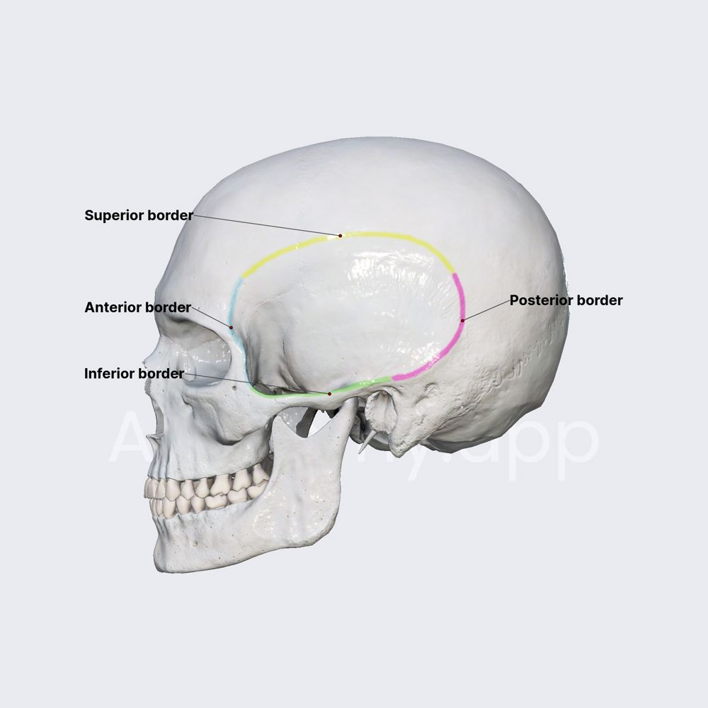
Describe the Temporal Fossa.
The Temporal Fossa
Shape: Fan-shaped depression on the lateral skull
Primary Occupant: Temporalis Muscle (a major muscle of mastication)
The “Box” Boundaries
Superior and Posterior: Superior temporal line
Inferior: Zygomatic arch
Anterior: Frontal bone and Zygomatic bone
Lateral: Temporal fascia
Medial: The pterion (where the frontal, parietal, temporal and sphenoid bones meet)
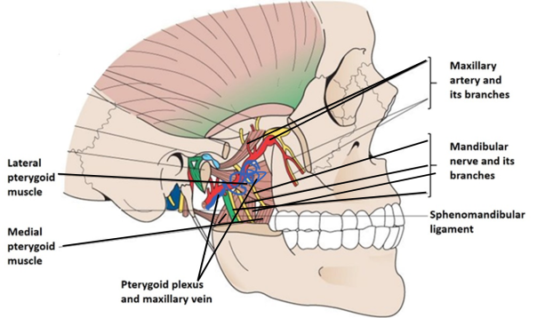
Describe the Infratemporal Fossa.
Where is it?
Medial to: Jawbone (Ramus of the Mandible)
Deep to: Cheekbone (Zygomatic Arch)
What’s inside? (The “3 M’s”)
Muscles: The Lateral and Medial Pterygoids and the bottom of the Temporalis
Mandibular Nerve (V3): The nerve that gives sensation to lower teeth and chin
Maxillary Artery: The main artery supplying blood to the face and teeth
How do things get in?
The Ceiling: Foramen Ovale (hole where the V3 nerve drops down from the brain)
The Side Door: Pterygomaxillary Fissure