Looks like no one added any tags here yet for you.
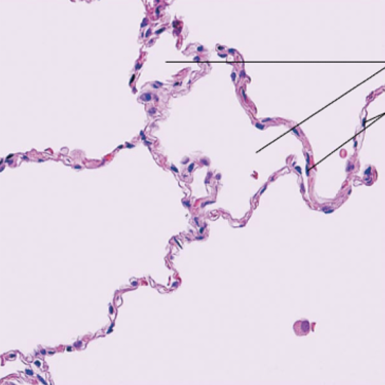
simple squamous epithelia (lymphatic)
description:
-single layer of flat, disc-like cells
-flattened nuclei
-functions as surface for filtration and diffusion
Location:
-air sacs of lungs→alveoli
-glomeruli
-blood vessel and capillary lumens
-body cavity linings
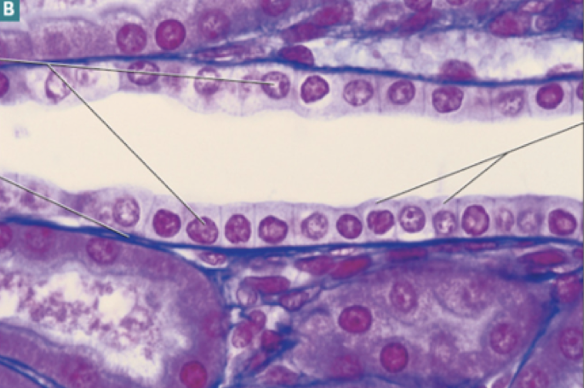
simple cuboidal epithelia
description:
-single layer of cube-like cells
-large, round nuclei
-responsible for secretion and absorption
location:
-ducts and glands
-kidney tubules
-ovary
-thyroid
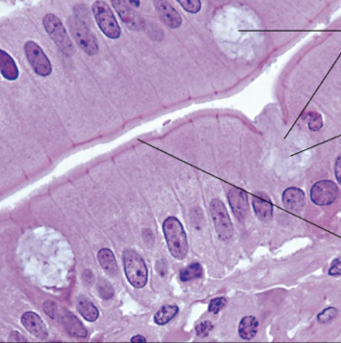
simple columnar epithelia (intestinal)
description:
-single layer of tall, rectangular cells
-elongated nuclei
-microvilli (absorption) and cilia (propulsion) common on apical surface
-function in absorption (digestive tract) and secretion (mucous, reproduction)
location:
-digestive tract lining
-respiratory tract lining
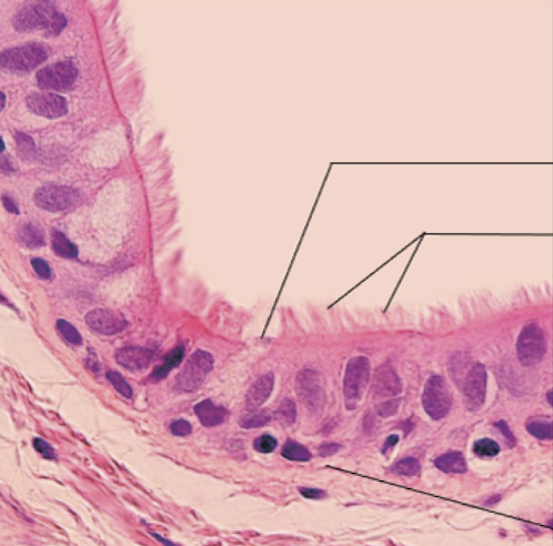
psuedostratified columnar epithelia
description:
-single layer of columnar cells of different heights
-nuclei appear at different heights
-have appearance of multiple layers
-function in secretion and propulsion (can be ciliated)
location:
-male sperm duct
-respiratory tract
-large glands
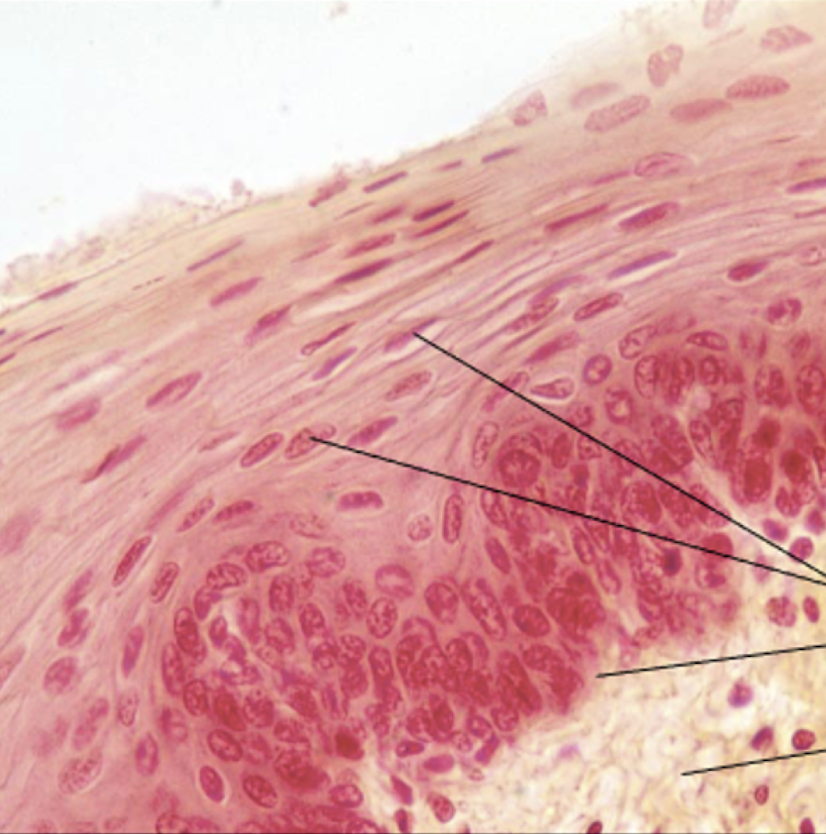
stratified squamous epithelia (keratinized and nonkeratinized)
description:
-multiple layers of squamous cells on a basement membrane
-surface cells are dead, flat and keratinized
-basal cells are cube-shaped and mitotically active
-form protection against abrasion and damage for tissue underneath
location:
-skin (keratinized)
-lining of esophagus
-lining of vagina
-lining of mouth
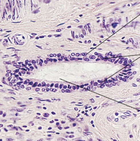
stratified cuboidal epithelia
description:
-generally two layers of cubelike cells
-protection
location:
-largest ducts of sweat glands, mammary glands, and salivary glands
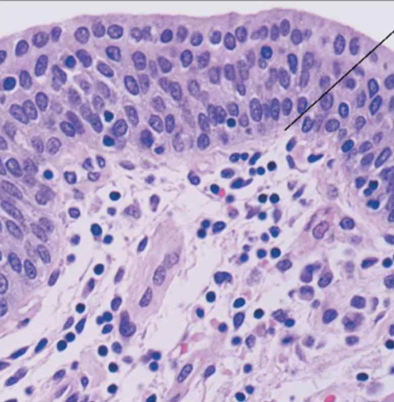
stratified columnar epithelia
description:
-several cell layers, basal cells usually cuboidal
-superficial cells elongated and columnar
-protection; secretion
location:
-rare in the body; small amounts in the male urethra
-in large ducts of some glands
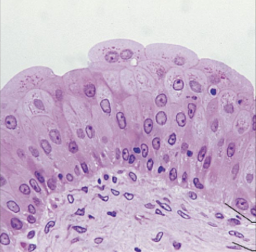
transitional epithelia
description:
-appears to be stratified squamous or cuboidal
-basal cells are cuboidal
-surface cells are rounded/squamous
-stretch permitted, enabling storage of urine
location:
-lining of urinary tracts such as:
-ureter
-bladder
-urethra
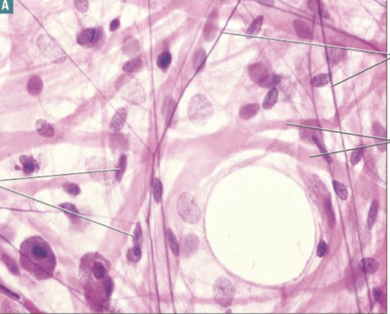
areolar loose connective tissue
description:
-loosely-packed assembly of all fiber types, fibroblasts, and immune cells
-main function: cushions organs
-also contains key immune mediators to fight off infections
location:
-supporting under epithelial layers and surrounding organs
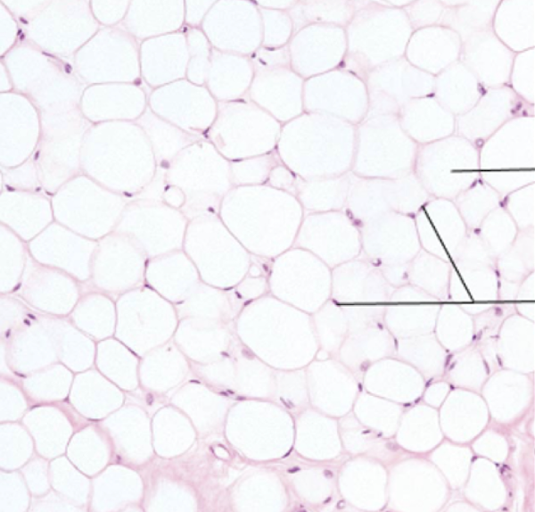
adipose loose connective tissue
description:
-matrix crowded by tightly-packed adipocytes
-serves to insulate and protect organs
-provides energy storage depot
location:
-around major organs
-within subcutaneous layer of skin (under dermis)
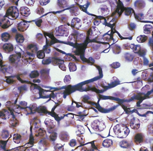
reticular loose connective tissue
description:
-meshwork of reticular fibers loosely-organized
-forms a flexible meshwork
-supports tissue and immune cells
location:
-lymph nodes
-bone marrow
-splenic pulp
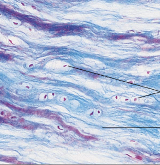
regular dense connective tissue
description:
-fibroblasts embedded within a regularly-ordered assembly of collagen fibers
-functions to resist pulling stress
location:
-attaches muscle to bone (tendon)
-attaches bone to bone (ligament)
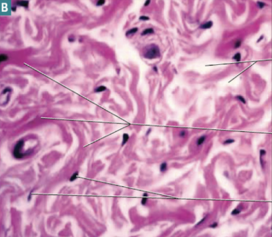
irregular dense connective tissue
description:
-irregularly-arranged collagen fibers with fibroblasts embedded within
-resists force in many directions
location:
-dermis
-joint capsules
-underlying epithelial linings such as in the digestive tract
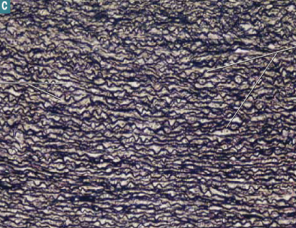
elastic dense connective tissue
description:
-dense regular connective tissue enriched with elastin fibers
-ordered arrangement of elastin
-allows for stretch and recoil of tissue
location:
-walls of arteries
-walls of bronchial tubes
-some ligaments
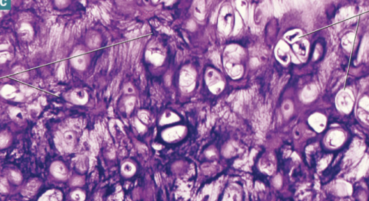
hyaline cartilage connective tissue
description:
-flexible, firm gel matrix secreted by chondroblasts
-this becomes embedded in lacunae (chondrocytes)
-functions to cushion, support and reinforce other tissues and organs
location:
-nose
-trachea
-ribs
-ends of long bones
-embryonic skeleton
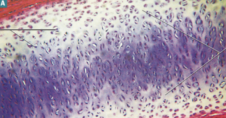
elastic cartilage connective tissue
description:
-same organization as hyaline cartilage
-enriched with elastin fibers
-has greater degree of flexibility and stretch
-allows for tissue to return to original shape
location:
-outer ear
-epiglottis
fibrocartilage connective tissue
description:
-firm hyaline matrix, with more organized collagen fibers
-resists compressive force
location:
-components of joints with limited flexibility, like intervertebral discs
-menisci
-pubic symphysis
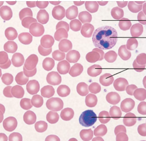
blood connective tissue
description:
-erythrocytes, leukocytes, and platelets within a plasma matrix
-primary function: transport of oxygen and carbon dioxide, nutrients and other dissolved molecules throughout the body
location:
-contained within arteries, veins and capillaries
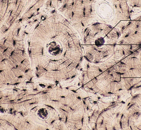
bone connective tissue
description:
-calcified collagen matrix embedded osteocytes
-cross talk among osteocytes
-high degree of vascularity
-functions in calcium storage, attachment for muscles
-spongy bone contains marrow, stem cells for blood formation
location:
-skeleton
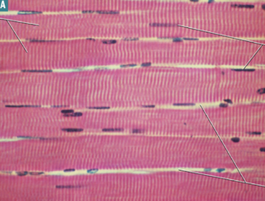
skeletal muscle tissue
description:
-elongated myotubes formed from fused cells
-multinucleated
-striations visible
-function in voluntary skeletal movement
location:
-attached to bones
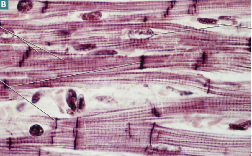
cardiac muscle tissue
description:
-elongated myotubes with greater degree of branching
-specialized cell junctions (intercalated discs) seen as deep red margins between cells
-individual nuclei evident
-striations visible
-ensure flow of blood to all body tissues
location:
-heart wall only
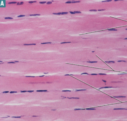
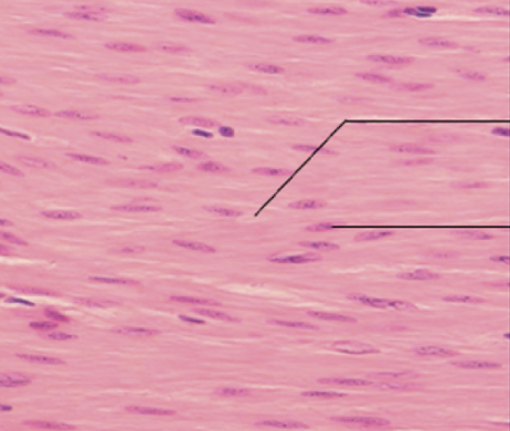
smooth muscle tissue
description:
-spindle-shaped cells with central nucleus
-found in layers that form sheets
-can provide force (e.g. during uterine contractions)
-can accommodate stretch (e.g. digestive system)
location:
-walls of hollow organs
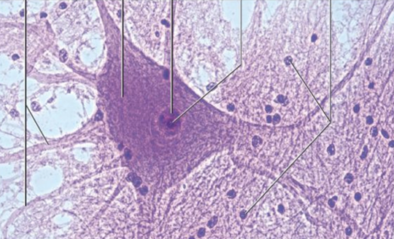
nervous tissue
description:
-neurons receive sensory information and carry it to the brain, transmit motor impulses from brain to effector organs, and form synapses with one another
-glial cells provide structural support, protection and nourishment for neurons in the brain and spinal cord
location:
-brain
-spinal cord
-cranial and peripheral nerves